Advertisement

Main menu

  • Home
  • Content
    • Current Issue
    • Accepted Manuscripts
    • Article Preview
    • Past Issue Archive
    • Video Articles
    • AJNR Case Collection
    • Case of the Week Archive
    • Case of the Month Archive
    • Classic Case Archive
  • Special Collections
    • AJNR Awards
    • Low-Field MRI
    • Alzheimer Disease
    • ASNR Foundation Special Collection
    • Photon-Counting CT
    • View All
  • Multimedia
    • AJNR Podcasts
    • AJNR SCANtastic
    • Trainee Corner
    • MRI Safety Corner
    • Imaging Protocols
  • For Authors
    • Submit a Manuscript
    • Submit a Video Article
    • Submit an eLetter to the Editor/Response
    • Manuscript Submission Guidelines
    • Statistical Tips
    • Fast Publishing of Accepted Manuscripts
    • Graphical Abstract Preparation
    • Imaging Protocol Submission
    • Author Policies
  • About Us
    • About AJNR
    • Editorial Board
    • Editorial Board Alumni
  • More
    • Become a Reviewer/Academy of Reviewers
    • Subscribers
    • Permissions
    • Alerts
    • Feedback
    • Advertisers
    • ASNR Home

User menu

  • Alerts
  • Log in

Search

  • Advanced search
American Journal of Neuroradiology

American Journal of Neuroradiology

ASHNR American Society of Functional Neuroradiology ASHNR American Society of Pediatric Neuroradiology ASSR
  • Alerts
  • Log in

Advanced Search

  • Home
  • Content
    • Current Issue
    • Accepted Manuscripts
    • Article Preview
    • Past Issue Archive
    • Video Articles
    • AJNR Case Collection
    • Case of the Week Archive
    • Case of the Month Archive
    • Classic Case Archive
  • Special Collections
    • AJNR Awards
    • Low-Field MRI
    • Alzheimer Disease
    • ASNR Foundation Special Collection
    • Photon-Counting CT
    • View All
  • Multimedia
    • AJNR Podcasts
    • AJNR SCANtastic
    • Trainee Corner
    • MRI Safety Corner
    • Imaging Protocols
  • For Authors
    • Submit a Manuscript
    • Submit a Video Article
    • Submit an eLetter to the Editor/Response
    • Manuscript Submission Guidelines
    • Statistical Tips
    • Fast Publishing of Accepted Manuscripts
    • Graphical Abstract Preparation
    • Imaging Protocol Submission
    • Author Policies
  • About Us
    • About AJNR
    • Editorial Board
    • Editorial Board Alumni
  • More
    • Become a Reviewer/Academy of Reviewers
    • Subscribers
    • Permissions
    • Alerts
    • Feedback
    • Advertisers
    • ASNR Home
  • Follow AJNR on Twitter
  • Visit AJNR on Facebook
  • Follow AJNR on Instagram
  • Join AJNR on LinkedIn
  • RSS Feeds

AJNR Awards, New Junior Editors, and more. Read the latest AJNR updates

LetterLetter

MR Spectroscopy of a Bright Cerebellar Lesion in a Patient with Langerhans Cell Histiocytosis

A. Munoz, R. Simon and G. Carmen
American Journal of Neuroradiology April 2008, 29 (4) e14; DOI: https://doi.org/10.3174/ajnr.A0911
A. Munoz
  • Find this author on Google Scholar
  • Find this author on PubMed
  • Search for this author on this site
R. Simon
  • Find this author on Google Scholar
  • Find this author on PubMed
  • Search for this author on this site
G. Carmen
  • Find this author on Google Scholar
  • Find this author on PubMed
  • Search for this author on this site
  • Article
  • Figures & Data
  • Info & Metrics
  • Responses
  • References
  • PDF
Loading

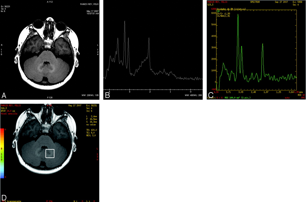
We have read with interest the paper by Prosch et al1 published in the American Journal of Neuroradiology concerning the long-term MR imaging course of 9 patients diagnosed with Langerhans cell histiocytosis (LCH). We were particularly interested in the evolution of MR abnormalities of cerebellum, pons, and basal ganglia structures. The authors observed no correlation between the progression of such abnormalities and clinical neurologic deterioration, and stated that the signal intensity persistence and progression were compatible with a neurodegenerative process, as reported by previous histopathologic studies. Furthermore, symmetric hyperintensities on T1-weighted images observed in these patients were first reported in other diseases, such a chronic liver failure. Such abnormalities could be found in up to 10% of patients with LCH. We have had the opportunity to study a patient similar to those presented in their report, a 3-year-old boy who was diagnosed with LCH because of diabetes insipidus and was treated only with antidiuretic hormone replacement and followed, both clinically and radiologically, over a course of 7 years. In the first MR imaging study, abnormalities encountered in our patient were those typically found in the pituitary-hypothalamic area, in patients with LCH and positive granulomata involvement, consisting of a lack of a hyperintense neurohypophyseal spot on noncontrast T1-weighted images and thickening and enhancement of the pituitary stalk and adjacent hypothalamic area. During this period of time, the MR enhancing pituitary lesions resolved with almost complete resolution. However, new progressive different lesions appeared, mainly in the basal ganglia and cerebellum, similar to those described in the report by Prosch et al.1 In the last study, we also performed monovoxel (TE, 35 ms) and multivoxel (TE, 144 ms) 1H-MR spectroscopy on the cerebellar lesions (point-resolved spectroscopy sequence acquisition; Fig 1). The spectrum showed a marked decrease of the N-acetylaspartate peak and an increase of the choline, as the most important findings. These findings would correlate with the histopathologic results found in other patients referred with similar lesions2 and would fit with the histopathologic findings of loss of neurons, demyelination, gliosis, and inflammation. Such a histopathologic-spectroscopic correlation has not been reported previously.

  Fig 1.
  • Download figure
  • Open in new tab
  • Download powerpoint
Fig 1.

A, Axial noncontrast T1-weighted (500/12, TR/TE) MR image through the cerebellum shows hyperintense abnormal signal intensity lesions on both dentate nuclei of cerebellum. B–D, Monovoxel (B) 1H-MRS spectrum (TE, 35 ms) and multivoxel (C; TE, 144 ms) taken from the left dentate nucleus (D) shows a prominent Cho peak and decrease of the NAA peak.

References

  1. ↵
    Prosch H, Grois N, Wnorowski M, et al. Long-term MR imaging course of neurodegenerative Langerhans cell histiocytosis. AJNR Am J Neuroradiol 2007;28:1022–28
    Abstract/FREE Full Text
  2. ↵
    Gros N, Prayer D, Prosch H, et al. Neuropathology of CNS disease in Langerhans cell hystiocytosis. Brain 2005;128:829–38
    Abstract/FREE Full Text
  • Copyright © American Society of Neuroradiology
PreviousNext
Back to top

In this issue

American Journal of Neuroradiology: 29 (4)
American Journal of Neuroradiology
Vol. 29, Issue 4
April 2008
  • Table of Contents
  • Index by author
Advertisement
Print
Download PDF
Email Article

Thank you for your interest in spreading the word on American Journal of Neuroradiology.

NOTE: We only request your email address so that the person you are recommending the page to knows that you wanted them to see it, and that it is not junk mail. We do not capture any email address.

Enter multiple addresses on separate lines or separate them with commas.
MR Spectroscopy of a Bright Cerebellar Lesion in a Patient with Langerhans Cell Histiocytosis
(Your Name) has sent you a message from American Journal of Neuroradiology
(Your Name) thought you would like to see the American Journal of Neuroradiology web site.
CAPTCHA
This question is for testing whether or not you are a human visitor and to prevent automated spam submissions.
Cite this article
A. Munoz, R. Simon, G. Carmen
MR Spectroscopy of a Bright Cerebellar Lesion in a Patient with Langerhans Cell Histiocytosis
American Journal of Neuroradiology Apr 2008, 29 (4) e14; DOI: 10.3174/ajnr.A0911

Citation Manager Formats

  • BibTeX
  • Bookends
  • EasyBib
  • EndNote (tagged)
  • EndNote 8 (xml)
  • Medlars
  • Mendeley
  • Papers
  • RefWorks Tagged
  • Ref Manager
  • RIS
  • Zotero
0 Responses
Respond to this article
Share
Bookmark this article
MR Spectroscopy of a Bright Cerebellar Lesion in a Patient with Langerhans Cell Histiocytosis
A. Munoz, R. Simon, G. Carmen
American Journal of Neuroradiology Apr 2008, 29 (4) e14; DOI: 10.3174/ajnr.A0911
del.icio.us logo Twitter logo Facebook logo Mendeley logo
  • Tweet Widget
  • Facebook Like
  • Google Plus One
Purchase

Jump to section

  • Article
    • References
  • Figures & Data
  • Info & Metrics
  • Responses
  • References
  • PDF

Related Articles

  • No related articles found.
  • PubMed
  • Google Scholar

Cited By...

  • No citing articles found.
  • Crossref
  • Google Scholar

This article has not yet been cited by articles in journals that are participating in Crossref Cited-by Linking.

More in this TOC Section

  • Letter to the Editor regarding “Automated Volumetric Software in Dementia: Help or Hindrance to the Neuroradiologist?”
  • Reply:
  • Brain AVM’s Nidus: What if We Hadn’t Understood Anything?
Show more LETTERS

Similar Articles

Advertisement

Indexed Content

  • Current Issue
  • Accepted Manuscripts
  • Article Preview
  • Past Issues
  • Editorials
  • Editor's Choice
  • Fellows' Journal Club
  • Letters to the Editor
  • Video Articles

Cases

  • Case Collection
  • Archive - Case of the Week
  • Archive - Case of the Month
  • Archive - Classic Case

More from AJNR

  • Trainee Corner
  • Imaging Protocols
  • MRI Safety Corner
  • Book Reviews

Multimedia

  • AJNR Podcasts
  • AJNR Scantastics

Resources

  • Turnaround Time
  • Submit a Manuscript
  • Submit a Video Article
  • Submit an eLetter to the Editor/Response
  • Manuscript Submission Guidelines
  • Statistical Tips
  • Fast Publishing of Accepted Manuscripts
  • Graphical Abstract Preparation
  • Imaging Protocol Submission
  • Evidence-Based Medicine Level Guide
  • Publishing Checklists
  • Author Policies
  • Become a Reviewer/Academy of Reviewers
  • News and Updates

About Us

  • About AJNR
  • Editorial Board
  • Editorial Board Alumni
  • Alerts
  • Permissions
  • Not an AJNR Subscriber? Join Now
  • Advertise with Us
  • Librarian Resources
  • Feedback
  • Terms and Conditions
  • AJNR Editorial Board Alumni

American Society of Neuroradiology

  • Not an ASNR Member? Join Now

© 2025 by the American Society of Neuroradiology All rights, including for text and data mining, AI training, and similar technologies, are reserved.
Print ISSN: 0195-6108 Online ISSN: 1936-959X

Powered by HighWire