Advertisement

Main menu

  • Home
  • Content
    • Current Issue
    • Accepted Manuscripts
    • Article Preview
    • Past Issue Archive
    • Video Articles
    • AJNR Case Collection
    • Case of the Week Archive
    • Case of the Month Archive
    • Classic Case Archive
  • Special Collections
    • AJNR Awards
    • Low-Field MRI
    • Alzheimer Disease
    • ASNR Foundation Special Collection
    • Photon-Counting CT
    • View All
  • Multimedia
    • AJNR Podcasts
    • AJNR SCANtastic
    • Trainee Corner
    • MRI Safety Corner
    • Imaging Protocols
  • For Authors
    • Submit a Manuscript
    • Submit a Video Article
    • Submit an eLetter to the Editor/Response
    • Manuscript Submission Guidelines
    • Statistical Tips
    • Fast Publishing of Accepted Manuscripts
    • Graphical Abstract Preparation
    • Imaging Protocol Submission
    • Author Policies
  • About Us
    • About AJNR
    • Editorial Board
    • Editorial Board Alumni
  • More
    • Become a Reviewer/Academy of Reviewers
    • Subscribers
    • Permissions
    • Alerts
    • Feedback
    • Advertisers
    • ASNR Home

User menu

  • Alerts
  • Log in

Search

  • Advanced search
American Journal of Neuroradiology

American Journal of Neuroradiology

ASHNR American Society of Functional Neuroradiology ASHNR American Society of Pediatric Neuroradiology ASSR
  • Alerts
  • Log in

Advanced Search

  • Home
  • Content
    • Current Issue
    • Accepted Manuscripts
    • Article Preview
    • Past Issue Archive
    • Video Articles
    • AJNR Case Collection
    • Case of the Week Archive
    • Case of the Month Archive
    • Classic Case Archive
  • Special Collections
    • AJNR Awards
    • Low-Field MRI
    • Alzheimer Disease
    • ASNR Foundation Special Collection
    • Photon-Counting CT
    • View All
  • Multimedia
    • AJNR Podcasts
    • AJNR SCANtastic
    • Trainee Corner
    • MRI Safety Corner
    • Imaging Protocols
  • For Authors
    • Submit a Manuscript
    • Submit a Video Article
    • Submit an eLetter to the Editor/Response
    • Manuscript Submission Guidelines
    • Statistical Tips
    • Fast Publishing of Accepted Manuscripts
    • Graphical Abstract Preparation
    • Imaging Protocol Submission
    • Author Policies
  • About Us
    • About AJNR
    • Editorial Board
    • Editorial Board Alumni
  • More
    • Become a Reviewer/Academy of Reviewers
    • Subscribers
    • Permissions
    • Alerts
    • Feedback
    • Advertisers
    • ASNR Home
  • Follow AJNR on Twitter
  • Visit AJNR on Facebook
  • Follow AJNR on Instagram
  • Join AJNR on LinkedIn
  • RSS Feeds

AJNR Awards, New Junior Editors, and more. Read the latest AJNR updates

Research ArticleBRAIN

Quantitative Estimation of Permeability Surface-Area Product in Astroglial Brain Tumors Using Perfusion CT and Correlation with Histopathologic Grade

R. Jain, S.K. Ellika, L. Scarpace, L.R. Schultz, J.P. Rock, J. Gutierrez, S.C. Patel, J. Ewing and T. Mikkelsen
American Journal of Neuroradiology April 2008, 29 (4) 694-700; DOI: https://doi.org/10.3174/ajnr.A0899
R. Jain
  • Find this author on Google Scholar
  • Find this author on PubMed
  • Search for this author on this site
S.K. Ellika
  • Find this author on Google Scholar
  • Find this author on PubMed
  • Search for this author on this site
L. Scarpace
  • Find this author on Google Scholar
  • Find this author on PubMed
  • Search for this author on this site
L.R. Schultz
  • Find this author on Google Scholar
  • Find this author on PubMed
  • Search for this author on this site
J.P. Rock
  • Find this author on Google Scholar
  • Find this author on PubMed
  • Search for this author on this site
J. Gutierrez
  • Find this author on Google Scholar
  • Find this author on PubMed
  • Search for this author on this site
S.C. Patel
  • Find this author on Google Scholar
  • Find this author on PubMed
  • Search for this author on this site
J. Ewing
  • Find this author on Google Scholar
  • Find this author on PubMed
  • Search for this author on this site
T. Mikkelsen
  • Find this author on Google Scholar
  • Find this author on PubMed
  • Search for this author on this site
  • Article
  • Figures & Data
  • Info & Metrics
  • Responses
  • References
  • PDF
Loading

Article Figures & Data

Figures

  • Tables
  • Fig 1.
    • Download figure
    • Open in new tab
    • Download powerpoint
    Fig 1.

    Box and whisker graph showing PS (A) and CBV (B) for low-versus-high-grade astroglial tumors.

  • Fig 2.
    • Download figure
    • Open in new tab
    • Download powerpoint
    Fig 2.

    A and B, Postcontrast T1-weighted (TR/TE, 3059/6.35 ms) axial image (A) and base image from perfusion CT study (B) in 32-year-old woman with WHO grade II astrocytoma showing a nonenhancing right frontal tumor with no surrounding perilesional edema. C and D, PS (C) and CBV (D) perfusion CT maps showing low permeability (PS = 0.7 mL/100 g per minute) and low blood volume (CBV = 1.01 mL/100 g) within the tumor.
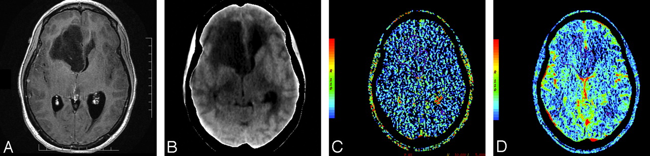
  • Fig 3.
    • Download figure
    • Open in new tab
    • Download powerpoint
    Fig 3.

    A, Postcontrast T1-weighted (TR/TE, 3218/5.71 ms) axial image in a 55-year-old man with glioblastoma multiforme showing a heterogeneously enhancing mass with irregular central necrosis in the right peritrigonal region. B, Base image from the perfusion CT study showing the tumor in the right peritrigonal region. C, Perfusion CT PS maps (C) showing very high permeability (PS = 5.14 mL/100 g per minute) along the enhancing nodular margins of the tumor and CBV maps (D) showing high blood volume (CBV = 3.49 mL/100 g) within the enhancing periphery of the tumor.
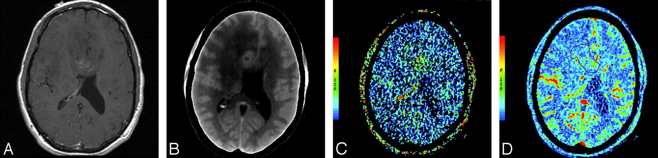
  • Fig 4.
    • Download figure
    • Open in new tab
    • Download powerpoint
    Fig 4.

    A and B, Postcontrast T1-weighted (TR/TE, 650/13 ms) axial image (A) and base image from perfusion CT study (B) in a 46-year-old man with a heterogeneously enhancing mass lesion predominantly involving the right frontal lobe with extension across the genu of the corpus callosum. C and D, Perfusion CT PS (C) and CBV (D) maps showing high permeability (PS = 2.04 mL/100 g per minute) and tumor blood volume (CBV = 2.21 mL/100 g). Histopathology revealed a grade III anaplastic astrocytoma.

  • Fig 5.
    • Download figure
    • Open in new tab
    • Download powerpoint
    Fig 5.

    A and B, Postcontrast T1-weighted (TR/TE, 3103/6.35 ms) image (A) and base image from perfusion CT study (B) in a 31-year-old man who presented with memory impairment and speech problems showing a nonenhancing mass in the left temporal lobe with minimal mass effect and perilesional edema. C and D, PS (C) and CBV (D) maps showing low permeability (PSA = 0.61 mL/100 g per minute) and low blood volume (CBV = 0.92 mL/100 g) within the mass. Histopathology revealed grade III anaplastic astrocytoma.

Tables

  • Figures
    • View popup
    Table 1:

    Perfusion parameters and ROC analysis for low- and high-grade gliomas

    Group (No. of patients)PS (mL/100 g/min) Mean (SD)CBV (mL/100 g) Mean (SD)CBF (mL/100 g/min) Mean (SD)MTT (s) Mean (SD)
    Low grade (8)0.52 (0.15)0.95 (0.22)27.0 (8.3)4.24 (1.42)
    High grade (24)2.37 (1.40)2.79 (1.20)82.0 (73.7)4.67 (1.37)
    P value<.001<.001.0024.564
    C-statistic0.9270.9300.8490.573
    • ROC indicates receiver operating characteristic; PS, permeability surface-area product; CBV, cerebral blood volume; CBF, cerebral blood flow; MTT, mean transit time.

    • View popup
    Table 2:

    Perfusion parameters and ROC analysis for WHO grades III and grade IV

    Group (No. of patients)PS (mL/100 g/min) Mean (SD)CBV (mL/100 g) Mean (SD)CBF (mL/100 g/min) Mean (SD)MTT (s) Mean (SD)
    WHO grade III (6)1.04 (0.73)1.82 (1.41)38.9 (19.3)4.60 (1.62)
    WHO grade IV (18)2.81 (1.28)3.12 (0.97)96.3 (79.8)4.69 (1.32)
    P value<.001.039.014.922
    C-statistic0.9260.7870.8330.509
    • ROC indicates receiver operating characteristic; PS, permeability surface-area product; CBV, cerebral blood volume; CBF, cerebral blood flow; MIT, mean transit time.

    • View popup
    Table 3:

    Perfusion parameters and P values for nonenhancing versus enhancing grade III, nonenhancing grade III versus low grade, and enhancing grade III versus grade IV gliomas

    PS (mL/100 g/min) Mean (SD)CBV (mL/100 g) Mean (SD)CBF (mL/100 g/min) Mean (SD)MTT (s) Mean (SD)
    Low grade (8)0.52 (0.15)0.95 (0.22)27.0 (8.3)4.24 (1.42)
    Nonenhancing grade III (3)0.46 (0.13)0.88 (0.16)26.1 (3.4)3.23 (0.64)
    Enhancing grade III (3)1.61 (0.57)2.77 (1.52)51.7 (20.8)5.96 (0.75)
    WHO grade IV (18)2.81 (1.28)3.12 (0.97)96.3 (79.8)4.69 (1.32)
    P values for nonenhancing vs. enhancing grade III.011.029.055.012
    P values for low grade vs. nonenhancing grade III.623.699.971.330
    P values for enhancing grade III vs. grade IV.052.438.322.128
    • PS indicates permeability surface-area product; CBV, cerebral blood volume; CBF, cerebral blood flow; MTT, mean transit time.

PreviousNext
Back to top

In this issue

American Journal of Neuroradiology: 29 (4)
American Journal of Neuroradiology
Vol. 29, Issue 4
April 2008
  • Table of Contents
  • Index by author
Advertisement
Print
Download PDF
Email Article

Thank you for your interest in spreading the word on American Journal of Neuroradiology.

NOTE: We only request your email address so that the person you are recommending the page to knows that you wanted them to see it, and that it is not junk mail. We do not capture any email address.

Enter multiple addresses on separate lines or separate them with commas.
Quantitative Estimation of Permeability Surface-Area Product in Astroglial Brain Tumors Using Perfusion CT and Correlation with Histopathologic Grade
(Your Name) has sent you a message from American Journal of Neuroradiology
(Your Name) thought you would like to see the American Journal of Neuroradiology web site.
CAPTCHA
This question is for testing whether or not you are a human visitor and to prevent automated spam submissions.
Cite this article
R. Jain, S.K. Ellika, L. Scarpace, L.R. Schultz, J.P. Rock, J. Gutierrez, S.C. Patel, J. Ewing, T. Mikkelsen
Quantitative Estimation of Permeability Surface-Area Product in Astroglial Brain Tumors Using Perfusion CT and Correlation with Histopathologic Grade
American Journal of Neuroradiology Apr 2008, 29 (4) 694-700; DOI: 10.3174/ajnr.A0899

Citation Manager Formats

  • BibTeX
  • Bookends
  • EasyBib
  • EndNote (tagged)
  • EndNote 8 (xml)
  • Medlars
  • Mendeley
  • Papers
  • RefWorks Tagged
  • Ref Manager
  • RIS
  • Zotero
0 Responses
Respond to this article
Share
Bookmark this article
Quantitative Estimation of Permeability Surface-Area Product in Astroglial Brain Tumors Using Perfusion CT and Correlation with Histopathologic Grade
R. Jain, S.K. Ellika, L. Scarpace, L.R. Schultz, J.P. Rock, J. Gutierrez, S.C. Patel, J. Ewing, T. Mikkelsen
American Journal of Neuroradiology Apr 2008, 29 (4) 694-700; DOI: 10.3174/ajnr.A0899
del.icio.us logo Twitter logo Facebook logo Mendeley logo
  • Tweet Widget
  • Facebook Like
  • Google Plus One
Purchase

Jump to section

  • Article
    • Abstract
    • Materials and Methods
    • Results
    • Discussion
    • Conclusions
    • References
  • Figures & Data
  • Info & Metrics
  • Responses
  • References
  • PDF

Related Articles

  • No related articles found.
  • PubMed
  • Google Scholar

Cited By...

  • Glioma Angiogenesis and Perfusion Imaging: Understanding the Relationship between Tumor Blood Volume and Leakiness with Increasing Glioma Grade
  • Evaluating Blood-Brain Barrier Permeability in Delayed Cerebral Infarction after Aneurysmal Subarachnoid Hemorrhage
  • Effects of Microvascular Permeability Changes on Contrast-Enhanced T1 and Pharmacokinetic MR Imagings After Ischemia
  • Correlation of Perfusion Parameters with Genes Related to Angiogenesis Regulation in Glioblastoma: A Feasibility Study
  • Imaging biomarkers of angiogenesis and the microvascular environment in cerebral tumours
  • Perfusion CT Imaging of Brain Tumors: An Overview
  • Permeability Estimates in Histopathology-Proved Treatment-Induced Necrosis Using Perfusion CT: Can These Add to Other Perfusion Parameters in Differentiating from Recurrent/Progressive Tumors?
  • In Vivo Correlation of Tumor Blood Volume and Permeability with Histologic and Molecular Angiogenic Markers in Gliomas
  • Crossref (114)
  • Google Scholar

This article has been cited by the following articles in journals that are participating in Crossref Cited-by Linking.

  • Differentiating tumor recurrence from treatment necrosis: a review of neuro-oncologic imaging strategies
    Nishant Verma, Matthew C. Cowperthwaite, Mark G. Burnett, Mia K. Markey
    Neuro-Oncology 2013 15 5
  • Correlation of volume transfer coefficient Ktrans with histopathologic grades of gliomas
    Na Zhang, Lijuan Zhang, Bensheng Qiu, Li Meng, Xiaoyi Wang, Bob L. Hou
    Journal of Magnetic Resonance Imaging 2012 36 2
  • In vivo quantitative imaging of tumor pH by nanosonophore assisted multispectral photoacoustic imaging
    Janggun Jo, Chang H. Lee, Raoul Kopelman, Xueding Wang
    Nature Communications 2017 8 1
  • Model selection in measures of vascular parameters using dynamic contrast‐enhanced MRI: experimental and clinical applications
    James R. Ewing, Hassan Bagher‐Ebadian
    NMR in Biomedicine 2013 26 8
  • In Vivo Correlation of Tumor Blood Volume and Permeability with Histologic and Molecular Angiogenic Markers in Gliomas
    R. Jain, J. Gutierrez, J. Narang, L. Scarpace, L.R. Schultz, N. Lemke, S.C. Patel, T. Mikkelsen, J.P. Rock
    American Journal of Neuroradiology 2011 32 2
  • Targeted delivery of antibody-based therapeutic and imaging agents to CNS tumors: crossing the blood–brain barrier divide
    Ann-Marie Chacko, Chunsheng Li, Daniel A Pryma, Steven Brem, George Coukos, Vladimir Muzykantov
    Expert Opinion on Drug Delivery 2013 10 7
  • Clinical Utility of Quantitative Imaging
    Andrew B. Rosenkrantz, Mishal Mendiratta-Lala, Brian J. Bartholmai, Dhakshinamoorthy Ganeshan, Richard G. Abramson, Kirsteen R. Burton, John-Paul J. Yu, Ernest M. Scalzetti, Thomas E. Yankeelov, Rathan M. Subramaniam, Leon Lenchik
    Academic Radiology 2015 22 1
  • Measurements of tumor vascular leakiness using DCE in brain tumors: clinical applications
    Rajan Jain
    NMR in Biomedicine 2013 26 8
  • Perfusion CT Imaging of Brain Tumors: An Overview
    R. Jain
    American Journal of Neuroradiology 2011 32 9
  • Diagnostic examination performance by using microvascular leakage, cerebral blood volume, and blood flow derived from 3-T dynamic susceptibility-weighted contrast-enhanced perfusion MR imaging in the differentiation of glioblastoma multiforme and brain metastasis
    Andrés Server, Tone E. Døli Orheim, Bjørn A. Graff, Roger Josefsen, Theresa Kumar, Per H. Nakstad
    Neuroradiology 2011 53 5

More in this TOC Section

  • Multimodal CT Provides Improved Performance for Lacunar Infarct Detection
  • Optimal MRI Sequence for Identifying Occlusion Location in Acute Stroke: Which Value of Time-Resolved Contrast-Enhanced MRA?
  • Evaluating the Effects of White Matter Multiple Sclerosis Lesions on the Volume Estimation of 6 Brain Tissue Segmentation Methods
Show more BRAIN

Similar Articles

Advertisement

Indexed Content

  • Current Issue
  • Accepted Manuscripts
  • Article Preview
  • Past Issues
  • Editorials
  • Editor's Choice
  • Fellows' Journal Club
  • Letters to the Editor
  • Video Articles

Cases

  • Case Collection
  • Archive - Case of the Week
  • Archive - Case of the Month
  • Archive - Classic Case

More from AJNR

  • Trainee Corner
  • Imaging Protocols
  • MRI Safety Corner

Multimedia

  • AJNR Podcasts
  • AJNR Scantastics

Resources

  • Turnaround Time
  • Submit a Manuscript
  • Submit a Video Article
  • Submit an eLetter to the Editor/Response
  • Manuscript Submission Guidelines
  • Statistical Tips
  • Fast Publishing of Accepted Manuscripts
  • Graphical Abstract Preparation
  • Imaging Protocol Submission
  • Evidence-Based Medicine Level Guide
  • Publishing Checklists
  • Author Policies
  • Become a Reviewer/Academy of Reviewers
  • News and Updates

About Us

  • About AJNR
  • Editorial Board
  • Editorial Board Alumni
  • Alerts
  • Permissions
  • Not an AJNR Subscriber? Join Now
  • Advertise with Us
  • Librarian Resources
  • Feedback
  • Terms and Conditions
  • AJNR Editorial Board Alumni

American Society of Neuroradiology

  • Not an ASNR Member? Join Now

© 2025 by the American Society of Neuroradiology All rights, including for text and data mining, AI training, and similar technologies, are reserved.
Print ISSN: 0195-6108 Online ISSN: 1936-959X

Powered by HighWire