
Article Figures & Data
Figures
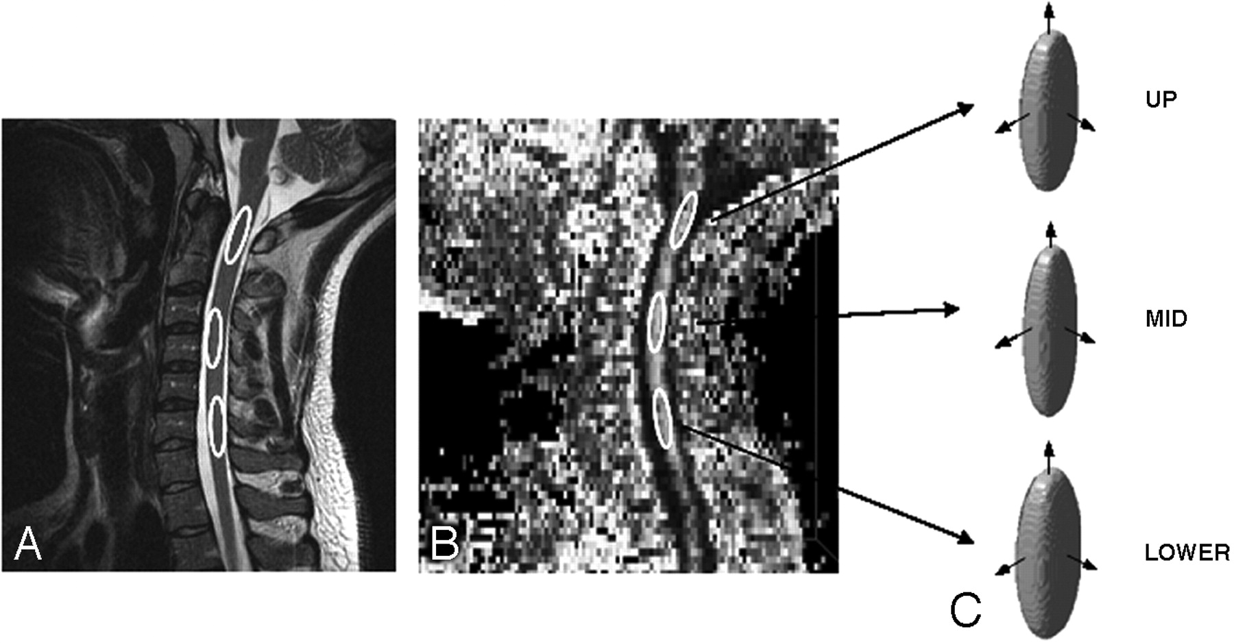
- Fig 1.
Images showing the placement of regions of interest in the sagittal T2-weighted image (A), FA image (B), and the corresponding ellipsoid representation of the tensor at each level of the cord (C).
- Fig 2.
A 56-year-old man admitted following a fall, with central cord syndrome. A, T2-weighted image shows a cord contusion (arrowhead) posterior to the C4-5 disk space. Ellipsoid representation of the tensor of the axial cervical cord of the patient and healthy volunteer at corresponding anatomic levels of the upper cord (B), adjacent to the contusion (C), cord contusion (D), and the lower cord (E) shows abnormal ellipsoid representation of the cord at and away from the cord contusion, where the conventional MR imaging shows no signal-intensity abnormality
- Fig 3.
Comparison of whole spine control (n = 8) DTI parameters with the patient population (n = 16), patients exhibiting hemorrhagic (Hem) injury (n = 6), patients with quadriplegia (Quads, n = 7), and site of injury (n = 11).


Tables
Regional DTI parameters of the normal cord in control individuals and patients
Parameters Control Patient Upper Mid Lower Upper Mid Lower ADC (*10−3 mm2/s) 0.83 ± 0.07 0.97 ± 0.11 0.97 ± 0.11 0.71 ± 0.13 0.77 ± 0.13 0.74 ± 0.13 P value .013 .0005 .00013 FA 0.64 ± 0.08 0.69 ± 0.06 0.63 ± 0.06 0.63 ± 0.09 0.64 ± 0.08 0.60 ± 0.08 P value .378 .075 .187 RA 0.63 ± 0.12 0.68 ± 0.09 0.59 ± 0.08 0.59 ± 0.12 0.60 ± 0.1 0.53 ± 0.1 P value .248 .037 .099 VR 0.53 ± 0.15 0.49 ± 0.11 0.59 ± 0.09 0.53 ± 0.15 0.55 ± 0.12 0.61 ± 0.13 P value .49 .135 .381 E1 (*10−3 mm2/s) 1.54 ± 0.15 1.88 ± 0.2 1.75 ± 0.15 1.25 ± 0.23 1.40 ± 0.27 1.27 ± 0.23 P value .0018 .0001 .000105 E2 (*10−3 mm2/s) 0.63 ± 0.09 0.68 ± 0.10 0.74 ± 0.12 0.62 ± 0.14 0.64 ± 0.11 0.67 ± 0.1 P value .393 .199 .063 E3 (*10−3 mm2/s) 0.32 ± 0.1 0.36 ± 0.11 0.42 ± 0.11 0.25 ± 0.11 0.29 ± 0.08 0.29 ± 0.13 P value .09 .039 .01 Note:—Bold indicates P value < .05; ADC, apparent diffusion coefficient; FA, fractional anisotropy; RA, relative anisotropy; VR, volume ratio; E1, eigenvalue E1.
In this issue


Jump to section
Related Articles
Cited By...
- Cervical Cordotomy for Intractable Pain: Do Postoperative Imaging Features Correlate with Pain Outcomes and Mirror Pain?
- Local BDNF Delivery to the Injured Cervical Spinal Cord using an Engineered Hydrogel Enhances Diaphragmatic Respiratory Function
- Cervical Spinal Cord DTI Is Improved by Reduced FOV with Specific Balance between the Number of Diffusion Gradient Directions and Averages
- Pulse-Triggered DTI Sequence with Reduced FOV and Coronal Acquisition at 3T for the Assessment of the Cervical Spinal Cord in Patients with Myelitis
- Diffusion tensor imaging of the spinal cord and its clinical applications
- Diffusion Tensor Imaging of the Pediatric Spinal Cord at 1.5T: Preliminary Results