Advertisement

Main menu

  • Home
  • Content
    • Current Issue
    • Accepted Manuscripts
    • Article Preview
    • Past Issue Archive
    • Video Articles
    • AJNR Case Collection
    • Case of the Week Archive
    • Case of the Month Archive
    • Classic Case Archive
  • Special Collections
    • AJNR Awards
    • Low-Field MRI
    • Alzheimer Disease
    • ASNR Foundation Special Collection
    • Photon-Counting CT
    • View All
  • Multimedia
    • AJNR Podcasts
    • AJNR SCANtastic
    • Trainee Corner
    • MRI Safety Corner
    • Imaging Protocols
  • For Authors
    • Submit a Manuscript
    • Submit a Video Article
    • Submit an eLetter to the Editor/Response
    • Manuscript Submission Guidelines
    • Statistical Tips
    • Fast Publishing of Accepted Manuscripts
    • Graphical Abstract Preparation
    • Imaging Protocol Submission
    • Author Policies
  • About Us
    • About AJNR
    • Editorial Board
    • Editorial Board Alumni
  • More
    • Become a Reviewer/Academy of Reviewers
    • Subscribers
    • Permissions
    • Alerts
    • Feedback
    • Advertisers
    • ASNR Home

User menu

  • Alerts
  • Log in

Search

  • Advanced search
American Journal of Neuroradiology

American Journal of Neuroradiology

ASHNR American Society of Functional Neuroradiology ASHNR American Society of Pediatric Neuroradiology ASSR
  • Alerts
  • Log in

Advanced Search

  • Home
  • Content
    • Current Issue
    • Accepted Manuscripts
    • Article Preview
    • Past Issue Archive
    • Video Articles
    • AJNR Case Collection
    • Case of the Week Archive
    • Case of the Month Archive
    • Classic Case Archive
  • Special Collections
    • AJNR Awards
    • Low-Field MRI
    • Alzheimer Disease
    • ASNR Foundation Special Collection
    • Photon-Counting CT
    • View All
  • Multimedia
    • AJNR Podcasts
    • AJNR SCANtastic
    • Trainee Corner
    • MRI Safety Corner
    • Imaging Protocols
  • For Authors
    • Submit a Manuscript
    • Submit a Video Article
    • Submit an eLetter to the Editor/Response
    • Manuscript Submission Guidelines
    • Statistical Tips
    • Fast Publishing of Accepted Manuscripts
    • Graphical Abstract Preparation
    • Imaging Protocol Submission
    • Author Policies
  • About Us
    • About AJNR
    • Editorial Board
    • Editorial Board Alumni
  • More
    • Become a Reviewer/Academy of Reviewers
    • Subscribers
    • Permissions
    • Alerts
    • Feedback
    • Advertisers
    • ASNR Home
  • Follow AJNR on Twitter
  • Visit AJNR on Facebook
  • Follow AJNR on Instagram
  • Join AJNR on LinkedIn
  • RSS Feeds

AJNR Awards, New Junior Editors, and more. Read the latest AJNR updates

OtherBRAIN

Subthalamic Lesion on MR Imaging in a Patient with Nonketotic Hyperglycemia-Induced Hemiballism

H.-J. Kim, W.J. Moon, J. Oh, I.K. Lee, H.Y. Kim and S.-H. Han
American Journal of Neuroradiology March 2008, 29 (3) 526-527; DOI: https://doi.org/10.3174/ajnr.A0927
H.-J. Kim
  • Find this author on Google Scholar
  • Find this author on PubMed
  • Search for this author on this site
W.J. Moon
  • Find this author on Google Scholar
  • Find this author on PubMed
  • Search for this author on this site
J. Oh
  • Find this author on Google Scholar
  • Find this author on PubMed
  • Search for this author on this site
I.K. Lee
  • Find this author on Google Scholar
  • Find this author on PubMed
  • Search for this author on this site
H.Y. Kim
  • Find this author on Google Scholar
  • Find this author on PubMed
  • Search for this author on this site
S.-H. Han
  • Find this author on Google Scholar
  • Find this author on PubMed
  • Search for this author on this site
  • Article
  • Figures & Data
  • Info & Metrics
  • Responses
  • References
  • PDF
Loading

Abstract

SUMMARY: Hemiballism with corresponding striatal T1 hyperintensity on MR imaging has occasionally been reported in patients with nonketotic hyperglycemia. However, the subthalamic nucleus lesion, which is believed to be pathogenetically related to hemiballism, is rarely documented in a living patient with nonketotic hyperglycemia. We describe a patient with nonketotic hyperglycemia-induced hemiballism, whose responsible lesion (ie, the subthalamus) was demonstrated by MR imaging.

Hemiballism-hemichorea (HB-HC) is an involuntary, irregular, wide-amplitude, and poorly patterned movement disorder. HB-HC can mostly result from a focal vascular lesion in the contralateral basal ganglia.1 In the absence of an identifiable focal vascular lesion, metabolic derangements (eg, nonketotic hyperglycemia or hyperthyroidism), brain neoplasm, and infectious diseases of the central nervous system (eg, human immunodeficiency virus infection) are considered to be the more common causes.1 Recent studies have shown that striatal hyperintensity, seen on the T1-weighted MR imaging of the brain, can occur in the acute stage of HB-HC, caused by nonketotic hyperglycemia.2 These radiologic images provided the clue for the early diagnosis of this dyskinesia. Animal studies and subsequent human studies supported the finding that the subthalamus is strongly related to hemiballism.3 There are only scanty reports documenting the subthalamic involvement of nonketotic hyperglycemia in a living patient, which prompted us to report a case of nonketotic hyperglycemia-induced hemiballism, with the responsible subthalamic lesion confirmed by MR imaging.

Case Report

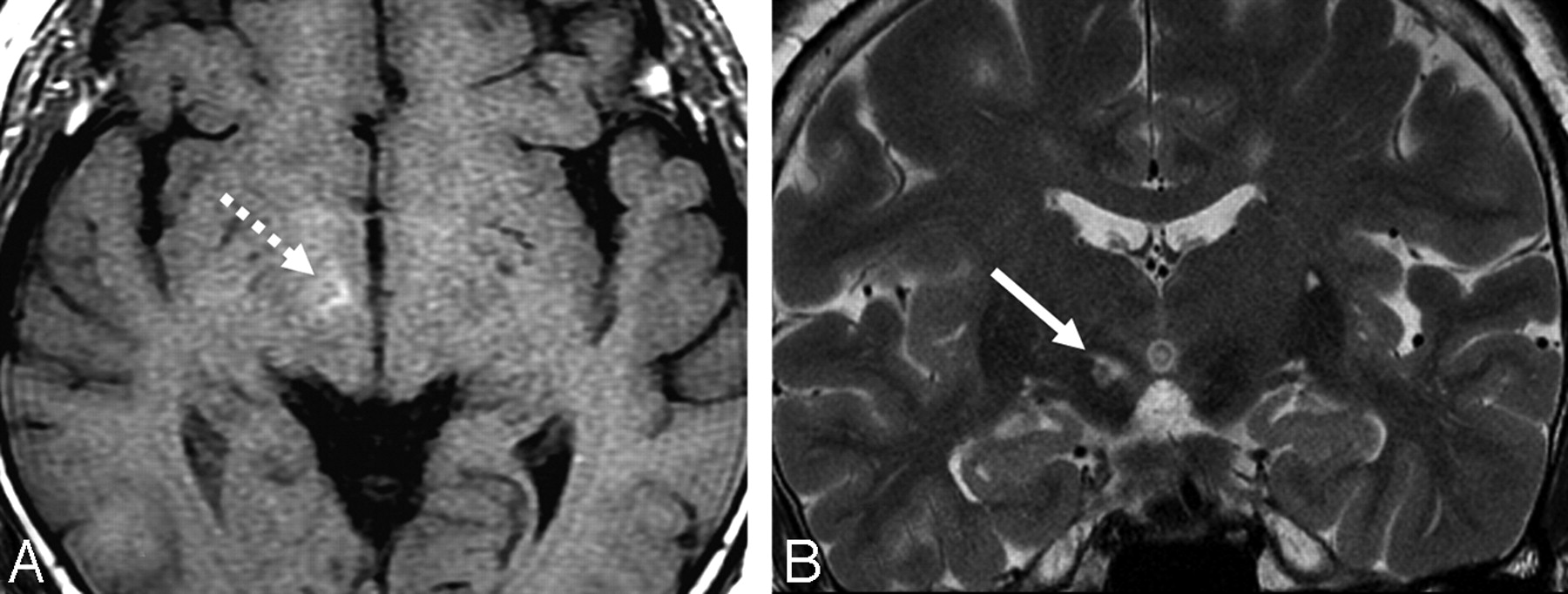
A 53-year-old woman was admitted for sudden onset of abnormal movement on the left side. This jerky irregular involuntary movement was first noted on the left leg 3 days before admission. Her symptoms worsened during the last 3 days. She had no history of hypertension, diabetes, headache, parkinsonism, or other neurologic diseases. At admission, her initial blood glucose level was 242 mg/dL and her hemoglobin A1c concentration was 11.7% (normal values are 70–110 mg/dL and 4.7–6.4%, respectively). The urine specimen was negative for ketones, indicating nonketotic hyperglycemia. A neurologic examination showed her sensorium to be normal. Deep tendon reflexes were symmetrically hypoactive. The remaining results of the examinations were unremarkable, except for left hemiballism. Brain CT, performed on the first hospital day, revealed no abnormalities. The MR image, obtained 4 days after her initial presentation, showed high signal intensities in the right subthalamic nucleus (STN) on T1-weighted and T2-weighted images (Fig 1) and subtle high signal intensities on diffusion-weighted images (DWIs, not shown). Gradient-echo images and B0 images of DWI showed no susceptibility artifact in the right STN. Contrast-enhanced MR images showed no additional findings. The caudate and putamen appeared normal in all sequences of MR images. Hemiballism was improved with clonazepam (6 mg/day) after 14 days. The serum glucose level was normalized with insulin administration.

  Fig 1.
  • Download figure
  • Open in new tab
  • Download powerpoint
Fig 1.

The T1-weighted axial image (A) shows increased signal intensity (broken arrow), whereas the T2-weighted coronal image clearly demonstrates a high-signal-intensity lesion (arrow) in the right STN.

Discussion

Hyperglycemia-associated chorea and ballismus have been reported predominantly in older patients with diabetes and nonketotic hyperglycemia.4 The T1-weighted MR images have shown unilateral hyperintensity in the putamen and caudate, but the findings on the T2-weighted MR images have varied.5 Although a significant association between hyperglycemia and hyperintensity on T1-weighted MR images has been reported, the mechanism underlying the imaging findings has been unknown. Protein dissociation in wallerian degeneration secondary to acute putaminal dysfunction has been proposed as one of the plausible mechanisms behind the imaging findings.6 Increased viscosity, the presence of myelin-breakdown products, the presence of blood products, or deposition of calcium and other minerals have been suggested as possible explanations as well.7–9

As to the MR signal intensities alone, our imaging findings confirmed results from previous reports. In this case, however, MR imaging revealed the sole involvement of the contralateral STN without a putaminal lesion. Although subthalamic involvement has never been reported in hyperglycemia-induced hemiballism, acute hemiballism was reported in a small cavernous angioma or infarction confined to the STN.10,11

The STN serves as the main excitatory input to the medial part of the globus pallidus, which inhibits, by using gamma amino butyric acid, the activity of the ventromedial thalamus. The destruction of the STN causes a decrease of this inhibitory function and increases thalamic excitation of the motor and premotor cortex, resulting in involuntary movement.12 This mechanism is supported by findings during deep brain stimulation of the subthalamus in Parkinson disease.13

Possible differential diagnosis in this case includes acute infarction by vascular insult. Unlike T1 hyperintensity due to nonketotic hyperglycemia, acute infarction of 3 days duration shows bright signal intensity on DWIs and low signal intensity on T1-weighted images. When T1 hyperintensity is noted in acute infarction at 3 days, it should be hemorrhagic transformation and should, therefore, show dark signal intensity on T2*-weighted images.14 Acute hemorrhage is also unlikely because there is no evidence of magnetic susceptibility artifacts on T2*-weighted images as well as on DWIs with a b-value of 0 s/mm2.

In conclusion, unilateral subthalamic involvement can be the sole imaging finding in nonketotic hyperglycemia-induced hemiballism. This case suggests a pathophysiologic explanation for hemiballism with its strategic localization.

Footnotes

  • This work was supported by the Second-Phase of BK (Brain Korea) 21 Project in 2007.

References

  1. ↵
    Dewey B, Jankovic J. Hemiballism-hemichorea: clinical and pharmacologic findings in 21 patients. Arch Neurol 1989;46:862–67
    CrossRefPubMed
  2. ↵
    Yahikozawa H, Hanyu N, Yamamoto K, et al. Hemiballism with striatal hyperintensity on T1- weighted MRI in diabetes patients; a unique syndrome. J Neurol Sci 1994;124:208–14
    CrossRefPubMed
  3. ↵
    Postuma RB, Lang AE. Hemiballism: revisiting a classic disorder. Lancet Neurol 2003;2:661–68
    CrossRefPubMed
  4. ↵
    Ifergane G, Masalha R, Herishanu YO. Transient hemichorea/hemiballismus associated with new onset hyperglycemia. Can J Neurol Sci 2001;28:365–68
    PubMed
  5. ↵
    Lee EJ, Choi JY, Lee SH, et al. Hemichorea-hemiballism in primary diabetic patients: MR correlation. J Comput Assist Tomogr 2002;26:905–11
    CrossRefPubMed
  6. ↵
    Wintermark M, Fischbein NJ, Mukherjee P, et al. Unilateral putaminal CT, MR, and diffusion abnormalities secondary to nonketotic hyperglycemia in the setting of acute neurologic symptoms mimicking stroke. AJNR Am J Neuroradiol 2004;25:975–76
    Abstract/FREE Full Text
  7. ↵
    Chu K, Kang DW, Kim ED, et al. Diffusion-weighted and gradient echo magnetic resonance findings of hemichorea-hemiballismus associated with diabetic hyperglycemia. Arch Neurol 2002;59:448–52
    CrossRefPubMed
  8. Lin JJ, Chang MK. Hemiballism-hemichorea and nonketotic hyperglycemia. J Neurol Neurosurg Psychiatry 1994;57:748–50
    Abstract/FREE Full Text
  9. ↵
    McCall AL. The impact of diabetes on the CNS. Diabetes 1992;41:557–70
    Abstract/FREE Full Text
  10. ↵
    Absher JR, Vogt BA, Clark DG, et al. Hypersexuality and hemiballism due to subthalamic infarction. Neuropsychiatry Neuropsychol Behav Neurol 2000;13:220–29
    PubMed
  11. ↵
    Linn JJ, Seelos KC, Botzel K. Hemiballism caused by a small cavernoma in the subthalamic nucleus. Mov Disord 2006;21:2266–67
    CrossRefPubMed
  12. ↵
    Guisado R, Arieff A. Neurologic manifestations of diabetic comas: correlation with biochemical alterations in the brain. Metabolism 1975;24:665–79
    CrossRefPubMed
  13. ↵
    Doshi P, Bhatt M. Hemiballism during subthalamic nucleus lesioning. Mov Disord 2002;17:848–49
    CrossRefPubMed
  14. ↵
    Chang MH, Chiang HT, Lai PH, et al. Putaminal petechial haemorrhage as the cause of chorea: a neuroimaging study. J Neurol Neurosurg Psychiatry 1997;63:300–03
    Abstract/FREE Full Text
  • Received October 23, 2007.
  • Accepted after revision October 30, 2007.
  • Copyright © American Society of Neuroradiology
View Abstract
PreviousNext
Back to top

In this issue

American Journal of Neuroradiology: 29 (3)
American Journal of Neuroradiology
Vol. 29, Issue 3
March 2008
  • Table of Contents
  • Index by author
Advertisement
Print
Download PDF
Email Article

Thank you for your interest in spreading the word on American Journal of Neuroradiology.

NOTE: We only request your email address so that the person you are recommending the page to knows that you wanted them to see it, and that it is not junk mail. We do not capture any email address.

Enter multiple addresses on separate lines or separate them with commas.
Subthalamic Lesion on MR Imaging in a Patient with Nonketotic Hyperglycemia-Induced Hemiballism
(Your Name) has sent you a message from American Journal of Neuroradiology
(Your Name) thought you would like to see the American Journal of Neuroradiology web site.
CAPTCHA
This question is for testing whether or not you are a human visitor and to prevent automated spam submissions.
Cite this article
H.-J. Kim, W.J. Moon, J. Oh, I.K. Lee, H.Y. Kim, S.-H. Han
Subthalamic Lesion on MR Imaging in a Patient with Nonketotic Hyperglycemia-Induced Hemiballism
American Journal of Neuroradiology Mar 2008, 29 (3) 526-527; DOI: 10.3174/ajnr.A0927

Citation Manager Formats

  • BibTeX
  • Bookends
  • EasyBib
  • EndNote (tagged)
  • EndNote 8 (xml)
  • Medlars
  • Mendeley
  • Papers
  • RefWorks Tagged
  • Ref Manager
  • RIS
  • Zotero
0 Responses
Respond to this article
Share
Bookmark this article
Subthalamic Lesion on MR Imaging in a Patient with Nonketotic Hyperglycemia-Induced Hemiballism
H.-J. Kim, W.J. Moon, J. Oh, I.K. Lee, H.Y. Kim, S.-H. Han
American Journal of Neuroradiology Mar 2008, 29 (3) 526-527; DOI: 10.3174/ajnr.A0927
del.icio.us logo Twitter logo Facebook logo Mendeley logo
  • Tweet Widget
  • Facebook Like
  • Google Plus One
Purchase

Jump to section

  • Article
    • Abstract
    • Case Report
    • Discussion
    • Footnotes
    • References
  • Figures & Data
  • Info & Metrics
  • Responses
  • References
  • PDF

Related Articles

  • No related articles found.
  • PubMed
  • Google Scholar

Cited By...

  • 3T MRI Whole-Brain Microscopy Discrimination of Subcortical Anatomy, Part 2: Basal Forebrain
  • Neuroimaging in Patients with Abnormal Blood Glucose Levels
  • Activated microglia in the subthalamic nucleus in hyperglycaemic hemiballism: a case report
  • Crossref (7)
  • Google Scholar

This article has been cited by the following articles in journals that are participating in Crossref Cited-by Linking.

  • Neuroimaging in Patients with Abnormal Blood Glucose Levels
    G. Bathla, B. Policeni, A. Agarwal
    American Journal of Neuroradiology 2014 35 5
  • MRI and CT appearances in metabolic encephalopathies due to systemic diseases in adults
    G. Bathla, A.N. Hegde
    Clinical Radiology 2013 68 6
  • 3T MRI Whole-Brain Microscopy Discrimination of Subcortical Anatomy, Part 2: Basal Forebrain
    M.J. Hoch, M.T. Bruno, A. Faustin, N. Cruz, A.Y. Mogilner, L. Crandall, T. Wisniewski, O. Devinsky, T.M. Shepherd
    American Journal of Neuroradiology 2019 40 7
  • T2*-based MR imaging of hyperglycemia-induced hemichorea-hemiballism
    Fang Yu, Andrew Steven, Lee Birnbaum, Wilson Altmeyer
    Journal of Neuroradiology 2017 44 1
  • Atypical neurologic presentations of new onset type 1 diabetes mellitus in pediatric age group: a report of five unusual cases and review of the literature
    Laurel Mulder, Ozlem Onur, Lora Kleis, Heather Borders, Ayse P. Cemeroglu
    Journal of Pediatric Endocrinology and Metabolism 2014 27 7-8
  • A Case of Type 2 Diabetes Mellitus Initially Presented as Monochorea Associated with Ketotic Hyperglycemia
    Si Hyeong Lee, Dong Woo Lee, Yeong Min Lee, Tae Kyun Kim, Min Jeong Kwon, Soon Hee Lee, Jeong Hyun Park
    The Journal of Korean Diabetes 2014 15 4
  • RADIOLOGINĖ PAMATO BRANDUOLIŲ POKYČIŲ DIFERENCINĖ DIAGNOSTIKA
    Eglė Janiūnienė, Agnė Ulytė, Jūratė Dementavičienė, Jurgita Valaikienė
    Medicinos teorija ir praktika 2016 22 1

More in this TOC Section

  • White Matter Alterations in the Brains of Patients with Active, Remitted, and Cured Cushing Syndrome: A DTI Study
  • Qualitative and Quantitative Analysis of MR Imaging Findings in Patients with Middle Cerebral Artery Stroke Implanted with Mesenchymal Stem Cells
  • Fast Contrast-Enhanced 4D MRA and 4D Flow MRI Using Constrained Reconstruction (HYPRFlow): Potential Applications for Brain Arteriovenous Malformations
Show more BRAIN

Similar Articles

Advertisement

Indexed Content

  • Current Issue
  • Accepted Manuscripts
  • Article Preview
  • Past Issues
  • Editorials
  • Editor's Choice
  • Fellows' Journal Club
  • Letters to the Editor
  • Video Articles

Cases

  • Case Collection
  • Archive - Case of the Week
  • Archive - Case of the Month
  • Archive - Classic Case

More from AJNR

  • Trainee Corner
  • Imaging Protocols
  • MRI Safety Corner
  • Book Reviews

Multimedia

  • AJNR Podcasts
  • AJNR Scantastics

Resources

  • Turnaround Time
  • Submit a Manuscript
  • Submit a Video Article
  • Submit an eLetter to the Editor/Response
  • Manuscript Submission Guidelines
  • Statistical Tips
  • Fast Publishing of Accepted Manuscripts
  • Graphical Abstract Preparation
  • Imaging Protocol Submission
  • Evidence-Based Medicine Level Guide
  • Publishing Checklists
  • Author Policies
  • Become a Reviewer/Academy of Reviewers
  • News and Updates

About Us

  • About AJNR
  • Editorial Board
  • Editorial Board Alumni
  • Alerts
  • Permissions
  • Not an AJNR Subscriber? Join Now
  • Advertise with Us
  • Librarian Resources
  • Feedback
  • Terms and Conditions
  • AJNR Editorial Board Alumni

American Society of Neuroradiology

  • Not an ASNR Member? Join Now

© 2025 by the American Society of Neuroradiology All rights, including for text and data mining, AI training, and similar technologies, are reserved.
Print ISSN: 0195-6108 Online ISSN: 1936-959X

Powered by HighWire