Advertisement

Main menu

  • Home
  • Content
    • Current Issue
    • Accepted Manuscripts
    • Article Preview
    • Past Issue Archive
    • Video Articles
    • AJNR Case Collection
    • Case of the Week Archive
    • Case of the Month Archive
    • Classic Case Archive
  • Special Collections
    • AJNR Awards
    • Low-Field MRI
    • Alzheimer Disease
    • ASNR Foundation Special Collection
    • Photon-Counting CT
    • View All
  • Multimedia
    • AJNR Podcasts
    • AJNR SCANtastic
    • Trainee Corner
    • MRI Safety Corner
    • Imaging Protocols
  • For Authors
    • Submit a Manuscript
    • Submit a Video Article
    • Submit an eLetter to the Editor/Response
    • Manuscript Submission Guidelines
    • Statistical Tips
    • Fast Publishing of Accepted Manuscripts
    • Graphical Abstract Preparation
    • Imaging Protocol Submission
    • Author Policies
  • About Us
    • About AJNR
    • Editorial Board
    • Editorial Board Alumni
  • More
    • Become a Reviewer/Academy of Reviewers
    • Subscribers
    • Permissions
    • Alerts
    • Feedback
    • Advertisers
    • ASNR Home

User menu

  • Alerts
  • Log in

Search

  • Advanced search
American Journal of Neuroradiology

American Journal of Neuroradiology

ASHNR American Society of Functional Neuroradiology ASHNR American Society of Pediatric Neuroradiology ASSR
  • Alerts
  • Log in

Advanced Search

  • Home
  • Content
    • Current Issue
    • Accepted Manuscripts
    • Article Preview
    • Past Issue Archive
    • Video Articles
    • AJNR Case Collection
    • Case of the Week Archive
    • Case of the Month Archive
    • Classic Case Archive
  • Special Collections
    • AJNR Awards
    • Low-Field MRI
    • Alzheimer Disease
    • ASNR Foundation Special Collection
    • Photon-Counting CT
    • View All
  • Multimedia
    • AJNR Podcasts
    • AJNR SCANtastic
    • Trainee Corner
    • MRI Safety Corner
    • Imaging Protocols
  • For Authors
    • Submit a Manuscript
    • Submit a Video Article
    • Submit an eLetter to the Editor/Response
    • Manuscript Submission Guidelines
    • Statistical Tips
    • Fast Publishing of Accepted Manuscripts
    • Graphical Abstract Preparation
    • Imaging Protocol Submission
    • Author Policies
  • About Us
    • About AJNR
    • Editorial Board
    • Editorial Board Alumni
  • More
    • Become a Reviewer/Academy of Reviewers
    • Subscribers
    • Permissions
    • Alerts
    • Feedback
    • Advertisers
    • ASNR Home
  • Follow AJNR on Twitter
  • Visit AJNR on Facebook
  • Follow AJNR on Instagram
  • Join AJNR on LinkedIn
  • RSS Feeds

AJNR Awards, New Junior Editors, and more. Read the latest AJNR updates

OtherPediatric Neuroimaging

Nearly Completely Reversible Brain Abnormalities in a Patient with Incontinentia Pigmenti

H. Lou, L. Zhang, W. Xiao, J. Zhang and M. Zhang
American Journal of Neuroradiology March 2008, 29 (3) 431-433; DOI: https://doi.org/10.3174/ajnr.A0890
H. Lou
  • Find this author on Google Scholar
  • Find this author on PubMed
  • Search for this author on this site
L. Zhang
  • Find this author on Google Scholar
  • Find this author on PubMed
  • Search for this author on this site
W. Xiao
  • Find this author on Google Scholar
  • Find this author on PubMed
  • Search for this author on this site
J. Zhang
  • Find this author on Google Scholar
  • Find this author on PubMed
  • Search for this author on this site
M. Zhang
  • Find this author on Google Scholar
  • Find this author on PubMed
  • Search for this author on this site
  • Article
  • Figures & Data
  • Info & Metrics
  • Responses
  • References
  • PDF
Loading

Abstract

SUMMARY: We report a case of incontinentia pigmenti with reversible cortex and subcortical white matter necrosis-like presentation by MR imaging. The reversible changes in follow-up imaging of the patient with incontinentia pigmenti suggest a course of natural repair of inflammation or cerebrovascular disease.

Incontinentia pigmenti (IP) is a rare X-linked dominant neurocutaneous syndrome, which primarily affects ectodermal tissues, such as the skin, eyes, teeth, and the central nervous system (CNS).1,2 Skin features are diagnostic and typically occur in 4 stages.3 We report a patient with IP with reversible extensive subcortical white matter involvement as seen on serial MR imaging, proton MR spectroscopy (1H-MR spectroscopy), and diffusion tensor imaging (DTI). Drawing from a literature review, we tried to address the possible etiology of the imaging findings.

Case Presentation

A full-term girl was born with a weight of 2550 g and Apgar scores of 10 at 1 and 5 minutes. There was no known family history of incontinentia pigmenti. A rash was found on her trunk at the twelfth day of life. Three days later, she developed seizures consisting of focal clonic jerking of her arms; each seizure lasted several minutes. She was referred to our neonatal unit because of this. Physical examination disclosed skin lesions and bilateral conjunctivitis. Her skin showed erythema, papulae, and bullae distributed on her lower limbs and trunk. Hyperpigmented macules along the Blaschko lines were present on both thighs. Hair and nails and results of routine laboratory examinations were normal. Skin biopsy demonstrated intraepidermal vesicles and spongiotic dermatitis with the presence of eosinophils and a superficial lymphocytic infiltrate. The findings supported the diagnosis of IP.

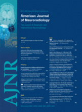
On day 19, the neonate underwent brain CT, with normal findings. MR imaging revealed a small focus of T1 signal-intensity abnormality in the left parietal lobe (Fig 1A). 1H-MR spectroscopy was performed from the right parietal lobe, which had a normal appearance on MR imaging. Single-voxel (SV) MR spectroscopy with TEs of 35 and 144 ms showed high choline (Cho), possible lactate (Lac) peak, and decreased N-acetylaspartate (NAA) (Fig 1B, -C).

Fig 1.
  • Download figure
  • Open in new tab
  • Download powerpoint
Fig 1.

MR imaging and spectroscopy at 19 days of age. T1-weighted imaging (TR = 400 ms, TE = 15 ms, section thickness = 6 mm) shows the foci signals of hemorrhage in the left parietal lobe (arrow, A). Spectroscopy with a 2 × 2 × 2 cm3 voxel located on the contralateral region of the parietal lobe, by using a point-resolved spectroscopic sequence technique (TE = 35 and 144 ms), shows a prominent Lac doublet and decreased NAA peak (B and C).

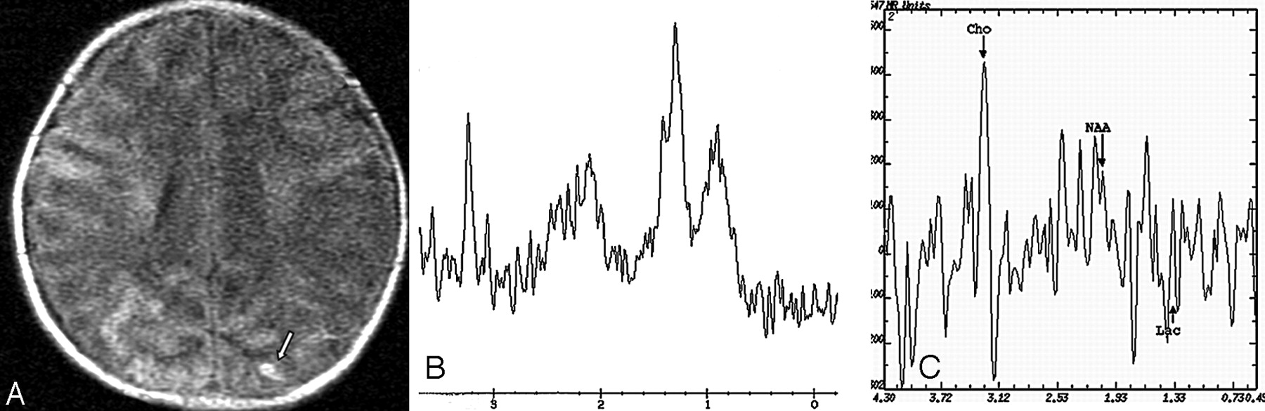
A second MR imaging was performed at day 34, when cutaneous pigmentation was most prominent (Fig 2A). This study showed extensive irregular patchy abnormalities in the periventricular and subcortical white matter in both hemispheres (Fig 2B). SV-MR spectroscopy, again in the right parietal lobe, showed findings similar to the previous one. Multivoxel MR spectroscopy with a long TE (144 ms) showed reduction of the NAA resonance and increased Cho peak (Fig 2C, -D).

Fig 2.
  • Download figure
  • Open in new tab
  • Download powerpoint
Fig 2.

MR imaging and spectroscopy at 34 days of age. Cutaneous hyperpigmented macules become more apparent along Blaschko lines in the inguinal regions (A). Fluid-attenuated inversion recovery image (B) demonstrates low signal intensity of multiple patchy softening of the subcortical white matter in the bilateral hemisphere. MR spectroscopy (multivoxel point-resolved spectroscopic sequence technique with TE = 144 ms) (C and D) in different regions shows evident reduction in NAA resonance peak amplitude at 2.02 ppm in cavities of white matter (D) (No. 4, 5 represented) and increase in the Cho peak in the adjacent parenchyma (D) (Nos. 2 and 3, 8 represented).

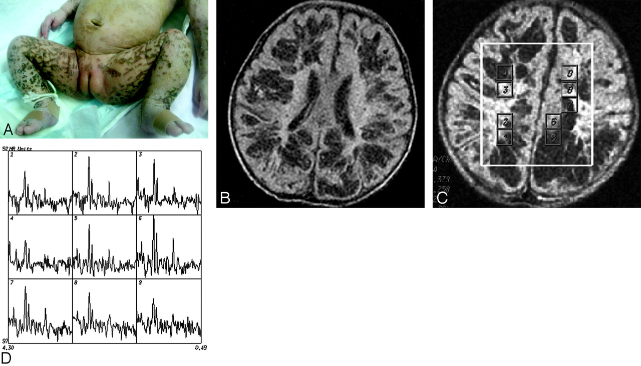
Follow-up MR imaging at 18 weeks of life revealed ventricular enlargement and multiple cystic lesions (Fig 3). To evaluate the development of white matter, we used DTI with b = 750 s/mm2 and 15 directions at day 34 and at 18 weeks. In comparison with the initial study, the latter showed fractional anisotropy (FA) values in the white matter of bilateral frontal, parietooccipital, and periventricular regions to be increased. One month later, the cutaneous lesions disappeared, and she experienced no further seizures. Mild-moderate global cognitive delay remains.

Fig 3.
  • Download figure
  • Open in new tab
  • Download powerpoint
Fig 3.

MR imaging at 4.5 months of age. Fluid-attenuated inversion recovery image reveals the disappearance of the previously observed abnormalities. However, multiple small cystic lesions exhibiting low intensity in the gray matter are present (arrow), and bilateral cerebral atrophy is also observed.

Discussion

The clinical expression of CNS involvement in IP includes mainly epilepsy and mental retardation.4,5 Ninety-two percent of patients have the characteristic skin rash by the age of 2 weeks.2 The patient in this study first presented with typical cutaneous and CNS manifestations of IP. Skin biopsy findings confirmed the diagnosis.

Our patient initially presented with a focus of high T1 signal intensity in a parietal lobe and MR spectroscopy changes suggesting ischemia. Two other articles report hemorrhagic infarcts in the periventricular white matter and associated MR angiographic findings of decreased branching of intracerebral arteries.6,7 Siemes et al8 demonstrated hemorrhagic necrosis with evidence of perivenous and capillary hemorrhage and vascular congestion in the cerebral white matter. We believe that these findings may be indicative of a microangiopathic process.

To the best of our knowledge, reversible extensive patchy abnormalities in the brain in a patient with IP as revealed by serial MR imaging, DTI, and MR spectroscopy have not been reported. In our patient, MR spectroscopy showed significant metabolic changes in these necrosis-like regions and the adjacent parenchyma. The NAA peak was decreased, and the Cho peak was increased; these changes suggested neuronal loss and possible glial proliferation. The FA values of these regions were lower when compared with normal-appearing parenchyma. When we reviewed the literature, we found that there are 2 different pathologic presentations. The first includes periventricular leukomalacia, encephalomalacia, or retinal and cerebral vascular involvement compatible with infarction.9–14 These findings support the hypothesis that vascular disease plays a major role in the pathogenesis of the CNS involvement in IP. This hypothesis is also supported by O’Doherty and Norman's neuropathologic reports.15

Different imaging features have also been recently reported.16,17 They include extensive cortical and subcortical white matter necrosis. Some lesions were cavities within the white matter accompanied by a patchy softening of the cortex. Their MR imaging appearance was similar to that of infants with acute infections of the CNS, suggesting that an inflammatory process may also contribute to the cerebral disease of IP. This hypothesis was verified by histopathology. Hauw et al18 described a diffuse inflammatory process involving the pia-arachnoid and brain, with perivascular cuffs of lymphocytes, histiocytes, and polymorphonuclear leukocytes in the white matter and cortex. Overt vascular abnormalities were absent. They argued that the brain may be involved by an inflammatory process that also results in vesicular skin eruptions. In our patient, the lesions were too extensive to be attributed to vascular occlusion. Although our patient had no signs of infection, we are unable to completely exclude encephalitis leading to encephalomalacia. The follow-up study showed an interesting appearance. MR imaging revealed the disappearance of the previously observed abnormalities. The brain looked normal except for a few residual cystic lesions in the gray matter as well as atrophy. FA values of the subcortical and centra semiovale white matter were increased compared with those in the initial study.

Yoshikawa et al19 reported a transient cavity in the white matter revealed by MR imaging in a patient with IP. They believed that the lesion was caused by a destructive process of vascular origin such as microinfarction or tissue necrosis. The reversible brain abnormalities in IP suggest a natural repair of underlying cerebrovascular disease or inflammation process.

Conclusion

Reversible extensive transient cavities in the cerebral parenchyma as seen on MR imaging, DTI, and MR spectroscopy in IP are rare. This Case Report emphasizes that the initial brain MR imaging findings in IP may simulate those caused by an infection or a microangiopathic process. IP should be included in the differential diagnosis of patients with cutaneous lesions and CNS involvement such as those seen in our patient. CNS involvement in IP might be more frequent than previously reported because of its transient nature.

Acknowledgments

We thank the patient and family for their contribution to this study. We appreciate the medical assistance of Dr. X.J. Wu of The Affiliated Children's Hospital of Zhejiang University.

References

  1. ↵
    Hadj-Rabia S, Froidevaux D, Bodak N, et al. Clinical study of 40 cases of incontinentia pigmenti. Arch Dermatol 2003;139:1163–70
    CrossRefPubMed
  2. ↵
    Carney RG. Incontinentia pigmenti: a world statistical analysis. Arch Dermatol 1976;112:535–42
    CrossRefPubMed
  3. ↵
    Shah SN, Gibbs S, Upton CJ, et al. Incontinentia pigmenti associated with cerebral palsy and cerebral leukomalacia: a case report and literature review. Pediatr Dermatol 2003;20:491–94
    CrossRefPubMed
  4. ↵
    Cohen PR. Incontinentia pigmenti: clinicopathologic characteristics and differential diagnosis. Cutis 1994;54:161–66
    PubMed
  5. ↵
    Nelson-Adesokan P, Mallory SB. Incontinentia pigmenti: the syndrome page. Pediatr Dermatol 1992;9:304–08
    PubMed
  6. ↵
    Hennel SJ, Ekert PG, Volpe JJ, et al. Insights into the pathogenesis of cerebral lesions in incontinentia pigmenti. Pediatr Neurol 2003;29:148–50
    CrossRefPubMed
  7. ↵
    Shuper A, Bryan RN, Singer HS. Destructive encephalopathy in incontinentia pigmenti: a primary disorder? Pediatr Neurol 1990;2:137–40
  8. ↵
    Siemes H, Schneider H, Dening D, et al. Encephalitis in two members of a family with incontinentia pigmenti (Bloch-Sulzberger syndrome): the possible role of inflammation in the pathogenesis of CNS involvement. Eur J Pediatr 1978;129:103–15
    CrossRefPubMed
  9. ↵
    Shah SN, Gibbs S, Upton CJ, et al. Incontinentia pigmenti associated with cerebral palsy and cerebral leukomalacia: a case report and literature review. Pediatr Dermatol 2003;20:491–94
    CrossRefPubMed
  10. Mangano S, Barbagallo A. Incontinentia pigmenti: clinical and neuroradiologic features. Brain Dev 1993;15:362–66
    CrossRefPubMed
  11. Fiorillo L, Sinclair DB, O’Byrne ML, et al. Bilateral cerebrovascular accidents in incontinentia pigmenti. Pediatr Neurol 2003;29:66–68
    CrossRefPubMed
  12. Lee AG, Goldberg MF, Gillard JH, et al. Intracranial assessment of incontinentia pigmenti using magnetic resonance imaging, angiography, and spectroscopic imaging. Arch Pediatr Adolesc Med 1995;149:573–80
    CrossRefPubMed
  13. Kasai T, Kato Z, Matsui E, et al. Cerebral infarction in incontinentia pigmenti: the first report of a case evaluated by single photon emission computed tomography. Acta Paediatr 1997;86:665–67
    PubMed
  14. ↵
    Pellegrino RJ, Shah AJ. Vascular occlusion associated with incontinentia pigmenti. Pediatr Neurol 1994;10:73–74
    CrossRefPubMed
  15. ↵
    O’Doherty NJ, Norman RM. Incontinentia pigmenti (Bloch-Sulzberger syndrome) with cerebral malformation. Dev Med Child Neurol 1968;10:168–74
    PubMed
  16. ↵
    Wolf NI, Kramer N, Harting I, et al. Diffuse cortical necrosis in a neonate with incontinentia pigmenti and an encephalitis-like presentation. AJNR Am J Neuroradiol 2005;26:1580–82
    Abstract/FREE Full Text
  17. ↵
    Porksen G, Pfeiffer C, Hahn G, et al. Neonatal seizures in two sisters with incontinentia pigmenti. Neuropediatrics 2004;35:139–42
    CrossRefPubMed
  18. ↵
    Hauw JJ, Perie G, Bonnette J, et al. [Neuropathological study of incontinentia pigmenti: anatomical case report] (author's translation). Acta Neuropathol (Berl) 1977;38:159–62
    CrossRefPubMed
  19. ↵
    Yoshikawa H, Uehara Y, Abe T, et al. Disappearance of a white matter lesion in incontinentia pigmenti. Pediatr Neurol 2000;23:364–67
    CrossRefPubMed
  • Received July 19, 2007.
  • Accepted after revision October 16, 2007.
  • Copyright © American Society of Neuroradiology
View Abstract
PreviousNext
Back to top

In this issue

American Journal of Neuroradiology: 29 (3)
American Journal of Neuroradiology
Vol. 29, Issue 3
March 2008
  • Table of Contents
  • Index by author
Advertisement
Print
Download PDF
Email Article

Thank you for your interest in spreading the word on American Journal of Neuroradiology.

NOTE: We only request your email address so that the person you are recommending the page to knows that you wanted them to see it, and that it is not junk mail. We do not capture any email address.

Enter multiple addresses on separate lines or separate them with commas.
Nearly Completely Reversible Brain Abnormalities in a Patient with Incontinentia Pigmenti
(Your Name) has sent you a message from American Journal of Neuroradiology
(Your Name) thought you would like to see the American Journal of Neuroradiology web site.
CAPTCHA
This question is for testing whether or not you are a human visitor and to prevent automated spam submissions.
Cite this article
H. Lou, L. Zhang, W. Xiao, J. Zhang, M. Zhang
Nearly Completely Reversible Brain Abnormalities in a Patient with Incontinentia Pigmenti
American Journal of Neuroradiology Mar 2008, 29 (3) 431-433; DOI: 10.3174/ajnr.A0890

Citation Manager Formats

  • BibTeX
  • Bookends
  • EasyBib
  • EndNote (tagged)
  • EndNote 8 (xml)
  • Medlars
  • Mendeley
  • Papers
  • RefWorks Tagged
  • Ref Manager
  • RIS
  • Zotero
0 Responses
Respond to this article
Share
Bookmark this article
Nearly Completely Reversible Brain Abnormalities in a Patient with Incontinentia Pigmenti
H. Lou, L. Zhang, W. Xiao, J. Zhang, M. Zhang
American Journal of Neuroradiology Mar 2008, 29 (3) 431-433; DOI: 10.3174/ajnr.A0890
del.icio.us logo Twitter logo Facebook logo Mendeley logo
  • Tweet Widget
  • Facebook Like
  • Google Plus One
Purchase

Jump to section

  • Article
    • Abstract
    • Case Presentation
    • Discussion
    • Conclusion
    • Acknowledgments
    • References
  • Figures & Data
  • Info & Metrics
  • Responses
  • References
  • PDF

Related Articles

  • No related articles found.
  • PubMed
  • Google Scholar

Cited By...

  • No citing articles found.
  • Crossref (10)
  • Google Scholar

This article has been cited by the following articles in journals that are participating in Crossref Cited-by Linking.

  • Neurological findings in incontinentia pigmenti; a review
    Marije E.C. Meuwissen, Grazia M.S. Mancini
    European Journal of Medical Genetics 2012 55 5
  • Incontinentia pigmenti
    Claudia Schermann Poziomczyk, Julia Kanaan Recuero, Luana Bringhenti, Fernanda Diffini Santa Maria, Carolina Wiltgen Campos, Giovanni Marcos Travi, Andre Moraes Freitas, Marcia Angelica Peter Maahs, Paulo Ricardo Gazzola Zen, Marilu Fiegenbaum, Sheila Tamanini de Almeida, Renan Rangel Bonamigo, Ana Elisa Kiszewski Bau
    Anais Brasileiros de Dermatologia 2014 89 1
  • Pediatric Neurology Part I
    Christine Bodemer
    2013 111
  • Neuroimaging Part I
    Nitasha Klar, Bernard Cohen, Doris D.M. Lin
    2016 135
  • The Rare Neurocutaneous Disorders
    Felipe S. Barros, Victor Hugo R. Marussi, Lázaro L.F. Amaral, Antônio José da Rocha, Christiane M.S. Campos, Leonardo F. Freitas, Thierry A.G.M. Huisman, Bruno P. Soares
    Topics in Magnetic Resonance Imaging 2018 27 6
  • Cerebral Arteriopathy in a Newborn With Incontinentia Pigmenti
    Sarah B. Mulkey, Raghu H. Ramakrishnaiah, Tonya M. Balmakund
    Pediatric Neurology 2016 54
  • The Forgotten Phacomatoses: A Neuroimaging Review of Rare Neurocutaneous Disorders
    Amjad Samara, Mariya Gusman, Loai Aker, Matthew S. Parsons, Ali Y. Mian, Rami W. Eldaya
    Current Problems in Diagnostic Radiology 2022 51 5
  • Neurocutaneous Disorders Phakomatoses and Hamartoneoplastic Syndromes
    Ignacio Pascual-Castroviejo, Martino Ruggieri
    2008
  • Magnetic resonance imaging for diagnosing a rare disease: incontinentia pigmenti (Bloch–Sulzberger syndrome) on the example of a clinical case
    Igor I. Yarmola, Anatoly V. Anikin, Dmitry A. Gankin, Lyubov E. Fomina, Natalia A. Kharitonova, Ilya S. Zhanin, Aleksandr A. Pushkov, Milana A. Basargina, Olga B. Kondakova
    Digital Diagnostics 2023 4 3
  • Pediatric Neuro-Ophthalmology
    Michael C. Brodsky
    2016

More in this TOC Section

  • Comparing MRI Perfusion in Pediatric Brain Tumors
  • Frontal Paraventricular Cysts
  • Sodium MRI in Pediatric Brain Tumors
Show more PEDIATRIC NEUROIMAGING

Similar Articles

Advertisement

Indexed Content

  • Current Issue
  • Accepted Manuscripts
  • Article Preview
  • Past Issues
  • Editorials
  • Editor's Choice
  • Fellows' Journal Club
  • Letters to the Editor
  • Video Articles

Cases

  • Case Collection
  • Archive - Case of the Week
  • Archive - Case of the Month
  • Archive - Classic Case

More from AJNR

  • Trainee Corner
  • Imaging Protocols
  • MRI Safety Corner
  • Book Reviews

Multimedia

  • AJNR Podcasts
  • AJNR Scantastics

Resources

  • Turnaround Time
  • Submit a Manuscript
  • Submit a Video Article
  • Submit an eLetter to the Editor/Response
  • Manuscript Submission Guidelines
  • Statistical Tips
  • Fast Publishing of Accepted Manuscripts
  • Graphical Abstract Preparation
  • Imaging Protocol Submission
  • Evidence-Based Medicine Level Guide
  • Publishing Checklists
  • Author Policies
  • Become a Reviewer/Academy of Reviewers
  • News and Updates

About Us

  • About AJNR
  • Editorial Board
  • Editorial Board Alumni
  • Alerts
  • Permissions
  • Not an AJNR Subscriber? Join Now
  • Advertise with Us
  • Librarian Resources
  • Feedback
  • Terms and Conditions
  • AJNR Editorial Board Alumni

American Society of Neuroradiology

  • Not an ASNR Member? Join Now

© 2025 by the American Society of Neuroradiology All rights, including for text and data mining, AI training, and similar technologies, are reserved.
Print ISSN: 0195-6108 Online ISSN: 1936-959X

Powered by HighWire