Advertisement

Main menu

  • Home
  • Content
    • Current Issue
    • Accepted Manuscripts
    • Article Preview
    • Past Issue Archive
    • Video Articles
    • AJNR Case Collection
    • Case of the Week Archive
    • Case of the Month Archive
    • Classic Case Archive
  • Special Collections
    • Advancing NeuroMRI with High-Relaxivity Contrast Agents
    • AJNR Awards
    • Low-Field MRI
    • Alzheimer Disease
    • ASNR Foundation Special Collection
    • Photon-Counting CT
    • View All
  • Multimedia
    • AJNR Podcasts
    • AJNR SCANtastic
    • Trainee Corner
    • MRI Safety Corner
    • Imaging Protocols
  • For Authors
    • Submit a Manuscript
    • Submit a Video Article
    • Submit an eLetter to the Editor/Response
    • Manuscript Submission Guidelines
    • Statistical Tips
    • Fast Publishing of Accepted Manuscripts
    • Graphical Abstract Preparation
    • Imaging Protocol Submission
    • Author Policies
  • About Us
    • About AJNR
    • Editorial Board
    • Editorial Board Alumni
  • More
    • Become a Reviewer/Academy of Reviewers
    • Subscribers
    • Permissions
    • Alerts
    • Feedback
    • Advertisers
    • ASNR Home

User menu

  • Alerts
  • Log in

Search

  • Advanced search
American Journal of Neuroradiology

American Journal of Neuroradiology

ASHNR American Society of Functional Neuroradiology ASHNR American Society of Pediatric Neuroradiology ASSR
  • Alerts
  • Log in

Advanced Search

  • Home
  • Content
    • Current Issue
    • Accepted Manuscripts
    • Article Preview
    • Past Issue Archive
    • Video Articles
    • AJNR Case Collection
    • Case of the Week Archive
    • Case of the Month Archive
    • Classic Case Archive
  • Special Collections
    • Advancing NeuroMRI with High-Relaxivity Contrast Agents
    • AJNR Awards
    • Low-Field MRI
    • Alzheimer Disease
    • ASNR Foundation Special Collection
    • Photon-Counting CT
    • View All
  • Multimedia
    • AJNR Podcasts
    • AJNR SCANtastic
    • Trainee Corner
    • MRI Safety Corner
    • Imaging Protocols
  • For Authors
    • Submit a Manuscript
    • Submit a Video Article
    • Submit an eLetter to the Editor/Response
    • Manuscript Submission Guidelines
    • Statistical Tips
    • Fast Publishing of Accepted Manuscripts
    • Graphical Abstract Preparation
    • Imaging Protocol Submission
    • Author Policies
  • About Us
    • About AJNR
    • Editorial Board
    • Editorial Board Alumni
  • More
    • Become a Reviewer/Academy of Reviewers
    • Subscribers
    • Permissions
    • Alerts
    • Feedback
    • Advertisers
    • ASNR Home
  • Follow AJNR on Twitter
  • Visit AJNR on Facebook
  • Follow AJNR on Instagram
  • Join AJNR on LinkedIn
  • RSS Feeds

AJNR Awards, New Junior Editors, and more. Read the latest AJNR updates


Improved Turnaround Times | Median time to first decision: 12 days

Research ArticleBrain

Thrombus Volume Comparison between Patients with and without Hyperattenuated Artery Sign on CT

E.Y. Kim, E. Yoo, H.Y. Choi, J.W. Lee and J.H. Heo
American Journal of Neuroradiology February 2008, 29 (2) 359-362; DOI: https://doi.org/10.3174/ajnr.A0800
E.Y. Kim
  • Find this author on Google Scholar
  • Find this author on PubMed
  • Search for this author on this site
E. Yoo
  • Find this author on Google Scholar
  • Find this author on PubMed
  • Search for this author on this site
H.Y. Choi
  • Find this author on Google Scholar
  • Find this author on PubMed
  • Search for this author on this site
J.W. Lee
  • Find this author on Google Scholar
  • Find this author on PubMed
  • Search for this author on this site
J.H. Heo
  • Find this author on Google Scholar
  • Find this author on PubMed
  • Search for this author on this site
  • Article
  • Figures & Data
  • Info & Metrics
  • Responses
  • References
  • PDF
Loading

Article Figures & Data

Figures

  • Tables
  • Fig 1.
    • Download figure
    • Open in new tab
    • Download powerpoint
    Fig 1.

    A representative case showing the procedure of thrombus volume measurement. A, Maximum intensity projection images of thin-section noncontrast CT in a patient with occlusion in both M1 and M2 segments on the right (arrows). B, All pixels are segmented with a threshold of 50–100 HU to select the thrombus (azure areas). After selecting the function of 3D region growing, we drew a region of interest around the thrombus on an axial image and then automatically segmented the whole thrombus (indigo areas). Note that the thrombus does not approximate the border of the probable thrombus. C, Pixel dilation with a single iteration for the segmented thrombus is performed at a threshold of 40–100 HU. The thrombus approximates its border more than does the thrombus on B (indigo areas). D, The volume and mean Hounsfield unit of the thrombus are calculated. MinValue indicates minimal value; MeanValue, mean value; MaxValue, maximal value.
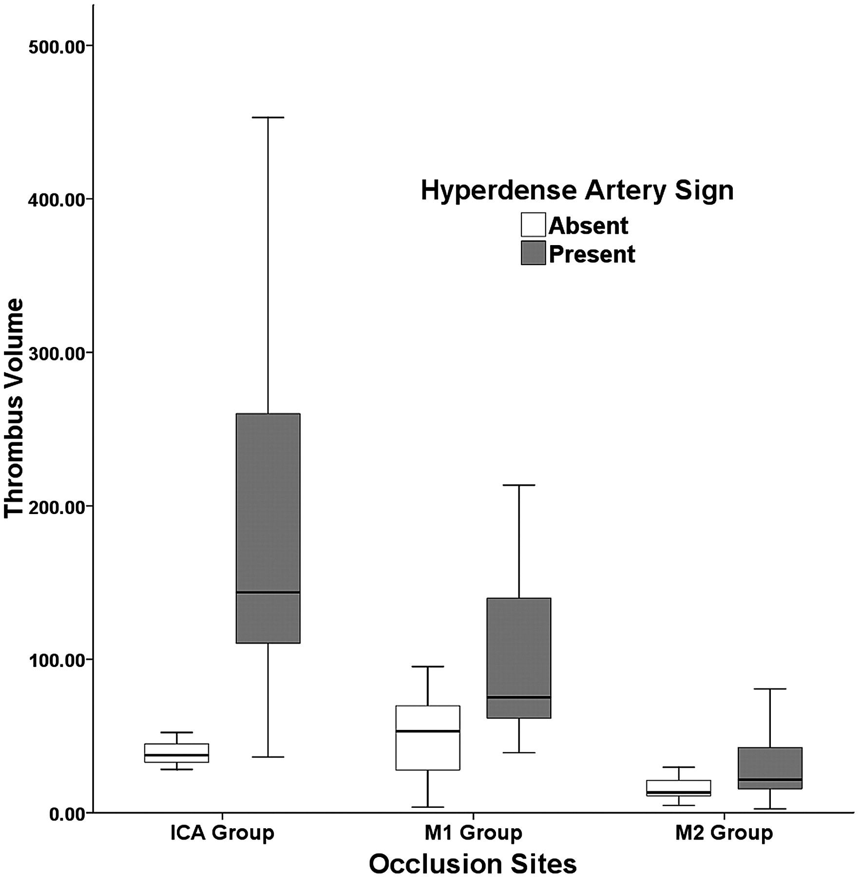
  • Fig 2.
    • Download figure
    • Open in new tab
    • Download powerpoint
    Fig 2.

    Thrombus volume comparison according to the presence or absence of hyperdense artery sign.

Tables

  • Figures
    • View popup
    Table 1:

    Thrombus volumes in patients with and without HAS

    ICA Group (n = 14)M1 Group (n = 42)M2 Group (n = 22)
    HAS (+) (n = 11)HAS (–) (n = 3)P*HAS (+) (n = 25)HAS (–) (n = 17)P*HAS (+) (n = 10)HAS (–) (n = 12)P*
    Mean thrombus volume (mm3)188.7 ± 122.539.4 ± 12.1.022128.1 ± 119.256.8 ± 32.5.00534.7 ± 32.220.0 ± 20.0.18
    • Note:—+ indicates presence; –indicates absence.

    • * Mann-Whitney U test.

    • View popup
    Table 2:

    Thrombus volume comparison according to the occlusion site

    ICA group (n = 14)M1 group (n = 42)M2 group (n = 22)P
    Mean thrombus volume (mm3)156.7 ± 125.094.1 ± 95.226.7 ± 26.7<.001*
    HAS (present/absent)11/3 (78.6%)25/17 (59.5%)10/12 (45.5%)>.05†
    • * Kruskal-Wallis one-way ANOVA test.

    • † χ2 test.

PreviousNext
Back to top

In this issue

American Journal of Neuroradiology: 29 (2)
American Journal of Neuroradiology
Vol. 29, Issue 2
February 2008
  • Table of Contents
  • Index by author
Advertisement
Print
Download PDF
Email Article

Thank you for your interest in spreading the word on American Journal of Neuroradiology.

NOTE: We only request your email address so that the person you are recommending the page to knows that you wanted them to see it, and that it is not junk mail. We do not capture any email address.

Enter multiple addresses on separate lines or separate them with commas.
Thrombus Volume Comparison between Patients with and without Hyperattenuated Artery Sign on CT
(Your Name) has sent you a message from American Journal of Neuroradiology
(Your Name) thought you would like to see the American Journal of Neuroradiology web site.
CAPTCHA
This question is for testing whether or not you are a human visitor and to prevent automated spam submissions.
Cite this article
E.Y. Kim, E. Yoo, H.Y. Choi, J.W. Lee, J.H. Heo
Thrombus Volume Comparison between Patients with and without Hyperattenuated Artery Sign on CT
American Journal of Neuroradiology Feb 2008, 29 (2) 359-362; DOI: 10.3174/ajnr.A0800

Citation Manager Formats

  • BibTeX
  • Bookends
  • EasyBib
  • EndNote (tagged)
  • EndNote 8 (xml)
  • Medlars
  • Mendeley
  • Papers
  • RefWorks Tagged
  • Ref Manager
  • RIS
  • Zotero
0 Responses
Respond to this article
Share
Bookmark this article
Thrombus Volume Comparison between Patients with and without Hyperattenuated Artery Sign on CT
E.Y. Kim, E. Yoo, H.Y. Choi, J.W. Lee, J.H. Heo
American Journal of Neuroradiology Feb 2008, 29 (2) 359-362; DOI: 10.3174/ajnr.A0800
del.icio.us logo Twitter logo Facebook logo Mendeley logo
  • Tweet Widget
  • Facebook Like
  • Google Plus One
Purchase

Jump to section

  • Article
    • Abstract
    • Materials and Methods
    • Results
    • Discussion
    • Conclusion
    • Footnotes
    • References
  • Figures & Data
  • Info & Metrics
  • Responses
  • References
  • PDF

Related Articles

  • No related articles found.
  • PubMed
  • Google Scholar

Cited By...

  • The Protective Effect of Middle Cerebral Artery Calcification on Symptomatic Middle Cerebral Artery Infarction
  • Effect of alteplase on the CT hyperdense artery sign and outcome after ischemic stroke
  • Sensitivity and Specificity of the Hyperdense Artery Sign for Arterial Obstruction in Acute Ischemic Stroke
  • Quantification of Thrombus Hounsfield Units on Noncontrast CT Predicts Stroke Subtype and Early Recanalization after Intravenous Recombinant Tissue Plasminogen Activator
  • The Importance of Size: Successful Recanalization by Intravenous Thrombolysis in Acute Anterior Stroke Depends on Thrombus Length
  • Assessment of Thrombus in Acute Middle Cerebral Artery Occlusion Using Thin-Slice Nonenhanced Computed Tomography Reconstructions
  • Crossref
  • Google Scholar

This article has not yet been cited by articles in journals that are participating in Crossref Cited-by Linking.

More in this TOC Section

  • Usefulness of Quantitative Susceptibility Mapping for the Diagnosis of Parkinson Disease
  • White Matter Alterations in the Brains of Patients with Active, Remitted, and Cured Cushing Syndrome: A DTI Study
  • Qualitative and Quantitative Analysis of MR Imaging Findings in Patients with Middle Cerebral Artery Stroke Implanted with Mesenchymal Stem Cells
Show more Brain

Similar Articles

Advertisement

Indexed Content

  • Current Issue
  • Accepted Manuscripts
  • Article Preview
  • Past Issues
  • Editorials
  • Editor's Choice
  • Fellows' Journal Club
  • Letters to the Editor
  • Video Articles

Cases

  • Case Collection
  • Archive - Case of the Week
  • Archive - Case of the Month
  • Archive - Classic Case

More from AJNR

  • Trainee Corner
  • Imaging Protocols
  • MRI Safety Corner
  • Book Reviews

Multimedia

  • AJNR Podcasts
  • AJNR Scantastics

Resources

  • Turnaround Time
  • Submit a Manuscript
  • Submit a Video Article
  • Submit an eLetter to the Editor/Response
  • Manuscript Submission Guidelines
  • Statistical Tips
  • Fast Publishing of Accepted Manuscripts
  • Graphical Abstract Preparation
  • Imaging Protocol Submission
  • Evidence-Based Medicine Level Guide
  • Publishing Checklists
  • Author Policies
  • Become a Reviewer/Academy of Reviewers
  • News and Updates

About Us

  • About AJNR
  • Editorial Board
  • Editorial Board Alumni
  • Alerts
  • Permissions
  • Not an AJNR Subscriber? Join Now
  • Advertise with Us
  • Librarian Resources
  • Feedback
  • Terms and Conditions
  • AJNR Editorial Board Alumni

American Society of Neuroradiology

  • Not an ASNR Member? Join Now

© 2025 by the American Society of Neuroradiology All rights, including for text and data mining, AI training, and similar technologies, are reserved.
Print ISSN: 0195-6108 Online ISSN: 1936-959X

Powered by HighWire