
Article Figures & Data
Figures
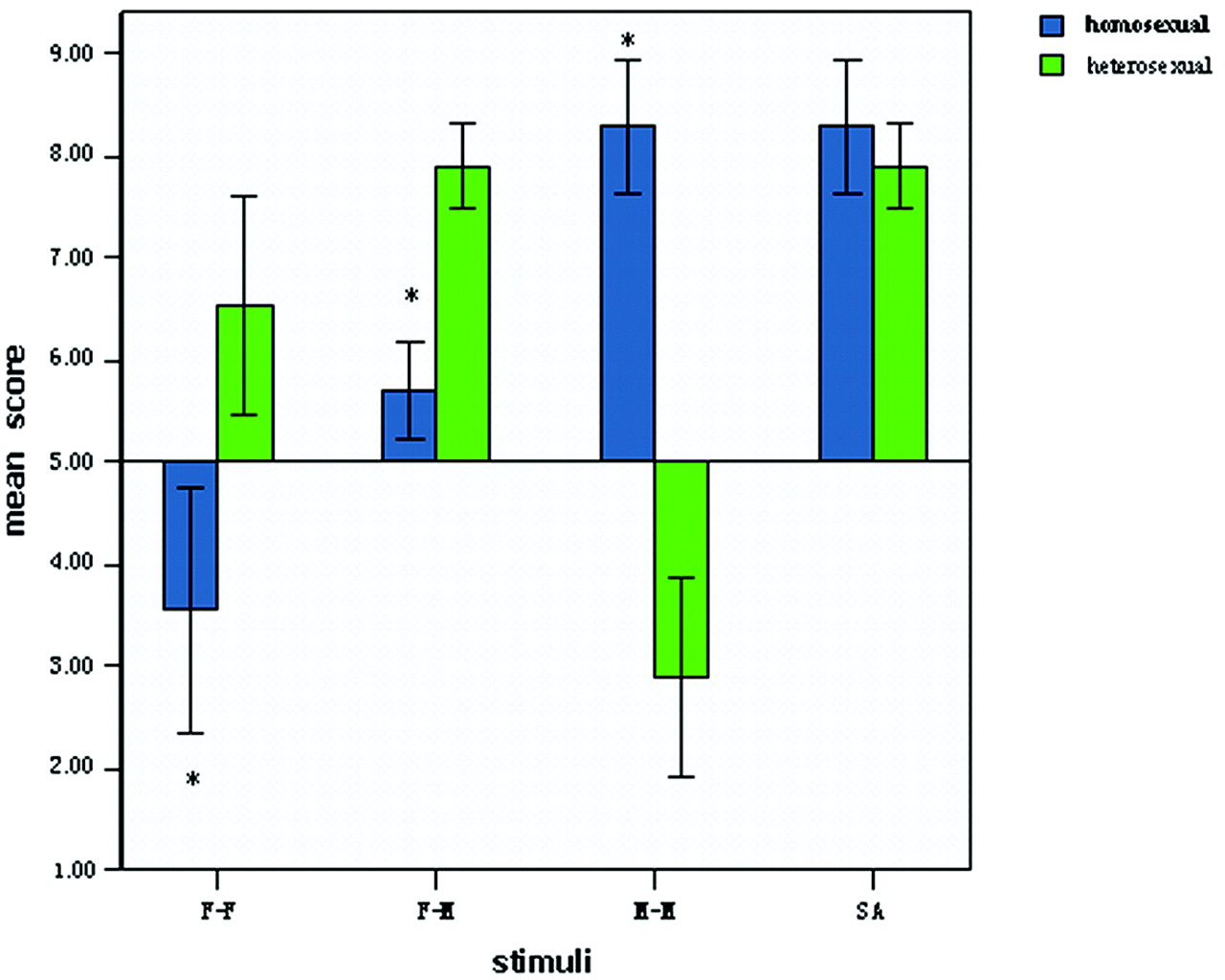
- Fig 1.
Mean scores of the sexual attractiveness of films showing F-F, F-M, and M-M and the maximal level of evoked sexual arousal (SA) in the 2 groups. In homosexual men, M-M stimuli induce maximal sexual arousal, whereas F-M stimuli are preferred in heterosexual men. Results of 2 independent samples test comparisons (homosexual versus heterosexual) are displayed. Blue indicates homosexual men; green, heterosexual men; the asterisk, P < .001. Error bars equal 1 SD.
- Fig 2.
Images showing brain activation in response to M-M stimuli in the homosexual group compared with the same regions in response to F-M stimuli in the heterosexual group, by 1-sample t test analysis. In each labeled panel of 6 images, the upper rows are from homosexual men and the lower rows are from heterosexual men. Significant activation occurs in the left angular gyrus (x = −42, y = −68, z = 48) (A), left caudate (x = −8, y = 0, z = 24) (B), and right pallidum (x = 14, y = 0, z = 6) (C). The bar shows the range of the t value. Numbers in the bottom row indicate the coordinates of the Montreal Neurological Institute brain.
- Fig 3.
Images showing brain activation in response to F-M stimuli in the heterosexual group compared with the same regions in response to M-M stimuli in the homosexual group, by 1-sample t test analysis. In each labeled panel of 8 images, the upper rows are from homosexual men and the lower rows from heterosexual men. Significant activation occurs in the left lingual gyrus (x = 2, y = −80, z = −2) (A), right lingual gyrus (x = 6, y = −70, z = 2) (B), right hippocampus (x = 20, y = −36, z = −2) (C), and right parahippocampus (x = 16, y = −36, z = −10) (D). The bar shows the range of the t value. Numbers in the bottom row indicate the coordinates of the Montreal Neurological Institute brain.


Tables
- Table 1:
Brain activation of the male homosexual group during visually evoked sexual arousal in M-M versus rest condition*
Brain Region L/R Coordinates SPM{t} Puncorr x y z Angular gyrus L −42 −68 48 10.24 <.001 Caudate L −8 0 24 6.3 <.001 Cerebellum L −12 −86 −28 9.21 <.001 Cerebellum L −6 −78 −48 8.36 <.001 Inferior frontal gyrus R 60 20 4 7.21 <.001 Inferior frontal gyrus R 54 30 20 6.49 <.001 Inferior frontal gyrus R 44 12 18 6.39 <.001 Inferior temporal gyrus L −52 −68 −10 17.25 <.001 Inferior temporal gyrus R 56 −66 −6 16.97 <.001 Inferior temporal gyrus L −50 −62 −18 16.19 <.001 Inferior temporal gyrus R 64 −30 −18 9.7 <.001 Insula L −38 0 6 5.45 <.001 Medial superior frontal gyrus R 8 72 12 7.86 <.001 Middle cingulate gyrus R 2 −6 38 9.26 <.001 Middle cingulate gyrus R 4 20 32 8.89 <.001 Middle frontal gyrus L −42 56 6 9.04 <.001 Middle frontal gyrus L −36 52 2 8.49 <.001 Middle frontal gyrus R 52 44 16 8.11 <.001 Middle occipital gyrus L −28 −84 32 9.68 <.001 Middle temporal gyrus L −64 −12 −18 8.01 <.001 Pallidum R 14 0 6 5.87 <.001 Postcentral gyrus R 32 −36 66 8.2 <.001 Postcentral gyrus L −64 −10 20 7.04 <.001 Posterior cingulate gyrus R 12 −36 6 7.48 <.001 Precentral gyrus R 54 2 30 7.38 <.001 Precuneus L −22 −42 18 7.6 <.001 Superior frontal gyrus L −16 72 6 10.67 <.001 Superior parietal gyrus L −26 −78 54 9.41 <.001 Thalamus L −8 −32 6 5.38 <.001 Thalamus R 12 −10 8 6.86 <.001 Vermis −2 −50 −30 4.98 <.001 Note:—Puncorr indicates P uncorrected; l, left; R, right; M-M, male homosexual couples.
* SPM2; 1-sample t test; P < 0.001 uncorrected; extent, 10 voxels.
- Table 2:
Brain activation of male heterosexual group during visually evoked sexual arousal in F-M versus rest condition*
Brain Region L/R Coordinates SPM{t} Puncorr x y z Anterior cingulate gyrus L −2 2 26 7.63 <.001 Cerebellum L −30 −90 −26 12.99 <.001 Cerebellum R 40 −60 −32 11.46 <.001 Hippocampus R 20 −36 −2 7.61 <.001 Inferior parietal gyrus L −32 −56 48 9.63 <.001 Inferior temporal gyrus R 48 −72 −10 20.21 <.001 Inferior frontal gyrus L −54 12 24 8.29 <.001 Inferior frontal gyrus R 54 20 10 10.7 <.001 Inferior frontal gyrus R 54 32 30 7.1 <.001 Inferior occipital gyrus L −54 −70 −8 19.07 <.001 Insula L −40 0 −4 5.97 <.001 Insula R 38 6 −10 6.77 <.001 Lingual gyrus L 2 −80 −2 8.97 <.001 Lingual gyrus R 6 −70 2 8.42 <.001 Middle frontal gyrus R 8 40 −6 13.65 <.001 Middle frontal gyrus L −38 58 16 11.13 <.001 Middle frontal gyrus R 52 42 20 11.73 <.001 Middle occipital gyrus L −52 −74 0 16.96 <.001 Middle occipital gyrus R 28 −92 2 7.67 <.001 Middle temporal gyrus R 62 −6 −26 8.53 <.001 Parahippocampus R 14 −14 −20 6.76 <.001 Postcentral gyrus L −60 −16 40 6.98 <.001 Precentral gyrus L −20 −16 62 7.72 <.001 Precentral gyrus R 48 10 36 10.14 <.001 Precuneus L −12 −40 4 8.84 <.001 Superior frontal gyrus L −20 70 8 9.07 <.001 Superior frontal gyrus R 32 64 16 10.97 <.001 Superior parietal gyrus R 28 −66 50 9.21 <.001 Superior parietal gyrus L −18 −52 66 11.9 <.001 Thalamus L −16 −18 12 6.19 <.001 Vermis 0 −26 −24 7.86 <.001 Vermis −6 −44 −30 5.74 <.001 Note:—Puncorr indicates P uncorrected; l, left; R, right; F-M, heterosexual couples.
* SPM2; 1-sample t test; P < .001 uncorrected; extent, 10 voxels.


