Advertisement

Main menu

  • Home
  • Content
    • Current Issue
    • Accepted Manuscripts
    • Article Preview
    • Past Issue Archive
    • Video Articles
    • AJNR Case Collection
    • Case of the Week Archive
    • Case of the Month Archive
    • Classic Case Archive
  • Special Collections
    • AJNR Awards
    • Low-Field MRI
    • Alzheimer Disease
    • ASNR Foundation Special Collection
    • Photon-Counting CT
    • View All
  • Multimedia
    • AJNR Podcasts
    • AJNR SCANtastic
    • Trainee Corner
    • MRI Safety Corner
    • Imaging Protocols
  • For Authors
    • Submit a Manuscript
    • Submit a Video Article
    • Submit an eLetter to the Editor/Response
    • Manuscript Submission Guidelines
    • Statistical Tips
    • Fast Publishing of Accepted Manuscripts
    • Graphical Abstract Preparation
    • Imaging Protocol Submission
    • Author Policies
  • About Us
    • About AJNR
    • Editorial Board
    • Editorial Board Alumni
  • More
    • Become a Reviewer/Academy of Reviewers
    • Subscribers
    • Permissions
    • Alerts
    • Feedback
    • Advertisers
    • ASNR Home

User menu

  • Alerts
  • Log in

Search

  • Advanced search
American Journal of Neuroradiology

American Journal of Neuroradiology

ASHNR American Society of Functional Neuroradiology ASHNR American Society of Pediatric Neuroradiology ASSR
  • Alerts
  • Log in

Advanced Search

  • Home
  • Content
    • Current Issue
    • Accepted Manuscripts
    • Article Preview
    • Past Issue Archive
    • Video Articles
    • AJNR Case Collection
    • Case of the Week Archive
    • Case of the Month Archive
    • Classic Case Archive
  • Special Collections
    • AJNR Awards
    • Low-Field MRI
    • Alzheimer Disease
    • ASNR Foundation Special Collection
    • Photon-Counting CT
    • View All
  • Multimedia
    • AJNR Podcasts
    • AJNR SCANtastic
    • Trainee Corner
    • MRI Safety Corner
    • Imaging Protocols
  • For Authors
    • Submit a Manuscript
    • Submit a Video Article
    • Submit an eLetter to the Editor/Response
    • Manuscript Submission Guidelines
    • Statistical Tips
    • Fast Publishing of Accepted Manuscripts
    • Graphical Abstract Preparation
    • Imaging Protocol Submission
    • Author Policies
  • About Us
    • About AJNR
    • Editorial Board
    • Editorial Board Alumni
  • More
    • Become a Reviewer/Academy of Reviewers
    • Subscribers
    • Permissions
    • Alerts
    • Feedback
    • Advertisers
    • ASNR Home
  • Follow AJNR on Twitter
  • Visit AJNR on Facebook
  • Follow AJNR on Instagram
  • Join AJNR on LinkedIn
  • RSS Feeds

AJNR Awards, New Junior Editors, and more. Read the latest AJNR updates

Research ArticleHead and Neck Imaging

The Hypodense Focus in the Petrous Apex: A Potential Pitfall on Multidetector CT Imaging of the Temporal Bone

T. Moser, F. Veillon, H. Sick and S. Riehm
American Journal of Neuroradiology January 2008, 29 (1) 35-39; DOI: https://doi.org/10.3174/ajnr.A0737
T. Moser
  • Find this author on Google Scholar
  • Find this author on PubMed
  • Search for this author on this site
F. Veillon
  • Find this author on Google Scholar
  • Find this author on PubMed
  • Search for this author on this site
H. Sick
  • Find this author on Google Scholar
  • Find this author on PubMed
  • Search for this author on this site
S. Riehm
  • Find this author on Google Scholar
  • Find this author on PubMed
  • Search for this author on this site
  • Article
  • Figures & Data
  • Info & Metrics
  • Responses
  • References
  • PDF
Loading

Article Figures & Data

Figures

  • Fig 1.
    • Download figure
    • Open in new tab
    • Download powerpoint
    Fig 1.

    Six-year-old girl with complicated bilateral acute otitis media. Transverse (A) and coronal (B) MDCT sections of the right temporal bone. Transverse (C) and coronal (D) MDCT sections of the left temporal bone. A well-defined hypoattenuated area (black arrow) is seen in the anterior aspect of the otic capsule on both sides. The hypoattenuated focus of the fissula ante fenestram (white arrow) is also apparent.

  • Fig 2.
    • Download figure
    • Open in new tab
    • Download powerpoint
    Fig 2.

    Thirty-four-year-old woman with dizziness. Transverse (A) and coronal (B) MDCT sections of the right temporal bone. Transverse (C) and coronal (D) MDCT sections of the left temporal bone. Bilateral hypoattenuated foci (black arrow) in the anterior otic capsules are particularly well underlined by complete pneumatization of the petrous apex. Otosclerosis was ruled out by other investigations, and these findings were considered as normal variants.

  • Fig 3.
    • Download figure
    • Open in new tab
    • Download powerpoint
    Fig 3.

    Photomicrograph of a parasagittal section of the head from an 8-month-old fetus. A, General view. Compared with other structures of the skull base, the otic capsule (frame) appears surprisingly large as it has already attained its adult size. Bony landmarks are F, frontal; Mx, maxillary; Md, mandible; S, sphenoid; C, clavicle; P, petrous; O, occipital; A, atlas. B, Close-up view of the otic capsule. The different layers (o indicates outer; m, middle; i, inner) of the otic capsule are well demonstrated around the basal turn of the cochlea (cbt). A focal dehiscence of the outer layer with protrusion of the middle layer is clearly seen (arrows). The protrusion of the middle layer is directed toward the petro-occipital fissure (pof) and imperceptibly merges with a cartilaginous cap (c) that is distinct from the fibrous tissue occupying the petro-occipital fissure.
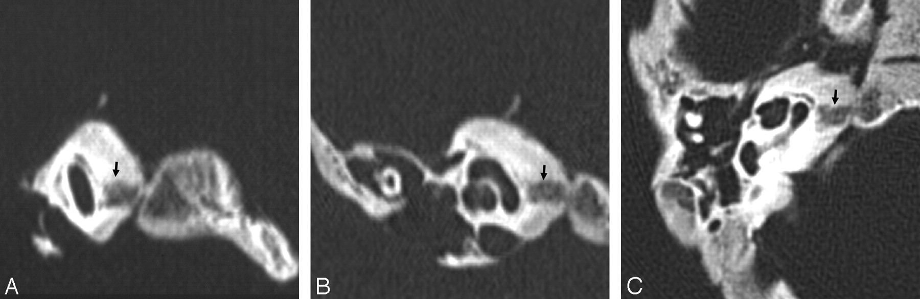
  • Fig 4.
    • Download figure
    • Open in new tab
    • Download powerpoint
    Fig 4.

    Sagittal oblique (A), coronal (B), and transverse (C) MDCT sections from a neonate specimen. The middle otic layer is seen as a smooth hypoattenuation around the cochlea, circumscribed by the attenuated inner and outer layers. A well-defined hypoattenuated focus (arrow) is seen in the anterior otic capsule, communicating laterally with the middle layer and medially with the petro-occipital fissure.

  • Fig 5.
    • Download figure
    • Open in new tab
    • Download powerpoint
    Fig 5.

    Transverse MDCT section from a 4-month-old fetal specimen. The middle otic layer is seen as a smooth hypoattenuation surrounding the cochlea, circumscribed by the attenuated inner and outer layers. The dehiscence of the outer layer of the otic capsule (arrows) allows broad communication between the middle layer and the petro-occipital fissure. The petrous apex is not yet developed.

  • Fig 6.
    • Download figure
    • Open in new tab
    • Download powerpoint
    Fig 6.

    Transverse MDCT image from a 2-month-old infant specimen. The middle otic layer persists, though tinier than in previous stages. The hypoattenuated focus (arrow) is well demonstrated, spanning the petrous apex, which is now developed and reaching the petro-occipital fissure through a focal cortical interruption.
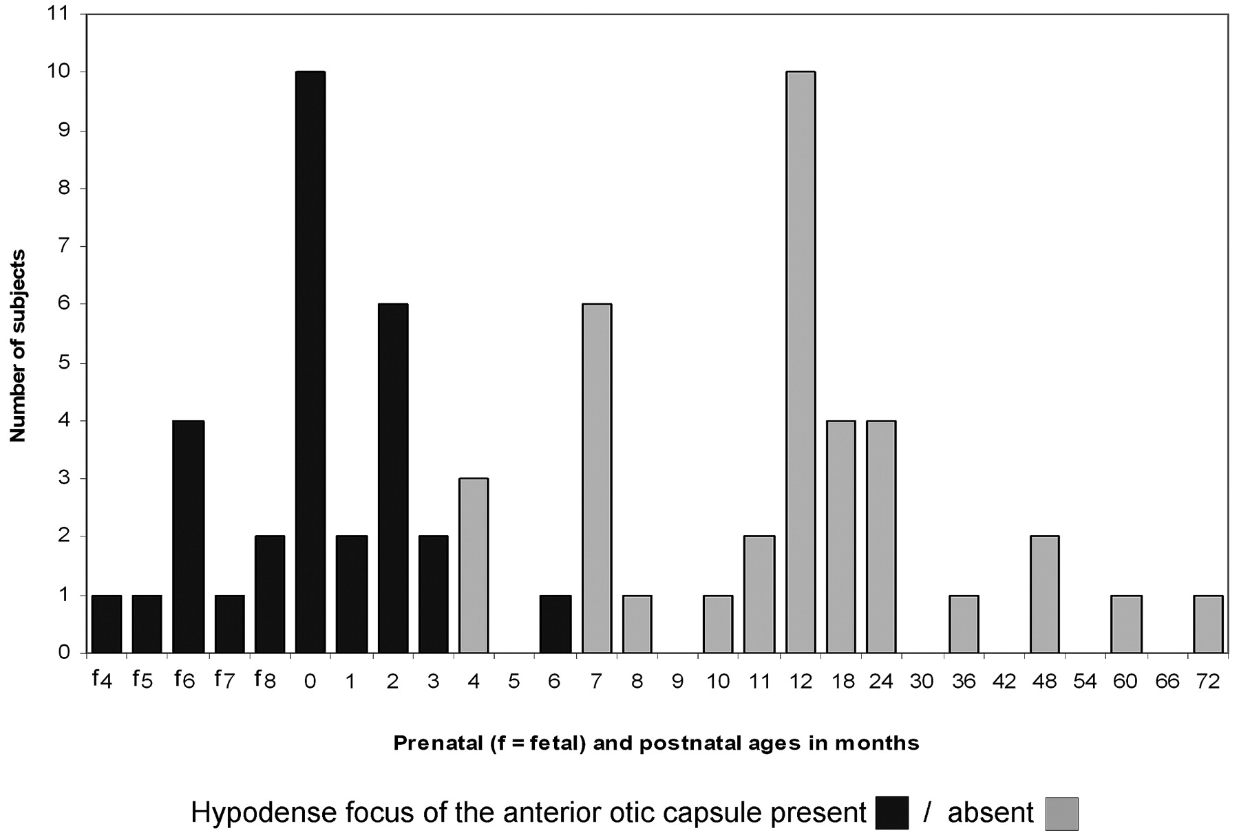
  • Fig 7.
    • Download figure
    • Open in new tab
    • Download powerpoint
    Fig 7.

    Summary of findings in anatomic specimens.

PreviousNext
Back to top

In this issue

American Journal of Neuroradiology: 29 (1)
American Journal of Neuroradiology
Vol. 29, Issue 1
January 2008
  • Table of Contents
  • Index by author
Advertisement
Print
Download PDF
Email Article

Thank you for your interest in spreading the word on American Journal of Neuroradiology.

NOTE: We only request your email address so that the person you are recommending the page to knows that you wanted them to see it, and that it is not junk mail. We do not capture any email address.

Enter multiple addresses on separate lines or separate them with commas.
The Hypodense Focus in the Petrous Apex: A Potential Pitfall on Multidetector CT Imaging of the Temporal Bone
(Your Name) has sent you a message from American Journal of Neuroradiology
(Your Name) thought you would like to see the American Journal of Neuroradiology web site.
CAPTCHA
This question is for testing whether or not you are a human visitor and to prevent automated spam submissions.
Cite this article
T. Moser, F. Veillon, H. Sick, S. Riehm
The Hypodense Focus in the Petrous Apex: A Potential Pitfall on Multidetector CT Imaging of the Temporal Bone
American Journal of Neuroradiology Jan 2008, 29 (1) 35-39; DOI: 10.3174/ajnr.A0737

Citation Manager Formats

  • BibTeX
  • Bookends
  • EasyBib
  • EndNote (tagged)
  • EndNote 8 (xml)
  • Medlars
  • Mendeley
  • Papers
  • RefWorks Tagged
  • Ref Manager
  • RIS
  • Zotero
0 Responses
Respond to this article
Share
Bookmark this article
The Hypodense Focus in the Petrous Apex: A Potential Pitfall on Multidetector CT Imaging of the Temporal Bone
T. Moser, F. Veillon, H. Sick, S. Riehm
American Journal of Neuroradiology Jan 2008, 29 (1) 35-39; DOI: 10.3174/ajnr.A0737
del.icio.us logo Twitter logo Facebook logo Mendeley logo
  • Tweet Widget
  • Facebook Like
  • Google Plus One
Purchase

Jump to section

  • Article
    • Abstract
    • Materials and Methods
    • Results
    • Discussion
    • Acknowledgments
    • References
  • Figures & Data
  • Info & Metrics
  • Responses
  • References
  • PDF

Related Articles

  • No related articles found.
  • PubMed
  • Google Scholar

Cited By...

  • Internal Auditory Canal Diverticula among Pediatric Patients: Prevalence and Assessment for Hearing Loss and Anatomic Associations
  • Incomplete Endochondral Ossification of the Otic Capsule, A Variation in Children: Evaluation of Its Prevalence and Extent in Children with and without Sensorineural Hearing Loss
  • Crossref
  • Google Scholar

This article has not yet been cited by articles in journals that are participating in Crossref Cited-by Linking.

More in this TOC Section

  • Diagnostic Performance of US in Neck Node NIRADS 2
  • Hydrops Herniation into the Semicircular Canals
  • ASL Sensitivity for Head and Neck Paraganglioma
Show more HEAD AND NECK IMAGING

Similar Articles

Advertisement

Indexed Content

  • Current Issue
  • Accepted Manuscripts
  • Article Preview
  • Past Issues
  • Editorials
  • Editor's Choice
  • Fellows' Journal Club
  • Letters to the Editor
  • Video Articles

Cases

  • Case Collection
  • Archive - Case of the Week
  • Archive - Case of the Month
  • Archive - Classic Case

More from AJNR

  • Trainee Corner
  • Imaging Protocols
  • MRI Safety Corner
  • Book Reviews

Multimedia

  • AJNR Podcasts
  • AJNR Scantastics

Resources

  • Turnaround Time
  • Submit a Manuscript
  • Submit a Video Article
  • Submit an eLetter to the Editor/Response
  • Manuscript Submission Guidelines
  • Statistical Tips
  • Fast Publishing of Accepted Manuscripts
  • Graphical Abstract Preparation
  • Imaging Protocol Submission
  • Evidence-Based Medicine Level Guide
  • Publishing Checklists
  • Author Policies
  • Become a Reviewer/Academy of Reviewers
  • News and Updates

About Us

  • About AJNR
  • Editorial Board
  • Editorial Board Alumni
  • Alerts
  • Permissions
  • Not an AJNR Subscriber? Join Now
  • Advertise with Us
  • Librarian Resources
  • Feedback
  • Terms and Conditions
  • AJNR Editorial Board Alumni

American Society of Neuroradiology

  • Not an ASNR Member? Join Now

© 2025 by the American Society of Neuroradiology All rights, including for text and data mining, AI training, and similar technologies, are reserved.
Print ISSN: 0195-6108 Online ISSN: 1936-959X

Powered by HighWire