
Article Figures & Data
Figures
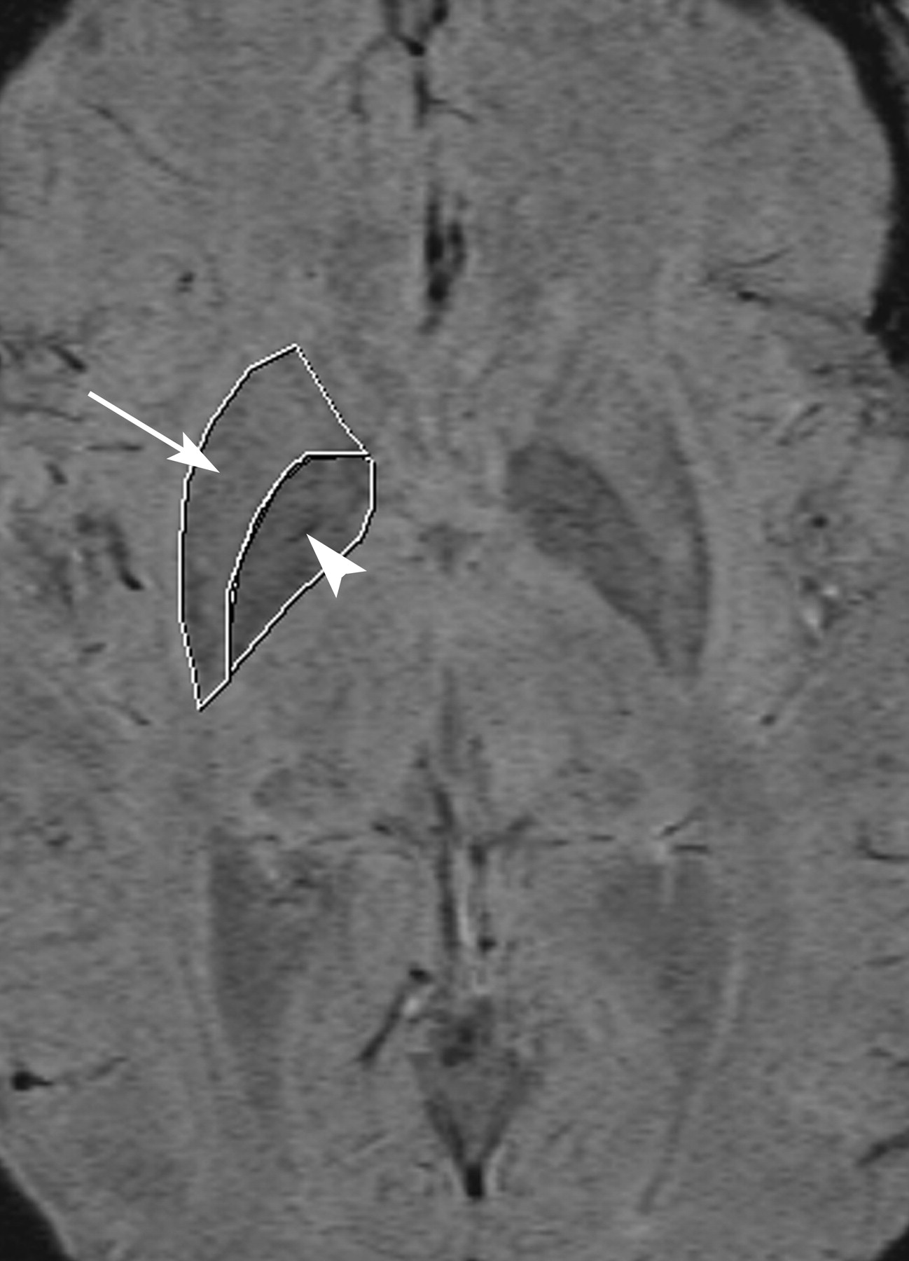
- Fig 1.
Axial SWI image demonstrating a region of interest drawn around the putamen (arrow) and globus pallidus (arrowhead).
- Fig 2.
A schematic drawing of the putamen (A) and corresponding axial SWI images (B) demonstrating the mSHIP scale (grades 0 to 4).
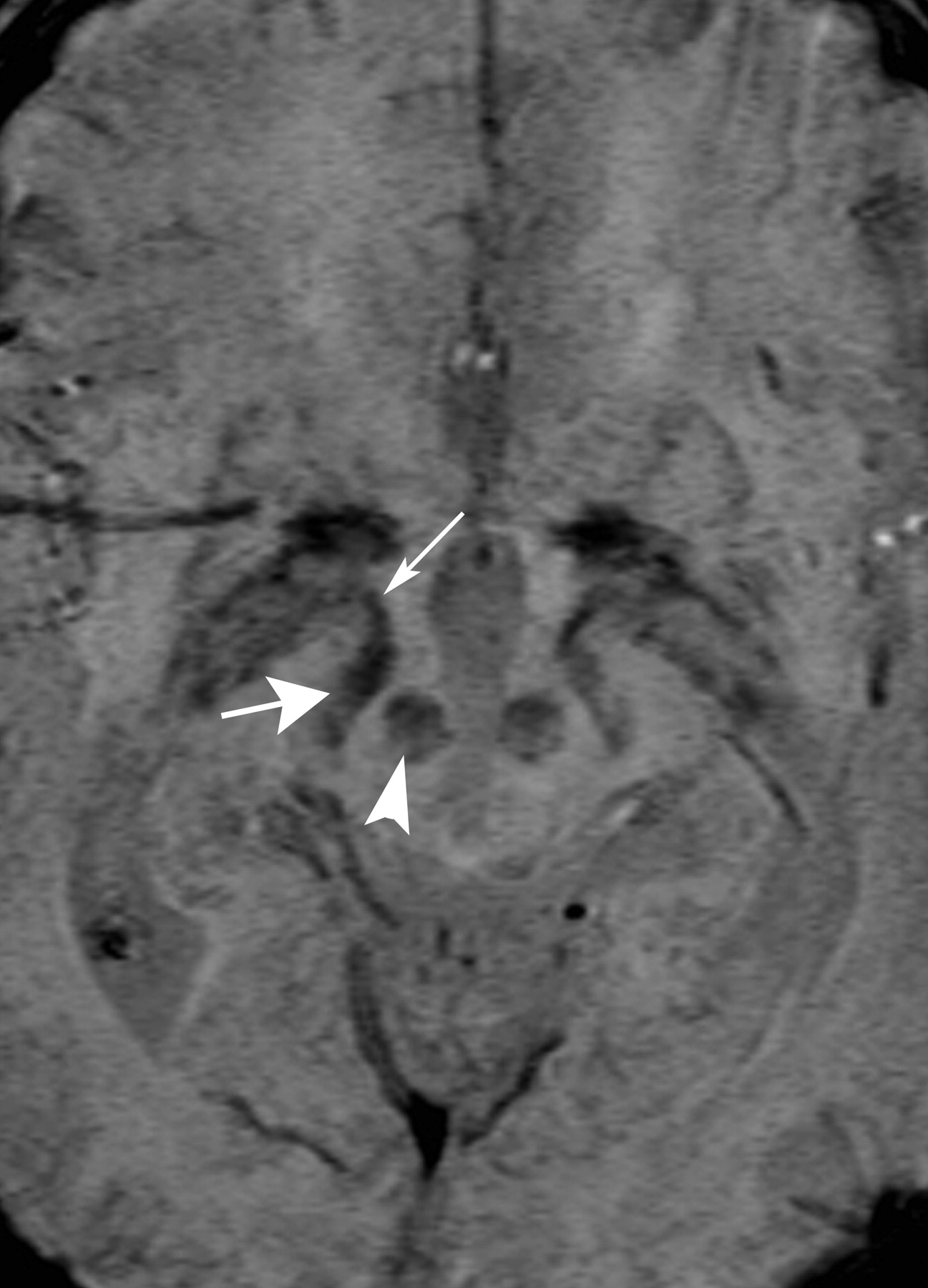
- Fig 3.
Axial SWI image through the globus pallidus demonstrates mineralization of the medial (arrow) and lateral (arrowhead) aspects of the nucleus.
- Fig 4.
Axial SWI images through the right globus pallidus. Images A to E demonstrate increasing numbers of waves with the final image (E), showing conglomerate waves.
- Fig 5.
Axial SWI image through the level of the midbrain demonstrating the red nucleus (arrowhead), substantia nigra (thick arrow), and fascicula nigrale (thin arrow).
- Fig 6.
Putaminal intensity per area decreases with age. The line represents Equation 1, with the parameter for the putamen as given in Table 4.
- Fig 7.
Intensity per area of the globus pallidus decreases with age. The line represents Equation 1, with the parameter for the globus pallidus as given in Table 4.
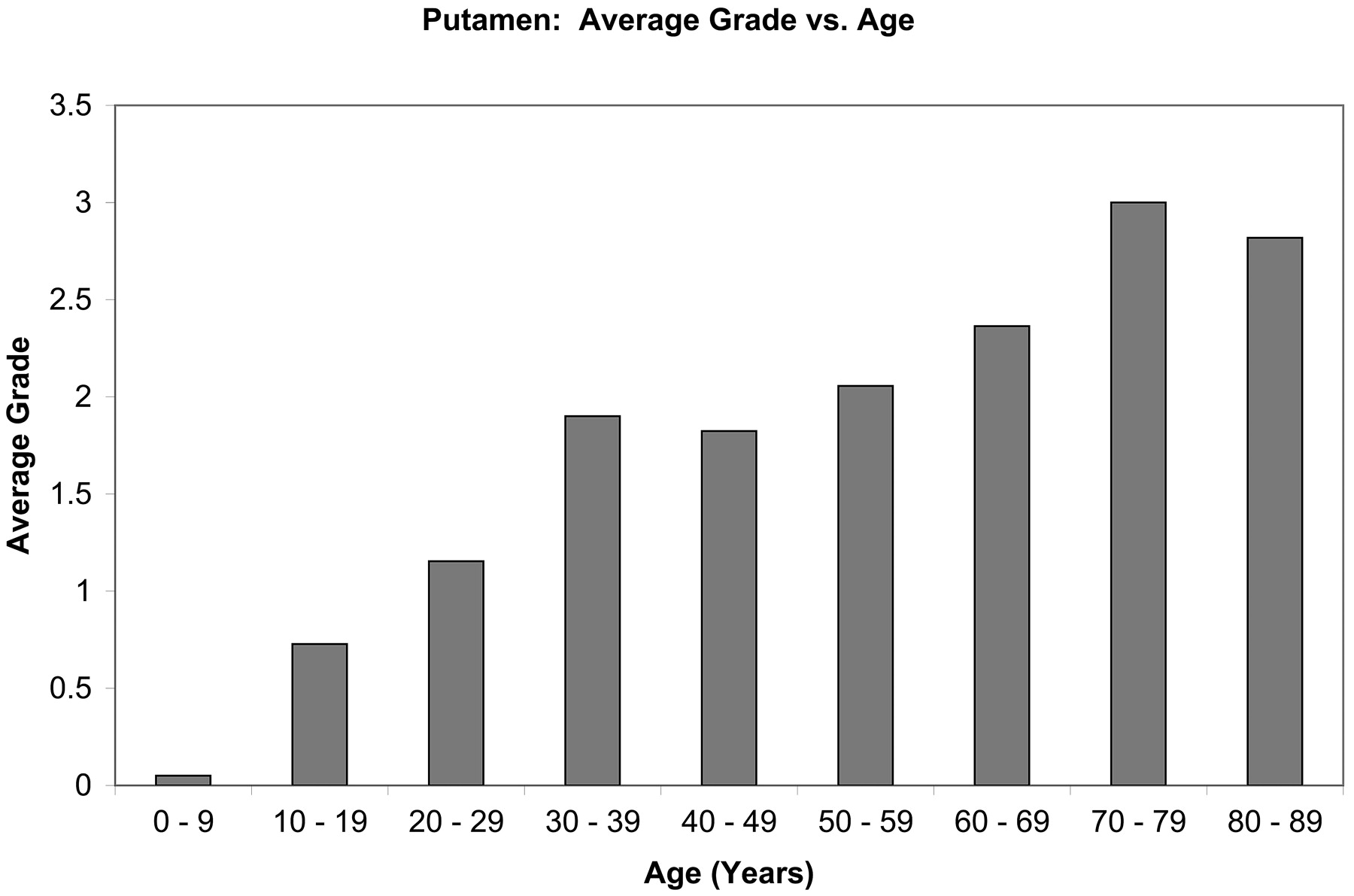
- Fig 8.
Putaminal grade is shown to increase with age.
- Fig 9.
Globus pallidus “waves” increase with age.


Tables
Decade No. of Subjects 1st 19 2nd 22 3rd 13 4th 10 5th 17 6th 18 7th 11 8th 13 9th 11 Grade Definition 0 No mineralization 1 Mineralization of the posterolateral aspect of the putamen 2 Mineralization of the lateral half of the putamen 3 Mineralization of the lateral half and the posteromedial aspect of the putamen 4 Mineralization of the entire putamen Score Definition 0 No waves 1 1 wave 2 2 waves 3 3 waves 4 4 waves 5 5 waves or confluent waves y0 A b Correlation, r Globus pallidus 0.020 0.018 0.09 0.8 Putamen 0.023 0.011 0.09 0.5 Substantia nigra 0.012 0.0065 0.04 0.5 * See Equation 1.
In this issue


Jump to section
Related Articles
Cited By...
- Susceptibility-Weighted Imaging of the Pediatric Brain after Repeat Doses of Gadolinium-Based Contrast Agent
- Unraveling Deep Gray Matter Atrophy and Iron and Myelin Changes in Multiple Sclerosis
- Estimating brain age from structural MRI and MEG data: Insights from dimensionality reduction techniques
- Looking Deep into the Eye-of-the-Tiger in Pantothenate Kinase-Associated Neurodegeneration
- Basal Ganglia Iron in Patients with Multiple Sclerosis Measured with 7T Quantitative Susceptibility Mapping Correlates with Inhibitory Control
- A Potential Biomarker in Amyotrophic Lateral Sclerosis: Can Assessment of Brain Iron Deposition with SWI and Corticospinal Tract Degeneration with DTI Help?
- Susceptibility-Weighted Imaging Improves the Diagnostic Accuracy of 3T Brain MRI in the Work-Up of Parkinsonism
- Distinctive MRI abnormalities in a man with dentatorubral-pallidoluysian atrophy
- Susceptibility-Weighted Imaging: Technical Aspects and Clinical Applications, Part 2
- Susceptibility-Weighted Imaging: Technical Aspects and Clinical Applications, Part 1