Advertisement

Main menu

  • Home
  • Content
    • Current Issue
    • Accepted Manuscripts
    • Article Preview
    • Past Issue Archive
    • Video Articles
    • AJNR Case Collection
    • Case of the Week Archive
    • Case of the Month Archive
    • Classic Case Archive
  • Special Collections
    • AJNR Awards
    • Low-Field MRI
    • Alzheimer Disease
    • ASNR Foundation Special Collection
    • Photon-Counting CT
    • View All
  • Multimedia
    • AJNR Podcasts
    • AJNR SCANtastic
    • Trainee Corner
    • MRI Safety Corner
    • Imaging Protocols
  • For Authors
    • Submit a Manuscript
    • Submit a Video Article
    • Submit an eLetter to the Editor/Response
    • Manuscript Submission Guidelines
    • Statistical Tips
    • Fast Publishing of Accepted Manuscripts
    • Graphical Abstract Preparation
    • Imaging Protocol Submission
    • Author Policies
  • About Us
    • About AJNR
    • Editorial Board
    • Editorial Board Alumni
  • More
    • Become a Reviewer/Academy of Reviewers
    • Subscribers
    • Permissions
    • Alerts
    • Feedback
    • Advertisers
    • ASNR Home

User menu

  • Alerts
  • Log in

Search

  • Advanced search
American Journal of Neuroradiology

American Journal of Neuroradiology

ASHNR American Society of Functional Neuroradiology ASHNR American Society of Pediatric Neuroradiology ASSR
  • Alerts
  • Log in

Advanced Search

  • Home
  • Content
    • Current Issue
    • Accepted Manuscripts
    • Article Preview
    • Past Issue Archive
    • Video Articles
    • AJNR Case Collection
    • Case of the Week Archive
    • Case of the Month Archive
    • Classic Case Archive
  • Special Collections
    • AJNR Awards
    • Low-Field MRI
    • Alzheimer Disease
    • ASNR Foundation Special Collection
    • Photon-Counting CT
    • View All
  • Multimedia
    • AJNR Podcasts
    • AJNR SCANtastic
    • Trainee Corner
    • MRI Safety Corner
    • Imaging Protocols
  • For Authors
    • Submit a Manuscript
    • Submit a Video Article
    • Submit an eLetter to the Editor/Response
    • Manuscript Submission Guidelines
    • Statistical Tips
    • Fast Publishing of Accepted Manuscripts
    • Graphical Abstract Preparation
    • Imaging Protocol Submission
    • Author Policies
  • About Us
    • About AJNR
    • Editorial Board
    • Editorial Board Alumni
  • More
    • Become a Reviewer/Academy of Reviewers
    • Subscribers
    • Permissions
    • Alerts
    • Feedback
    • Advertisers
    • ASNR Home
  • Follow AJNR on Twitter
  • Visit AJNR on Facebook
  • Follow AJNR on Instagram
  • Join AJNR on LinkedIn
  • RSS Feeds

AJNR Awards, New Junior Editors, and more. Read the latest AJNR updates

Research ArticleBRAIN

Improved Identification of Intracortical Lesions in Multiple Sclerosis with Phase-Sensitive Inversion Recovery in Combination with Fast Double Inversion Recovery MR Imaging

F. Nelson, A.H. Poonawalla, P. Hou, F. Huang, J.S. Wolinsky and P.A. Narayana
American Journal of Neuroradiology October 2007, 28 (9) 1645-1649; DOI: https://doi.org/10.3174/ajnr.A0645
F. Nelson
  • Find this author on Google Scholar
  • Find this author on PubMed
  • Search for this author on this site
A.H. Poonawalla
  • Find this author on Google Scholar
  • Find this author on PubMed
  • Search for this author on this site
P. Hou
  • Find this author on Google Scholar
  • Find this author on PubMed
  • Search for this author on this site
F. Huang
  • Find this author on Google Scholar
  • Find this author on PubMed
  • Search for this author on this site
J.S. Wolinsky
  • Find this author on Google Scholar
  • Find this author on PubMed
  • Search for this author on this site
P.A. Narayana
  • Find this author on Google Scholar
  • Find this author on PubMed
  • Search for this author on this site
  • Article
  • Figures & Data
  • Info & Metrics
  • Responses
  • References
  • PDF
Loading

Article Figures & Data

Figures

  • Tables
  • Fig 1.
    • Download figure
    • Open in new tab
    • Download powerpoint
    Fig 1.

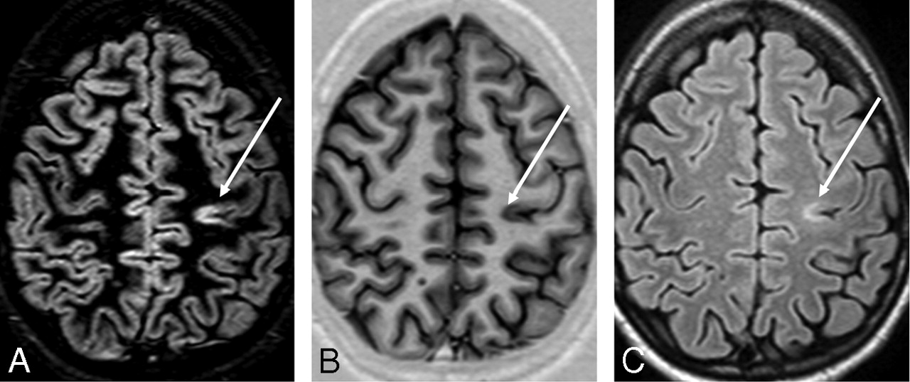
    DIR (A), PSIR (B), and FLAIR (C) images from a single patient with MS at the same section location. An intracortical lesion is evident in the left parietal area. Also note the excellent overall delineation of the gray-white matter border on PSIR.

  • Fig 2.
    • Download figure
    • Open in new tab
    • Download powerpoint
    Fig 2.

    Examples of cortical lesions on PSIR (left), DIR (center), and FLAIR (right). Lesions shown are (A) purely intracortical, (B) mixed, and (C) juxtacortical.

  • Fig 3.
    • Download figure
    • Open in new tab
    • Download powerpoint
    Fig 3.

    Example of flow artifact. On the DIR image (A), apparent cortical lesions (arrows) are visible. However, on the PSIR image (B), there are no corresponding hypointense signals. On PSIR, a flow artifact is seen more clearly (arrows), which is the likely source of the false-positive.

  • Fig 4.
    • Download figure
    • Open in new tab
    • Download powerpoint
    Fig 4.

    Example of RF inhomogeneity artifact. On the DIR image (A), an apparent cortical lesion is visible (white arrow). On the PSIR image (B), there is again no corresponding hypointensity. On DIR, the signal intensity is 38% higher for the apparent lesion than the contralateral area, compared with 14% for PSIR. The relative insensitivity of PSIR to the RF artifact allows rejection of the lesion on DIR as a false-positive.

Tables

  • Figures
    • View popup
    Table 1:

    Demographics of patients in the study

    Age/SexDisease TypeDisease Duration (years)EDSS Score
    44/MRR62
    50/MSP103
    59/FRR112
    50/FRR1.50
    55/FRR262
    53/FRR123
    49/FRR255
    25/FRR51
    64/FSP296.5
    62/FSP456.5
    24/FRR53
    27/FRR1.50
    48/FRR221
    35/FRR1.22
    68/MPP244
    39/FRR112
    • Note:—RR indicates relapsing-remitting; SP, secondary-progressive; PP, primary-progressive; EDSS, Expanded Disability Status Scale.

    • View popup
    Table 2:

    Acquisition parameters for MR imaging sequences in the MS protocol

    SequencePlaneTR (ms)TE (ms)TI (ms)Image MatrixFOV (mm)Section (mm)Scan Time (min)
    PSIRAxial430013400256 × 25624034.0
    DIRAxial15,000253400/325512 × 51224036.5
    Dual-FSEAxial680010/90–256 × 25624036.0
    FLAIRAxial10,000802600256 × 25624033.0
    • Note:—MS indicates multiple sclerosis; PSIR, phase-sensitive inversion recovery; DIR, double inversion recovery; FSE, fast spin-echo; FLAIR, fluid-attenuated inversion recovery; FOV, field of view.

    • View popup
    Table 3:

    Mean number of lesions detected by technique

    Type of LesionPSIR+DIRFLAIR% ImprovementP Value
    Intracortical7.8 ± 9.41.4 ± 1.8439.0002
    Mixed9 ± 111.8 ± 2.4414<.0001
    Juxtacortical2.5 ± 2.41.1 ± 1.5117.0078
    Overall19.2 ± 21.14.3 ± 4.3345<.0001
    • Note:—PSIR indicates phase-sensitive inversion recovery; DIR, double inversion recovery; FLAIR, fluid-attenuated inversion recovery.

PreviousNext
Back to top

In this issue

American Journal of Neuroradiology: 28 (9)
American Journal of Neuroradiology
Vol. 28, Issue 9
October 2007
  • Table of Contents
  • Index by author
Advertisement
Print
Download PDF
Email Article

Thank you for your interest in spreading the word on American Journal of Neuroradiology.

NOTE: We only request your email address so that the person you are recommending the page to knows that you wanted them to see it, and that it is not junk mail. We do not capture any email address.

Enter multiple addresses on separate lines or separate them with commas.
Improved Identification of Intracortical Lesions in Multiple Sclerosis with Phase-Sensitive Inversion Recovery in Combination with Fast Double Inversion Recovery MR Imaging
(Your Name) has sent you a message from American Journal of Neuroradiology
(Your Name) thought you would like to see the American Journal of Neuroradiology web site.
CAPTCHA
This question is for testing whether or not you are a human visitor and to prevent automated spam submissions.
Cite this article
F. Nelson, A.H. Poonawalla, P. Hou, F. Huang, J.S. Wolinsky, P.A. Narayana
Improved Identification of Intracortical Lesions in Multiple Sclerosis with Phase-Sensitive Inversion Recovery in Combination with Fast Double Inversion Recovery MR Imaging
American Journal of Neuroradiology Oct 2007, 28 (9) 1645-1649; DOI: 10.3174/ajnr.A0645

Citation Manager Formats

  • BibTeX
  • Bookends
  • EasyBib
  • EndNote (tagged)
  • EndNote 8 (xml)
  • Medlars
  • Mendeley
  • Papers
  • RefWorks Tagged
  • Ref Manager
  • RIS
  • Zotero
0 Responses
Respond to this article
Share
Bookmark this article
Improved Identification of Intracortical Lesions in Multiple Sclerosis with Phase-Sensitive Inversion Recovery in Combination with Fast Double Inversion Recovery MR Imaging
F. Nelson, A.H. Poonawalla, P. Hou, F. Huang, J.S. Wolinsky, P.A. Narayana
American Journal of Neuroradiology Oct 2007, 28 (9) 1645-1649; DOI: 10.3174/ajnr.A0645
del.icio.us logo Twitter logo Facebook logo Mendeley logo
  • Tweet Widget
  • Facebook Like
  • Google Plus One
Purchase

Jump to section

  • Article
    • Abstract
    • Materials and Methods
    • Results
    • Discussion
    • Conclusions
    • Acknowledgments
    • Footnotes
    • References
  • Figures & Data
  • Info & Metrics
  • Responses
  • References
  • PDF

Related Articles

  • No related articles found.
  • PubMed
  • Google Scholar

Cited By...

  • T1/T2 Ratio Imaging Improves Cortical Lesion Contrast in Multiple Sclerosis on 3T MRI
  • Evaluating Tissue Contrast and Detecting White Matter Injury in the Infant Brain: A Comparison Study of Synthetic Phase-Sensitive Inversion Recovery
  • Improving Detection of Multiple Sclerosis Lesions in the Posterior Fossa Using an Optimized 3D-FLAIR Sequence at 3T
  • Imaging outcome measures of neuroprotection and repair in MS: A consensus statement from NAIMS
  • Detection of Leukocortical Lesions in Multiple Sclerosis and Their Association with Physical and Cognitive Impairment: A Comparison of Conventional and Synthetic Phase-Sensitive Inversion Recovery MRI
  • Magnetic Resonance Imaging in Multiple Sclerosis
  • Improved Visualization of Cortical Lesions in Multiple Sclerosis Using 7T MP2RAGE
  • Current and Emerging Therapies in Multiple Sclerosis: Implications for the Radiologist, Part 1--Mechanisms, Efficacy, and Safety
  • Current and Emerging Therapies in Multiple Sclerosis: Implications for the Radiologist, Part 2--Surveillance for Treatment Complications and Disease Progression
  • Synthetic MRI in the Detection of Multiple Sclerosis Plaques
  • Evaluation of Focal Cervical Spinal Cord Lesions in Multiple Sclerosis: Comparison of White Matter-Suppressed T1 Inversion Recovery Sequence versus Conventional STIR and Proton Density-Weighted Turbo Spin-Echo Sequences
  • A longitudinal study of cortical grey matter lesion subtypes in relapse-onset multiple sclerosis
  • Double Inversion Recovery MR Sequence for the Detection of Subacute Subarachnoid Hemorrhage
  • Improved detection of cortical MS lesions with phase-sensitive inversion recovery MRI
  • Assessing Abnormal Iron Content in the Deep Gray Matter of Patients with Multiple Sclerosis versus Healthy Controls
  • Identification and Clinical Impact of Multiple Sclerosis Cortical Lesions as Assessed by Routine 3T MR Imaging
  • Consensus recommendations for MS cortical lesion scoring using double inversion recovery MRI
  • Imaging distribution and frequency of cortical lesions in patients with multiple sclerosis
  • MR Imaging of Gray Matter Involvement in Multiple Sclerosis: Implications for Understanding Disease Pathophysiology and Monitoring Treatment Efficacy
  • MRI criteria for MS in patients with clinically isolated syndromes
  • Quantitative Cervical Spinal Cord 3T Proton MR Spectroscopy in Multiple Sclerosis
  • In vivo imaging of cortical pathology in multiple sclerosis using ultra-high field MRI
  • Crossref (169)
  • Google Scholar

This article has been cited by the following articles in journals that are participating in Crossref Cited-by Linking.

  • Diagnostic criteria for multiple sclerosis: 2010 Revisions to the McDonald criteria
    Chris H. Polman, Stephen C. Reingold, Brenda Banwell, Michel Clanet, Jeffrey A. Cohen, Massimo Filippi, Kazuo Fujihara, Eva Havrdova, Michael Hutchinson, Ludwig Kappos, Fred D. Lublin, Xavier Montalban, Paul O'Connor, Magnhild Sandberg‐Wollheim, Alan J. Thompson, Emmanuelle Waubant, Brian Weinshenker, Jerry S. Wolinsky
    Annals of Neurology 2011 69 2
  • MRI criteria for the diagnosis of multiple sclerosis: MAGNIMS consensus guidelines
    Massimo Filippi, Maria A Rocca, Olga Ciccarelli, Nicola De Stefano, Nikos Evangelou, Ludwig Kappos, Alex Rovira, Jaume Sastre-Garriga, Mar Tintorè, Jette L Frederiksen, Claudio Gasperini, Jacqueline Palace, Daniel S Reich, Brenda Banwell, Xavier Montalban, Frederik Barkhof
    The Lancet Neurology 2016 15 3
  • Clinical and imaging assessment of cognitive dysfunction in multiple sclerosis
    Maria A Rocca, Maria P Amato, Nicola De Stefano, Christian Enzinger, Jeroen J Geurts, Iris-K Penner, Alex Rovira, James F Sumowski, Paola Valsasina, Massimo Filippi
    The Lancet Neurology 2015 14 3
  • Review of automatic segmentation methods of multiple sclerosis white matter lesions on conventional magnetic resonance imaging
    Daniel García-Lorenzo, Simon Francis, Sridar Narayanan, Douglas L. Arnold, D. Louis Collins
    Medical Image Analysis 2013 17 1
  • Cortical lesion load associates with progression of disability in multiple sclerosis
    M. Calabrese, V. Poretto, A. Favaretto, S. Alessio, V. Bernardi, C. Romualdi, F. Rinaldi, P. Perini, P. Gallo
    Brain 2012 135 10
  • The diagnosis of multiple sclerosis and the various related demyelinating syndromes: A critical review
    Dimitrios Karussis
    Journal of Autoimmunity 2014 48-49
  • Consensus recommendations for MS cortical lesion scoring using double inversion recovery MRI
    J.J.G. Geurts, S.D. Roosendaal, M. Calabrese, O. Ciccarelli, F. Agosta, D.T. Chard, A. Gass, E. Huerga, B. Moraal, D. Pareto, M.A. Rocca, M.P. Wattjes, T.A. Yousry, B.M.J. Uitdehaag, F. Barkhof
    Neurology 2011 76 5
  • Measurement and clinical effect of grey matter pathology in multiple sclerosis
    Jeroen JG Geurts, Massimiliano Calabrese, Elizabeth Fisher, Richard A Rudick
    The Lancet Neurology 2012 11 12
  • Cortical lesions in multiple sclerosis
    Massimiliano Calabrese, Massimo Filippi, Paolo Gallo
    Nature Reviews Neurology 2010 6 8
  • In vivo imaging of cortical pathology in multiple sclerosis using ultra-high field MRI
    C. Mainero, T. Benner, A. Radding, A. van der Kouwe, R. Jensen, B. R. Rosen, R. P. Kinkel
    Neurology 2009 73 12

More in this TOC Section

  • Predictors of Reperfusion in Patients with Acute Ischemic Stroke
  • Enhanced Axonal Metabolism during Early Natalizumab Treatment in Relapsing-Remitting Multiple Sclerosis
  • Progression of Microstructural Damage in Spinocerebellar Ataxia Type 2: A Longitudinal DTI Study
Show more BRAIN

Similar Articles

Advertisement

Indexed Content

  • Current Issue
  • Accepted Manuscripts
  • Article Preview
  • Past Issues
  • Editorials
  • Editor's Choice
  • Fellows' Journal Club
  • Letters to the Editor
  • Video Articles

Cases

  • Case Collection
  • Archive - Case of the Week
  • Archive - Case of the Month
  • Archive - Classic Case

More from AJNR

  • Trainee Corner
  • Imaging Protocols
  • MRI Safety Corner
  • Book Reviews

Multimedia

  • AJNR Podcasts
  • AJNR Scantastics

Resources

  • Turnaround Time
  • Submit a Manuscript
  • Submit a Video Article
  • Submit an eLetter to the Editor/Response
  • Manuscript Submission Guidelines
  • Statistical Tips
  • Fast Publishing of Accepted Manuscripts
  • Graphical Abstract Preparation
  • Imaging Protocol Submission
  • Evidence-Based Medicine Level Guide
  • Publishing Checklists
  • Author Policies
  • Become a Reviewer/Academy of Reviewers
  • News and Updates

About Us

  • About AJNR
  • Editorial Board
  • Editorial Board Alumni
  • Alerts
  • Permissions
  • Not an AJNR Subscriber? Join Now
  • Advertise with Us
  • Librarian Resources
  • Feedback
  • Terms and Conditions
  • AJNR Editorial Board Alumni

American Society of Neuroradiology

  • Not an ASNR Member? Join Now

© 2025 by the American Society of Neuroradiology All rights, including for text and data mining, AI training, and similar technologies, are reserved.
Print ISSN: 0195-6108 Online ISSN: 1936-959X

Powered by HighWire