Advertisement

Main menu

  • Home
  • Content
    • Current Issue
    • Accepted Manuscripts
    • Article Preview
    • Past Issue Archive
    • Video Articles
    • AJNR Case Collection
    • Case of the Week Archive
    • Case of the Month Archive
    • Classic Case Archive
  • Special Collections
    • Advancing NeuroMRI with High-Relaxivity Contrast Agents
    • AJNR Awards
    • Low-Field MRI
    • Alzheimer Disease
    • ASNR Foundation Special Collection
    • Photon-Counting CT
    • View All
  • Multimedia
    • AJNR Podcasts
    • AJNR SCANtastic
    • Trainee Corner
    • MRI Safety Corner
    • Imaging Protocols
  • For Authors
    • Submit a Manuscript
    • Submit a Video Article
    • Submit an eLetter to the Editor/Response
    • Manuscript Submission Guidelines
    • Statistical Tips
    • Fast Publishing of Accepted Manuscripts
    • Graphical Abstract Preparation
    • Imaging Protocol Submission
    • Author Policies
  • About Us
    • About AJNR
    • Editorial Board
    • Editorial Board Alumni
  • More
    • Become a Reviewer/Academy of Reviewers
    • Subscribers
    • Permissions
    • Alerts
    • Feedback
    • Advertisers
    • ASNR Home

User menu

  • Alerts
  • Log in

Search

  • Advanced search
American Journal of Neuroradiology

American Journal of Neuroradiology

ASHNR American Society of Functional Neuroradiology ASHNR American Society of Pediatric Neuroradiology ASSR
  • Alerts
  • Log in

Advanced Search

  • Home
  • Content
    • Current Issue
    • Accepted Manuscripts
    • Article Preview
    • Past Issue Archive
    • Video Articles
    • AJNR Case Collection
    • Case of the Week Archive
    • Case of the Month Archive
    • Classic Case Archive
  • Special Collections
    • Advancing NeuroMRI with High-Relaxivity Contrast Agents
    • AJNR Awards
    • Low-Field MRI
    • Alzheimer Disease
    • ASNR Foundation Special Collection
    • Photon-Counting CT
    • View All
  • Multimedia
    • AJNR Podcasts
    • AJNR SCANtastic
    • Trainee Corner
    • MRI Safety Corner
    • Imaging Protocols
  • For Authors
    • Submit a Manuscript
    • Submit a Video Article
    • Submit an eLetter to the Editor/Response
    • Manuscript Submission Guidelines
    • Statistical Tips
    • Fast Publishing of Accepted Manuscripts
    • Graphical Abstract Preparation
    • Imaging Protocol Submission
    • Author Policies
  • About Us
    • About AJNR
    • Editorial Board
    • Editorial Board Alumni
  • More
    • Become a Reviewer/Academy of Reviewers
    • Subscribers
    • Permissions
    • Alerts
    • Feedback
    • Advertisers
    • ASNR Home
  • Follow AJNR on Twitter
  • Visit AJNR on Facebook
  • Follow AJNR on Instagram
  • Join AJNR on LinkedIn
  • RSS Feeds

AJNR Awards, New Junior Editors, and more. Read the latest AJNR updates


Improved Turnaround Times | Median time to first decision: 12 days

Research ArticleINTERVENTIONAL

A New Covered Stent Designed for Intracranial Vasculature: Application in the Management of Pseudoaneurysms of the Cranial Internal Carotid Artery

M.-H. Li, Y.-D. Li, B.-L. Gao, C. Fang, Q.-Y. Luo, Y.-S. Cheng, Z.-Y. Xie, Y.-L. Wang, J.-G. Zhao, Y. Li, W. Wang, B.-L. Zhang and M. Li
American Journal of Neuroradiology September 2007, 28 (8) 1579-1585; DOI: https://doi.org/10.3174/ajnr.A0668
M.-H. Li
  • Find this author on Google Scholar
  • Find this author on PubMed
  • Search for this author on this site
Y.-D. Li
  • Find this author on Google Scholar
  • Find this author on PubMed
  • Search for this author on this site
B.-L. Gao
  • Find this author on Google Scholar
  • Find this author on PubMed
  • Search for this author on this site
C. Fang
  • Find this author on Google Scholar
  • Find this author on PubMed
  • Search for this author on this site
Q.-Y. Luo
  • Find this author on Google Scholar
  • Find this author on PubMed
  • Search for this author on this site
Y.-S. Cheng
  • Find this author on Google Scholar
  • Find this author on PubMed
  • Search for this author on this site
Z.-Y. Xie
  • Find this author on Google Scholar
  • Find this author on PubMed
  • Search for this author on this site
Y.-L. Wang
  • Find this author on Google Scholar
  • Find this author on PubMed
  • Search for this author on this site
J.-G. Zhao
  • Find this author on Google Scholar
  • Find this author on PubMed
  • Search for this author on this site
Y. Li
  • Find this author on Google Scholar
  • Find this author on PubMed
  • Search for this author on this site
W. Wang
  • Find this author on Google Scholar
  • Find this author on PubMed
  • Search for this author on this site
B.-L. Zhang
  • Find this author on Google Scholar
  • Find this author on PubMed
  • Search for this author on this site
M. Li
  • Find this author on Google Scholar
  • Find this author on PubMed
  • Search for this author on this site
  • Article
  • Figures & Data
  • Info & Metrics
  • Responses
  • References
  • PDF
Loading

Article Figures & Data

Figures

  • Tables
  • Fig 1.
    • Download figure
    • Open in new tab
    • Download powerpoint
    Fig 1.

    The Willis covered stent. A, The covered stent is attached to the balloon catheter, with the arrows demonstrating the 2 ends of the covered stent. The diameter of the entire system is 3.8F (1.27 mm) when it is not expanded. B, The covered stent is expanded completely against the wall of the model glass tube similar to the siphon segment of the internal carotid artery (arrows).

  • Fig 2.
    • Download figure
    • Open in new tab
    • Download powerpoint
    Fig 2.

    Case 8, a 11-year-old boy with a pseudoaneurysm secondary to a traumatic internal carotid artery. A,B, Anteroposterior and lateral cerebral angiograms show a narrow-necked pseudoaneurysm on the right C7 segment (arrow), with the stenosis of the parent artery. C, A plain film after stent placement clearly shows the position of the covered stent (arrow). D,E, Anteroposterior and lateral cerebral angiograms show complete resolution of the aneurysm immediately after stent placement, with obliteration of the stenosis of the parent artery.
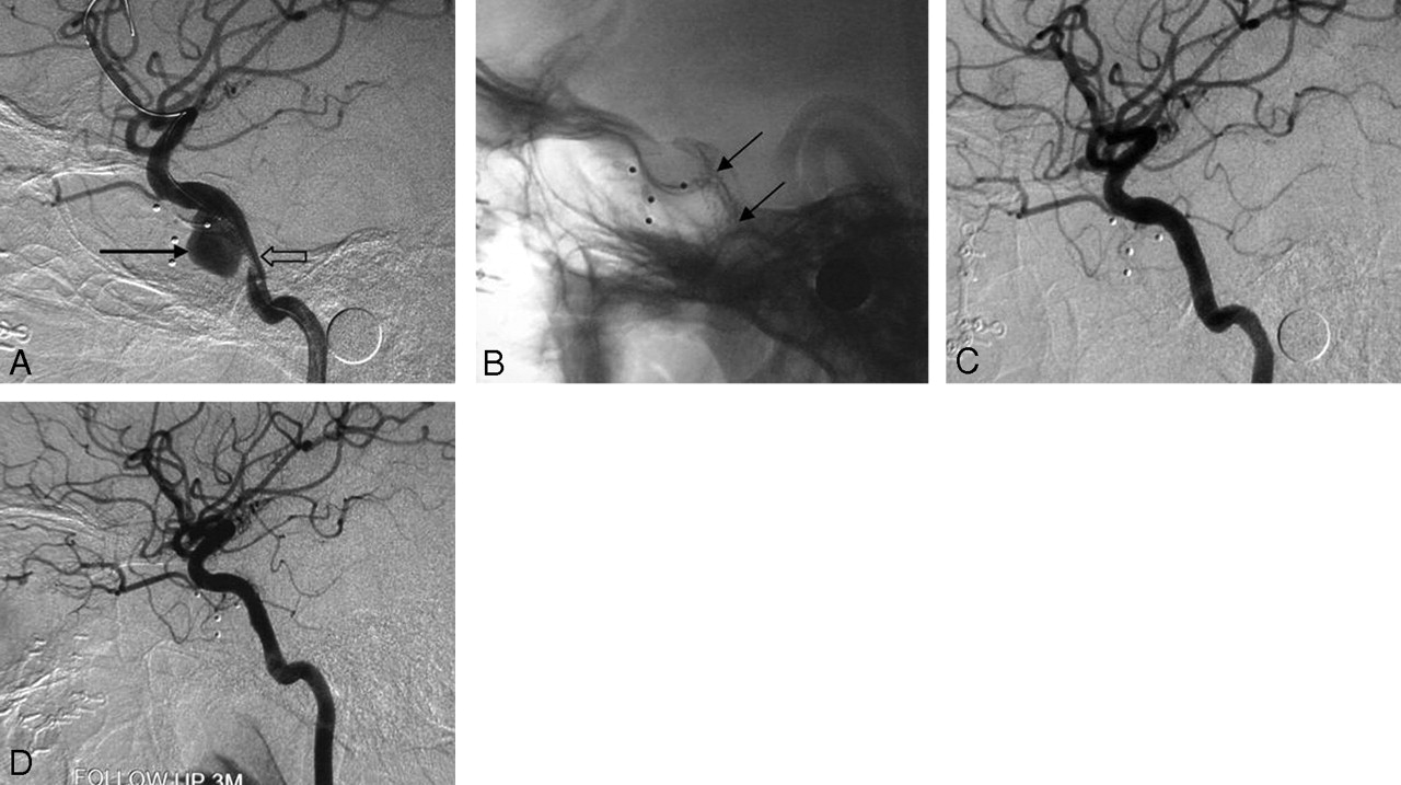
  • Fig 3.
    • Download figure
    • Open in new tab
    • Download powerpoint
    Fig 3.

    Case 6, a 23-year-old man with a pseudoaneurysm secondary to post-balloon embolization of a CCF. A, Lateral cerebral angiogram reveals a wide-necked pseudoaneurysm (black arrow) on the left C4 segment, with stenosis at the proximal part of the parent artery (empty arrow). B, Plain film after stent placement clearly shows the covered stent bridging the pseudoaneurysm and the stenosis (arrows). C, Lateral cerebral angiogram shows complete resolution of the aneurysm immediately after the stent placement. D, Cerebral angiography 3 months after the procedure shows total obliteration of the aneurysm with patency of the parent artery.
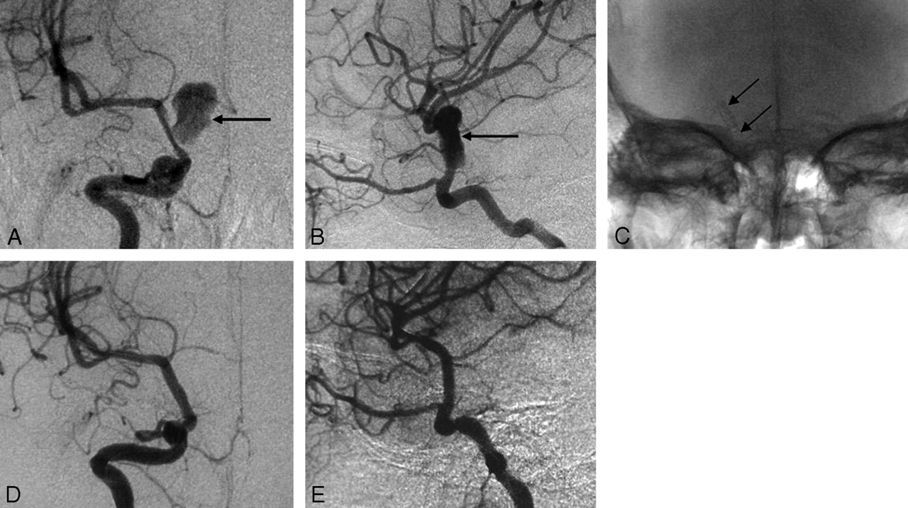
  • Fig 4.
    • Download figure
    • Open in new tab
    • Download powerpoint
    Fig 4.

    Case 5, a 35-year-old man with massive epistaxis. A, Lateral cerebral angiogram shows a giant pseudoaneurysm on the left C5 segment (arrow). B, The Willis covered stent can be clearly seen in the plain film (arrows) after stent placement. C, Cerebral angiogram immediately after stent placement demonstrates a minimal endoleak into the pseudoaneurysm (arrow) in the orifice of the ophthalmic artery (arrow). D, Follow-up cerebral angiogram 2 months after the procedure demonstrates that retention of contrast medium at the orifice of the ophthalmic artery is increased (arrow), which suggests the existence of a residual cavity. E, Follow-up cerebral angiogram 6 months after the procedure demonstrates obvious shrinkage of the residual cavity (arrow) with patency of the parent artery.

Tables

  • Figures
  • Summary of eight patients with intracranial pseudoaneurysms treated with the Willis covered stent

    Case No./Age /SexPresenting SymptomsCauseAneurysm LocationAneurysm Size (mm)Stent Size (mm)Resistance Reaching Target LesionImmediate Angiography, PoststentingFollow-Up (mos)Follow-Up AngiographyClinical Outcome
    1/48/MHeadache, diplopia, decreased visual acuityPost-balloon occlusion of CCFRight C310 × 84 × 10NonePA resolution, parent artery patency12PA resolution, parent artery patencyImprovement
    2/38/MLeft eye blindnessTraumaLeft C415 × 124 × 10No apparent resistancePA resolution, parent artery patency5PA resolution, parent artery patencyUnchanged
    3/34/MPalsyPost-balloon occlusion of CCFRight C415 × 125 × 13NonePA resolution, small CCF6PA resolution, CCF alleviationImprovement
    4/50/MPalsyPost-balloon occlusion of CCFRight C423 × 164 × 13NonePA resolution, parent artery patency6PA resolution, parent artery patencyFull recovery
    5/35/MEpistaxisTraumaLeft C522 × 184 × 10NoneMinimal endoleak into PA, parent artery patency6Residual cavity shrinkage, parent artery patencyFull recovery
    6/23/MNo symptomsPost-balloon occlusion of CCFLeft C415 × 124 × 13NonePA resolution, parent artery patency3PA resolution, parent artery patencyFull recovery
    7/60/MHeadache, PtosisTraumaLeft C630 × 153.5 × 13 3.5 × 10NoneMinimal endoleak into PA, parent artery patency3Residual cavity shrinkage, parent artery patencyImprovement
    8/11/MDecreased visual acuityTraumaRight C718 × 103.5 × 10NonePA resolution, parent artery patency3PA resolution, parent artery patencyFull recovery
    • Note:—CCF indicates carotid cavernous fistula; PA, pseudoaneurysm.

PreviousNext
Back to top

In this issue

American Journal of Neuroradiology: 28 (8)
American Journal of Neuroradiology
Vol. 28, Issue 8
September 2007
  • Table of Contents
  • Index by author
Advertisement
Print
Download PDF
Email Article

Thank you for your interest in spreading the word on American Journal of Neuroradiology.

NOTE: We only request your email address so that the person you are recommending the page to knows that you wanted them to see it, and that it is not junk mail. We do not capture any email address.

Enter multiple addresses on separate lines or separate them with commas.
A New Covered Stent Designed for Intracranial Vasculature: Application in the Management of Pseudoaneurysms of the Cranial Internal Carotid Artery
(Your Name) has sent you a message from American Journal of Neuroradiology
(Your Name) thought you would like to see the American Journal of Neuroradiology web site.
CAPTCHA
This question is for testing whether or not you are a human visitor and to prevent automated spam submissions.
Cite this article
M.-H. Li, Y.-D. Li, B.-L. Gao, C. Fang, Q.-Y. Luo, Y.-S. Cheng, Z.-Y. Xie, Y.-L. Wang, J.-G. Zhao, Y. Li, W. Wang, B.-L. Zhang, M. Li
A New Covered Stent Designed for Intracranial Vasculature: Application in the Management of Pseudoaneurysms of the Cranial Internal Carotid Artery
American Journal of Neuroradiology Sep 2007, 28 (8) 1579-1585; DOI: 10.3174/ajnr.A0668

Citation Manager Formats

  • BibTeX
  • Bookends
  • EasyBib
  • EndNote (tagged)
  • EndNote 8 (xml)
  • Medlars
  • Mendeley
  • Papers
  • RefWorks Tagged
  • Ref Manager
  • RIS
  • Zotero
0 Responses
Respond to this article
Share
Bookmark this article
A New Covered Stent Designed for Intracranial Vasculature: Application in the Management of Pseudoaneurysms of the Cranial Internal Carotid Artery
M.-H. Li, Y.-D. Li, B.-L. Gao, C. Fang, Q.-Y. Luo, Y.-S. Cheng, Z.-Y. Xie, Y.-L. Wang, J.-G. Zhao, Y. Li, W. Wang, B.-L. Zhang, M. Li
American Journal of Neuroradiology Sep 2007, 28 (8) 1579-1585; DOI: 10.3174/ajnr.A0668
del.icio.us logo Twitter logo Facebook logo Mendeley logo
  • Tweet Widget
  • Facebook Like
  • Google Plus One
Purchase

Jump to section

  • Article
    • Abstract
    • Materials and Methods
    • Results
    • Discussion
    • Acknowledgments
    • Footnotes
    • References
  • Figures & Data
  • Info & Metrics
  • Responses
  • References
  • PDF

Related Articles

  • No related articles found.
  • PubMed
  • Google Scholar

Cited By...

  • The use of PK Papyrus covered coronary stent for carotid reconstruction: an initial institutional experience
  • Safety of Non-invasive Brain Stimulation in Patients with Implants: A Computational Study
  • Post-traumatic pseudoaneurysm of internal carotid artery: a cause of intractable epistaxis
  • Endovascular treatment of recurrent intracranial aneurysms with re-coiling or covered stents
  • Placement of Covered Stents for the Treatment of Direct Carotid Cavernous Fistulas
  • Establishment of an Experimental Intracranial Internal Carotid Artery Model and the Application in Covered-Stent Navigability Testing
  • Advances in Interventional Neuroradiology 2007
  • Crossref
  • Google Scholar

This article has not yet been cited by articles in journals that are participating in Crossref Cited-by Linking.

More in this TOC Section

  • Safety, Efficacy, and Durability of Stent-Assisted Coiling Treatment of M2 (Insular) Segment MCA Aneurysms
  • Endovascular Management of Intracranial Dural AVFs: Transvenous Approach
  • A Meta-analysis of Combined Aspiration Catheter and Stent Retriever versus Stent Retriever Alone for Large-Vessel Occlusion Ischemic Stroke
Show more INTERVENTIONAL

Similar Articles

Advertisement

Indexed Content

  • Current Issue
  • Accepted Manuscripts
  • Article Preview
  • Past Issues
  • Editorials
  • Editor's Choice
  • Fellows' Journal Club
  • Letters to the Editor
  • Video Articles

Cases

  • Case Collection
  • Archive - Case of the Week
  • Archive - Case of the Month
  • Archive - Classic Case

More from AJNR

  • Trainee Corner
  • Imaging Protocols
  • MRI Safety Corner
  • Book Reviews

Multimedia

  • AJNR Podcasts
  • AJNR Scantastics

Resources

  • Turnaround Time
  • Submit a Manuscript
  • Submit a Video Article
  • Submit an eLetter to the Editor/Response
  • Manuscript Submission Guidelines
  • Statistical Tips
  • Fast Publishing of Accepted Manuscripts
  • Graphical Abstract Preparation
  • Imaging Protocol Submission
  • Evidence-Based Medicine Level Guide
  • Publishing Checklists
  • Author Policies
  • Become a Reviewer/Academy of Reviewers
  • News and Updates

About Us

  • About AJNR
  • Editorial Board
  • Editorial Board Alumni
  • Alerts
  • Permissions
  • Not an AJNR Subscriber? Join Now
  • Advertise with Us
  • Librarian Resources
  • Feedback
  • Terms and Conditions
  • AJNR Editorial Board Alumni

American Society of Neuroradiology

  • Not an ASNR Member? Join Now

© 2025 by the American Society of Neuroradiology All rights, including for text and data mining, AI training, and similar technologies, are reserved.
Print ISSN: 0195-6108 Online ISSN: 1936-959X

Powered by HighWire