
Article Figures & Data
Figures
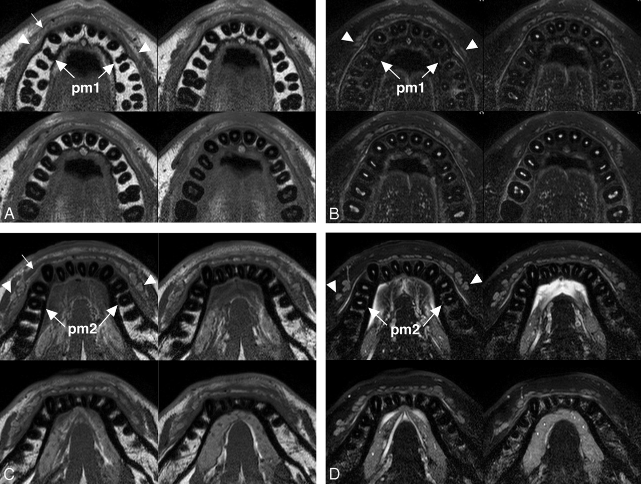
- Fig 1.
A 26-year-old man with healthy upper and lower labial glands. A, Sequential (upper left to lower right panels in a cephalocaudal direction) axial T1-weighted images using a 47-mm microscopy coil (TR/TE/number of signal-intensity acquisitions, 550/10 msec/3; section thickness, 2 mm; section gap, 0.2 mm) of the upper labial salivary gland (arrowheads) show gland clusters spanning the left and right first premolar (pm1) regions. Small arrow indicates orbicularis oris muscle. B, Sequential (upper left to lower right panels in a cephalocaudal direction) axial fat-suppressed T2-weighted images using a microscopy coil (TR/TE/number of signal-intensity acquisitions, 3140/90 msec/4) of the upper labial salivary gland (arrowheads) show hyperintense gland clusters consisting of 1 layer in the anterior part and, at most, 2 layers in the posterior part of the lip. pm1 indicates the first premolar. C, Sequential (upper left to lower right panels in a cephalocaudal direction) axial T1-weighted images using a microscopy coil (TR/TE/number of signal-intensity acquisitions, 550/10 msec/3) of the lower labial gland (arrowheads) show gland clusters spanning the left and right second premolars (pm2). Small arrow indicates the orbicularis oris muscle. D, Sequential (upper left to lower right panels in a cephalocaudal direction) axial fat-suppressed T2-weighted images using a microscopy coil (TR/TE/number of signal-intensity acquisitions, 3140/90 msec/4) of the lower labial salivary gland (arrowheads) show hyperintense gland clusters consisting of, at most, 2 layers in the anterior part and of, at most, 2 or 3 layers in the posterior part of the lip. pm2 indicates the second premolar.
- Fig 2.
A 22-year-old man with healthy lower labial glands. A, Nonenhanced fat-suppressed T2-weighted image using a 110-mm Synergy Flex S Coil (TR/TE/number of signal-intensity acquisitions, 4677/80 msec/4) shows the discrete contour of lower labial gland parenchyma (arrowheads). B, Nonenhanced axial T1-weighted image using an S Coil (TR/TE/number of signal-intensity acquisitions, 500/15 msec/4) of the lower labial salivary gland shows gland parenchyma (arrowheads) having a signal-intensity slightly higher than that of the muscles. C, Gadolinium-enhanced axial T1-weighted image using an S Coil (TR/TE/number of signal-intensity acquisitions, 500/15 msec/4) shows increased signal-intensity levels of the parenchyma of the lower glands (arrowheads), compared with those in B.
- Fig 3.
Box plot graph shows areas of healthy upper and lower labial salivary glands in men and women and of upper and lower labial salivary glands in patients with Sjögren syndrome (SS). The horizontal line in each box is a median (50th percentile) of the measured values; the top and bottom of the boxes represent the 25th and 75th percentiles, respectively; and whiskers indicate the range from the largest to smallest observed data points within a 1.5-interquartile range presented by the box (P value, Wilcoxon signed rank test).
- Fig 4.
A 55-year-old woman with mild Sjögren syndrome. A and B, Axial fat-suppressed T2-weighted images using a microscopy coil (TR/TE/number of signal-intensity acquisitions, 3140/90 msec/4) of the upper (A) and lower (B) labial salivary glands (arrowheads) show gland areas at a level similar to those in healthy glands.
- Fig 5.
A 53-year-old woman with severe Sjögren syndrome. A and B, Axial fat-suppressed T2-weighted images using a microscopy coil (TR/TE/number of signal-intensity acquisitions, 3140/90 msec/4) of the upper (A) and lower (B) labial salivary glands (arrowheads) show greatly decreased gland areas compared with those in healthy glands.
- Fig 6.
A 29-year-old man with pleomorphic adenoma of upper labial gland. A, Axial fat-suppressed T2-weighted image using an S Coil (TR/TE/number of signal-intensity acquisitions, 4677/80 msec/4) shows a heterogeneous mass (arrow) displacing the anterior portion of the upper labial gland. B, Sagittal fat-suppressed contrast-enhanced T1-weighted MR image using an S Coil (TR/TE/number of signal-intensity acquisitions, 500/15 msec/4) shows a heterogeneously enhanced tumor (arrow).
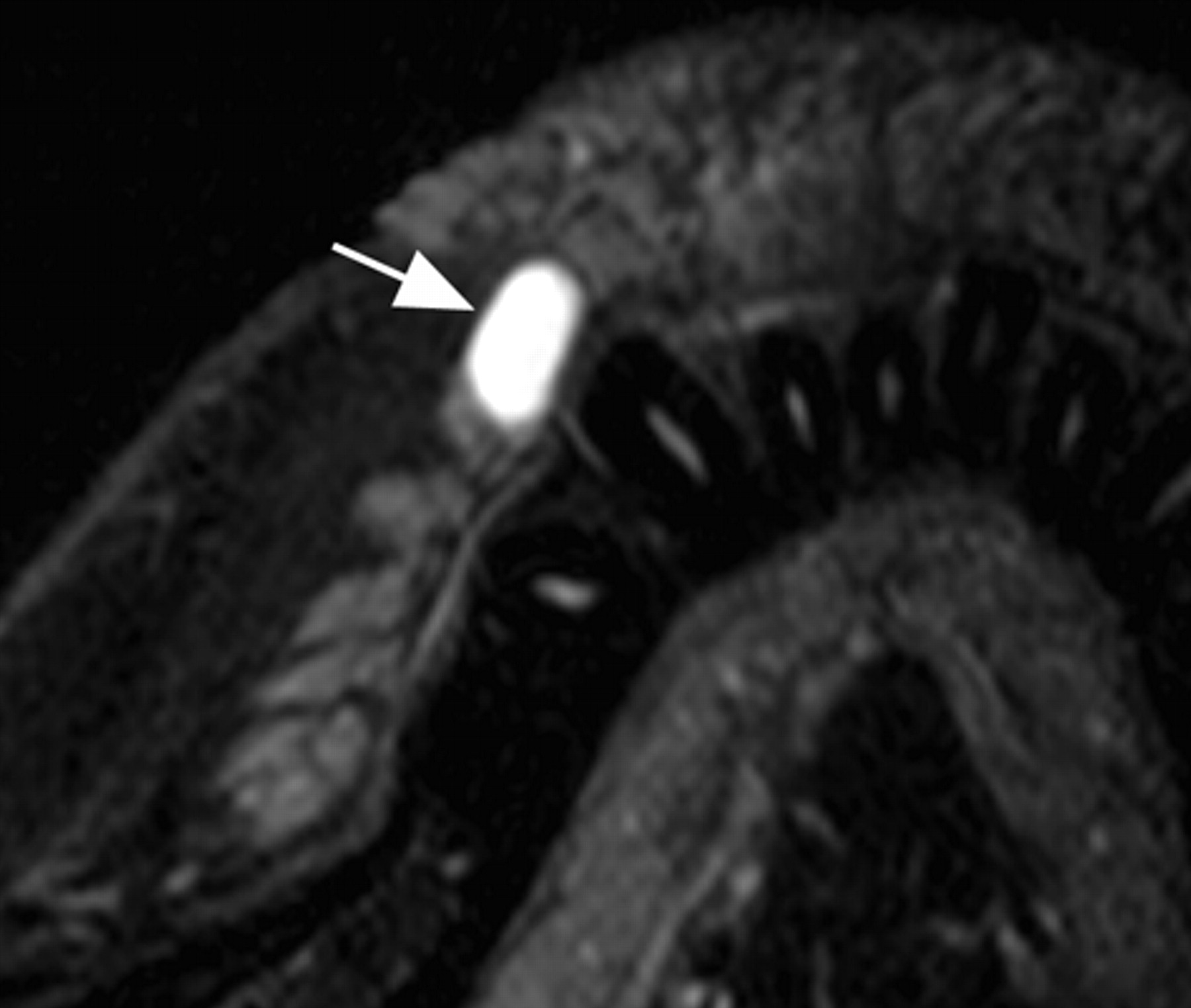
- Fig 7.
A 31-year-old woman with mucocele in the upper labial gland. Axial fat-suppressed T2-weighted image using a microscopy coil (TR/TE/number of signal-intensity acquisitions, 3140/90 msec/4) shows a cystic lesion (arrow) with hyperintense signals, deriving from the upper labial gland. The lesion was also imaged by using an S Coil, but this small lesion is better visualized by using the microscopy coil.


In this issue


Jump to section
Related Articles
Cited By...
- No citing articles found.