Advertisement

Main menu

  • Home
  • Content
    • Current Issue
    • Accepted Manuscripts
    • Article Preview
    • Past Issue Archive
    • Video Articles
    • AJNR Case Collection
    • Case of the Week Archive
    • Case of the Month Archive
    • Classic Case Archive
  • Special Collections
    • AJNR Awards
    • Low-Field MRI
    • Alzheimer Disease
    • ASNR Foundation Special Collection
    • Photon-Counting CT
    • View All
  • Multimedia
    • AJNR Podcasts
    • AJNR SCANtastic
    • Trainee Corner
    • MRI Safety Corner
    • Imaging Protocols
  • For Authors
    • Submit a Manuscript
    • Submit a Video Article
    • Submit an eLetter to the Editor/Response
    • Manuscript Submission Guidelines
    • Statistical Tips
    • Fast Publishing of Accepted Manuscripts
    • Graphical Abstract Preparation
    • Imaging Protocol Submission
    • Author Policies
  • About Us
    • About AJNR
    • Editorial Board
    • Editorial Board Alumni
  • More
    • Become a Reviewer/Academy of Reviewers
    • Subscribers
    • Permissions
    • Alerts
    • Feedback
    • Advertisers
    • ASNR Home

User menu

  • Alerts
  • Log in

Search

  • Advanced search
American Journal of Neuroradiology

American Journal of Neuroradiology

ASHNR American Society of Functional Neuroradiology ASHNR American Society of Pediatric Neuroradiology ASSR
  • Alerts
  • Log in

Advanced Search

  • Home
  • Content
    • Current Issue
    • Accepted Manuscripts
    • Article Preview
    • Past Issue Archive
    • Video Articles
    • AJNR Case Collection
    • Case of the Week Archive
    • Case of the Month Archive
    • Classic Case Archive
  • Special Collections
    • AJNR Awards
    • Low-Field MRI
    • Alzheimer Disease
    • ASNR Foundation Special Collection
    • Photon-Counting CT
    • View All
  • Multimedia
    • AJNR Podcasts
    • AJNR SCANtastic
    • Trainee Corner
    • MRI Safety Corner
    • Imaging Protocols
  • For Authors
    • Submit a Manuscript
    • Submit a Video Article
    • Submit an eLetter to the Editor/Response
    • Manuscript Submission Guidelines
    • Statistical Tips
    • Fast Publishing of Accepted Manuscripts
    • Graphical Abstract Preparation
    • Imaging Protocol Submission
    • Author Policies
  • About Us
    • About AJNR
    • Editorial Board
    • Editorial Board Alumni
  • More
    • Become a Reviewer/Academy of Reviewers
    • Subscribers
    • Permissions
    • Alerts
    • Feedback
    • Advertisers
    • ASNR Home
  • Follow AJNR on Twitter
  • Visit AJNR on Facebook
  • Follow AJNR on Instagram
  • Join AJNR on LinkedIn
  • RSS Feeds

AJNR Awards, New Junior Editors, and more. Read the latest AJNR updates

Research ArticleHead and Neck Imaging

Bone-Subtraction CT Angiography: Evaluation of Two Different Fully Automated Image-Registration Procedures for Interscan Motion Compensation

M.M. Lell, H. Ditt, C. Panknin, J.W. Sayre, S.G. Ruehm, E. Klotz, B.F. Tomandl and J.P. Villablanca
American Journal of Neuroradiology August 2007, 28 (7) 1362-1368; DOI: https://doi.org/10.3174/ajnr.A0558
M.M. Lell
  • Find this author on Google Scholar
  • Find this author on PubMed
  • Search for this author on this site
H. Ditt
  • Find this author on Google Scholar
  • Find this author on PubMed
  • Search for this author on this site
C. Panknin
  • Find this author on Google Scholar
  • Find this author on PubMed
  • Search for this author on this site
J.W. Sayre
  • Find this author on Google Scholar
  • Find this author on PubMed
  • Search for this author on this site
S.G. Ruehm
  • Find this author on Google Scholar
  • Find this author on PubMed
  • Search for this author on this site
E. Klotz
  • Find this author on Google Scholar
  • Find this author on PubMed
  • Search for this author on this site
B.F. Tomandl
  • Find this author on Google Scholar
  • Find this author on PubMed
  • Search for this author on this site
J.P. Villablanca
  • Find this author on Google Scholar
  • Find this author on PubMed
  • Search for this author on this site
  • Article
  • Figures & Data
  • Info & Metrics
  • Responses
  • References
  • PDF
Loading

Article Figures & Data

Figures

  • Tables
  • Fig 1.
    • Download figure
    • Open in new tab
    • Download powerpoint
    Fig 1.

    Comparison of BSCTA with rigid registration BSCTA, SB-BSCTA, and PR-BSCTA: CTA (MIP) shows a patient with high-grade stenosis of left ICA. Rigid registration leads to insufficient removal of the lower cervical spine (A, Lateral. B, Frontal projection). The edges of the individual registration slabs from SB-BSCTA (C and D) can be identified by the bone remnants from the mandible and cervical spine (arrows). Weighting of total bone mass within each slab is performed in this algorithm, explaining the distribution of the bone remnants. PR-BSCTA (E and F) demonstrates almost complete elimination of the bone pixels from the mandible and cervical spine. The thyroid cartilage remains misregistered with both approaches.
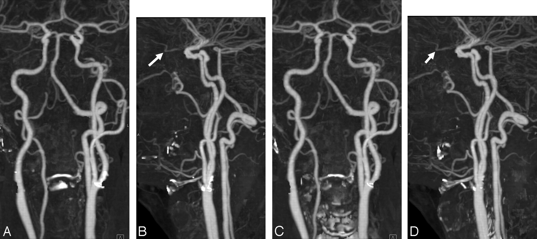
  • Fig 2.
    • Download figure
    • Open in new tab
    • Download powerpoint
    Fig 2.

    CTA (MIP, frontal [A and C] and lateral [B and D] views) shows a patient with atherosclerotic disease at the carotid bifurcation and siphon. Correct registration and successful removal of the calcified plaque at the carotid siphon is obtained with both registration techniques. Pulsation leads to incomplete removal at the left carotid bifurcation, more pronounced with SB-BSCTA (A and B), whereas removal of the calcifications at the right carotid bifurcation is almost perfect with both methods. Removal of the lower cervical spine is more complete with SB-BSCTA compared with PR-BSCTA (C and D). The OA (arrow) can be identified with both approaches. The right ECA is occluded.
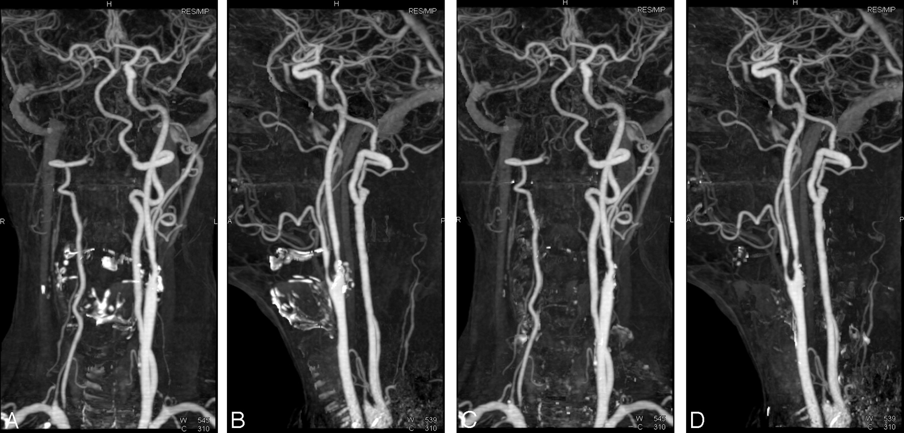
  • Fig 3.
    • Download figure
    • Open in new tab
    • Download powerpoint
    Fig 3.

    CTA (MIP, frontal [A and C] and lateral [B and D] views) shows a patient with occlusion of the right carotid artery and steno-occlusive disease at the left carotid bifurcation and right VA. Both registration techniques provide excellent image quality; more bone remnants can be depicted with SB-BSCTA (A and B) in comparison with PR-BSCTA (C and D) in this patient.
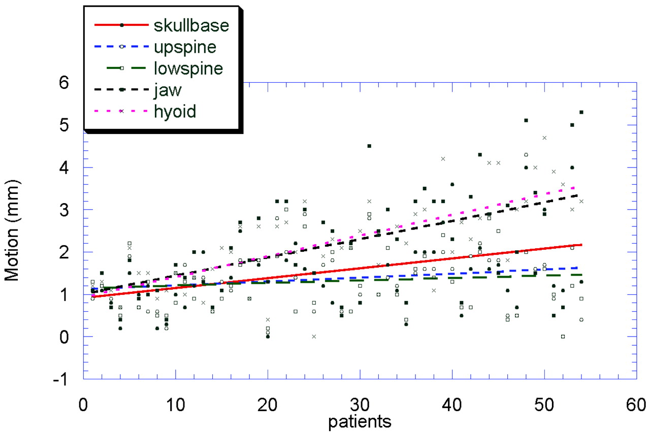
  • Fig 4.
    • Download figure
    • Open in new tab
    • Download powerpoint
    Fig 4.

    Graphic representation of the individual motion amplitudes for each patient. Note that the patients are ranked by increasing complexity of motion. Trend lines indicate the displacement of different anatomic landmarks.

  • Fig 5.
    • Download figure
    • Open in new tab
    • Download powerpoint
    Fig 5.

    Graphic representation (and trend lines) of the quality scores achieved with the different techniques for individual vessels (A, VA. B, Cervical ICA. C, PC-ICA). There is a trend toward decreased image quality with increasing complexity of motion, expressed by the SD of the motion amplitudes except for the PC-ICA.

Tables

  • Figures
    • View popup
    Table 1:

    Summary of quality scores for angiograms processed with pure rigid BSCTA, SB-BSCTA, and PR-BSCTA

    VesselBSCTASB-BSCTAPR-BSCTA
    Mean±SDMedRangeMean±SDMedRangeMean±SDMedRange
    CCA
        Reader 13.501.141–54.410.654.53–54.330.6443–5
        Reader 23.501.141–54.240.6643–54.370.5943–5
    ICA
        Reader 13.480.813.52–53.960.7942–54.260.6443–5
        Reader 23.480.813.52–53.890.7442–54.200.6543–5
    ECA
        Reader 13.430.8332–53.910.7543–54.300.6043–5
        Reader 23.460.883.52–53.780.7143–54.240.6143–5
    PC-ICA
        Reader 14.650.5153–54.920.2754–54.870.3454–5
        Reader 24.610.5653–54.810.3954–54.790.4154–5
    VA
        Reader 12.501.131–53.500.8342–53.500.8841–5
        Reader 22.521.131–53.610.7042–53.740.7542–5
    BA
        Reader 14.900.354–54.980.1454–54.960.1954–5
        Reader 24.880.3354–54.920.2754–54.960.1954–5
    OA
        Reader 14.800.6851–54.830.6751–54.830.6751–5
        Reader 24.600.9251–54.630.9151–54.700.8151–5
    • Note:—Med indicates median.

    • View popup
    Table 2:

    Influence of the 3 registration procedures on image quality of different vessels (P values, Wilcoxon test)*

    VesselReader 1Reader 2
    BSCTA vs SB-BSCTABSCTA vs PR-BSCTASB-BSCTA vs PR-BSCTABSCTA vs SB-BSCTABSCTA vs PR-BSCTASB-BSCTA vs PR-BSCTA
    CCA.000.000.353.000.000.052
    ICA.000.000.003.000.000.001
    ECA.000.000.000.002.000.000
    PC-ICA.001.007.180.050.104.564
    VA.000.000.870.000.000.108
    BA.103.180.564.414.103.157
    OA.317.5641.0.317.157.317
    • * Differences between bone subtraction without and with motion compensation are statistically significant for all vessels except for the BAs and OAs; differences between SB- and PR-BSCTA are statistically significant for the ECA and the cervical ICA.

    • View popup
    Table 3:

    Interobserver agreement for the assessment of the vascular segments with 3 registration algorithms*

    BSCTASB-BSCTAPR-BSCTA
    CCA0.950.730.80
    ICA0.970.910.93
    ECA0.950.810.72
    PC-ICA0.870.520.60
    VA0.980.820.69
    BA0.900.640.46
    OA0.680.670.70
    • * The kappa values indicate high overall interobserver agreement for the assessment of the different vascular segments.

PreviousNext
Back to top

In this issue

American Journal of Neuroradiology: 28 (7)
American Journal of Neuroradiology
Vol. 28, Issue 7
August 2007
  • Table of Contents
  • Index by author
Advertisement
Print
Download PDF
Email Article

Thank you for your interest in spreading the word on American Journal of Neuroradiology.

NOTE: We only request your email address so that the person you are recommending the page to knows that you wanted them to see it, and that it is not junk mail. We do not capture any email address.

Enter multiple addresses on separate lines or separate them with commas.
Bone-Subtraction CT Angiography: Evaluation of Two Different Fully Automated Image-Registration Procedures for Interscan Motion Compensation
(Your Name) has sent you a message from American Journal of Neuroradiology
(Your Name) thought you would like to see the American Journal of Neuroradiology web site.
CAPTCHA
This question is for testing whether or not you are a human visitor and to prevent automated spam submissions.
Cite this article
M.M. Lell, H. Ditt, C. Panknin, J.W. Sayre, S.G. Ruehm, E. Klotz, B.F. Tomandl, J.P. Villablanca
Bone-Subtraction CT Angiography: Evaluation of Two Different Fully Automated Image-Registration Procedures for Interscan Motion Compensation
American Journal of Neuroradiology Aug 2007, 28 (7) 1362-1368; DOI: 10.3174/ajnr.A0558

Citation Manager Formats

  • BibTeX
  • Bookends
  • EasyBib
  • EndNote (tagged)
  • EndNote 8 (xml)
  • Medlars
  • Mendeley
  • Papers
  • RefWorks Tagged
  • Ref Manager
  • RIS
  • Zotero
0 Responses
Respond to this article
Share
Bookmark this article
Bone-Subtraction CT Angiography: Evaluation of Two Different Fully Automated Image-Registration Procedures for Interscan Motion Compensation
M.M. Lell, H. Ditt, C. Panknin, J.W. Sayre, S.G. Ruehm, E. Klotz, B.F. Tomandl, J.P. Villablanca
American Journal of Neuroradiology Aug 2007, 28 (7) 1362-1368; DOI: 10.3174/ajnr.A0558
del.icio.us logo Twitter logo Facebook logo Mendeley logo
  • Tweet Widget
  • Facebook Like
  • Google Plus One
Purchase

Jump to section

  • Article
    • Abstract
    • Materials and Methods
    • Results
    • Discussion
    • Conclusion
    • References
  • Figures & Data
  • Info & Metrics
  • Responses
  • References
  • PDF

Related Articles

  • No related articles found.
  • PubMed
  • Google Scholar

Cited By...

  • Bone-Subtracted Spinal CT Angiography Using Nonrigid Registration for Better Visualization of Arterial Feeders in Spinal Arteriovenous Fistulas
  • 2011 ASA/ACCF/AHA/AANN/AANS/ACR/ASNR/CNS/SAIP/SCAI/SIR/SNIS/SVM/SVS Guideline on the Management of Patients With Extracranial Carotid and Vertebral Artery Disease: A Report of the American College of Cardiology Foundation/American Heart Association Task Force on Practice Guidelines, and the American Stroke Association, American Association of Neuroscience Nurses, American Association of Neurological Surgeons, American College of Radiology, American Society of Neuroradiology, Congress of Neurological Surgeons, Society of Atherosclerosis Imaging and Prevention, Society for Cardiovascular Angiography and Interventions, Society of Interventional Radiology, Society of NeuroInterventional Surgery, Society for Vascular Medicine, and Society for Vascular Surgery
  • 2011 ASA/ACCF/AHA/AANN/AANS/ACR/ASNR/CNS/SAIP/SCAI/SIR/SNIS/SVM/SVS Guideline on the Management of Patients With Extracranial Carotid and Vertebral Artery Disease: A Report of the American College of Cardiology Foundation/American Heart Association Task Force on Practice Guidelines, and the American Stroke Association, American Association of Neuroscience Nurses, American Association of Neurological Surgeons, American College of Radiology, American Society of Neuroradiology, Congress of Neurological Surgeons, Society of Atherosclerosis Imaging and Prevention, Society for Cardiovascular Angiography and Interventions, Society of Interventional Radiology, Society of NeuroInterventional Surgery, Society for Vascular Medicine, and Society for Vascular Surgery
  • 2011 ASA/ACCF/AHA/AANN/AANS/ACR/ASNR/CNS/SAIP/SCAI/SIR/SNIS/SVM/SVS Guideline on the Management of Patients With Extracranial Carotid and Vertebral Artery Disease: A Report of the American College of Cardiology Foundation/American Heart Association Task Force on Practice Guidelines, and the American Stroke Association, American Association of Neuroscience Nurses, American Association of Neurological Surgeons, American College of Radiology, American Society of Neuroradiology, Congress of Neurological Surgeons, Society of Atherosclerosis Imaging and Prevention, Society for Cardiovascular Angiography and Interventions, Society of Interventional Radiology, Society of NeuroInterventional Surgery, Society for Vascular Medicine, and Society for Vascular Surgery Developed in Collaboration With the American Academy of Neurology and Society of Cardiovascular Computed Tomography
  • Multi-Detector Row CT Angiography with Direct Intra-Arterial Contrast Injection for the Evaluation of Neurovascular Disease: Technique, Applications, and Initial Experience
  • Crossref
  • Google Scholar

This article has not yet been cited by articles in journals that are participating in Crossref Cited-by Linking.

More in this TOC Section

  • Hydrops Herniation into the Semicircular Canals
  • ASL Sensitivity for Head and Neck Paraganglioma
  • Post SRS Peritumoral Hyperintense Signal of VSs
Show more HEAD AND NECK IMAGING

Similar Articles

Advertisement

Indexed Content

  • Current Issue
  • Accepted Manuscripts
  • Article Preview
  • Past Issues
  • Editorials
  • Editor's Choice
  • Fellows' Journal Club
  • Letters to the Editor
  • Video Articles

Cases

  • Case Collection
  • Archive - Case of the Week
  • Archive - Case of the Month
  • Archive - Classic Case

More from AJNR

  • Trainee Corner
  • Imaging Protocols
  • MRI Safety Corner
  • Book Reviews

Multimedia

  • AJNR Podcasts
  • AJNR Scantastics

Resources

  • Turnaround Time
  • Submit a Manuscript
  • Submit a Video Article
  • Submit an eLetter to the Editor/Response
  • Manuscript Submission Guidelines
  • Statistical Tips
  • Fast Publishing of Accepted Manuscripts
  • Graphical Abstract Preparation
  • Imaging Protocol Submission
  • Evidence-Based Medicine Level Guide
  • Publishing Checklists
  • Author Policies
  • Become a Reviewer/Academy of Reviewers
  • News and Updates

About Us

  • About AJNR
  • Editorial Board
  • Editorial Board Alumni
  • Alerts
  • Permissions
  • Not an AJNR Subscriber? Join Now
  • Advertise with Us
  • Librarian Resources
  • Feedback
  • Terms and Conditions
  • AJNR Editorial Board Alumni

American Society of Neuroradiology

  • Not an ASNR Member? Join Now

© 2025 by the American Society of Neuroradiology All rights, including for text and data mining, AI training, and similar technologies, are reserved.
Print ISSN: 0195-6108 Online ISSN: 1936-959X

Powered by HighWire