Advertisement

Main menu

  • Home
  • Content
    • Current Issue
    • Accepted Manuscripts
    • Article Preview
    • Past Issue Archive
    • Video Articles
    • AJNR Case Collection
    • Case of the Week Archive
    • Case of the Month Archive
    • Classic Case Archive
  • Special Collections
    • AJNR Awards
    • Low-Field MRI
    • Alzheimer Disease
    • ASNR Foundation Special Collection
    • Photon-Counting CT
    • View All
  • Multimedia
    • AJNR Podcasts
    • AJNR SCANtastic
    • Trainee Corner
    • MRI Safety Corner
    • Imaging Protocols
  • For Authors
    • Submit a Manuscript
    • Submit a Video Article
    • Submit an eLetter to the Editor/Response
    • Manuscript Submission Guidelines
    • Statistical Tips
    • Fast Publishing of Accepted Manuscripts
    • Graphical Abstract Preparation
    • Imaging Protocol Submission
    • Author Policies
  • About Us
    • About AJNR
    • Editorial Board
    • Editorial Board Alumni
  • More
    • Become a Reviewer/Academy of Reviewers
    • Subscribers
    • Permissions
    • Alerts
    • Feedback
    • Advertisers
    • ASNR Home

User menu

  • Alerts
  • Log in

Search

  • Advanced search
American Journal of Neuroradiology

American Journal of Neuroradiology

ASHNR American Society of Functional Neuroradiology ASHNR American Society of Pediatric Neuroradiology ASSR
  • Alerts
  • Log in

Advanced Search

  • Home
  • Content
    • Current Issue
    • Accepted Manuscripts
    • Article Preview
    • Past Issue Archive
    • Video Articles
    • AJNR Case Collection
    • Case of the Week Archive
    • Case of the Month Archive
    • Classic Case Archive
  • Special Collections
    • AJNR Awards
    • Low-Field MRI
    • Alzheimer Disease
    • ASNR Foundation Special Collection
    • Photon-Counting CT
    • View All
  • Multimedia
    • AJNR Podcasts
    • AJNR SCANtastic
    • Trainee Corner
    • MRI Safety Corner
    • Imaging Protocols
  • For Authors
    • Submit a Manuscript
    • Submit a Video Article
    • Submit an eLetter to the Editor/Response
    • Manuscript Submission Guidelines
    • Statistical Tips
    • Fast Publishing of Accepted Manuscripts
    • Graphical Abstract Preparation
    • Imaging Protocol Submission
    • Author Policies
  • About Us
    • About AJNR
    • Editorial Board
    • Editorial Board Alumni
  • More
    • Become a Reviewer/Academy of Reviewers
    • Subscribers
    • Permissions
    • Alerts
    • Feedback
    • Advertisers
    • ASNR Home
  • Follow AJNR on Twitter
  • Visit AJNR on Facebook
  • Follow AJNR on Instagram
  • Join AJNR on LinkedIn
  • RSS Feeds

AJNR Awards, New Junior Editors, and more. Read the latest AJNR updates

Research ArticleBrain

Hippocampal Shape Analysis of Alzheimer Disease Based on Machine Learning Methods

S. Li, F. Shi, F. Pu, X. Li, T. Jiang, S. Xie and Y. Wang
American Journal of Neuroradiology August 2007, 28 (7) 1339-1345; DOI: https://doi.org/10.3174/ajnr.A0620
S. Li
  • Find this author on Google Scholar
  • Find this author on PubMed
  • Search for this author on this site
F. Shi
  • Find this author on Google Scholar
  • Find this author on PubMed
  • Search for this author on this site
F. Pu
  • Find this author on Google Scholar
  • Find this author on PubMed
  • Search for this author on this site
X. Li
  • Find this author on Google Scholar
  • Find this author on PubMed
  • Search for this author on this site
T. Jiang
  • Find this author on Google Scholar
  • Find this author on PubMed
  • Search for this author on this site
S. Xie
  • Find this author on Google Scholar
  • Find this author on PubMed
  • Search for this author on this site
Y. Wang
  • Find this author on Google Scholar
  • Find this author on PubMed
  • Search for this author on this site
  • Article
  • Figures & Data
  • Info & Metrics
  • Responses
  • References
  • PDF
Loading

Article Figures & Data

Figures

  • Tables
  • Fig 1.
    • Download figure
    • Open in new tab
    • Download powerpoint
    Fig 1.

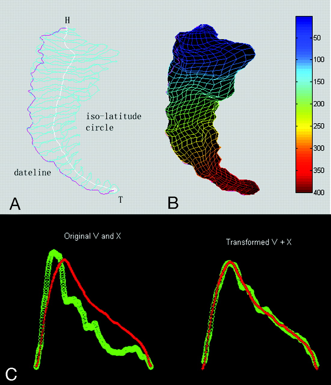
    The steps of parameterization and correspondence of hippocampus.

    A, Hippocampus shape characters; H and T represent the head and tail landmarks, respectively; the cyan shows isolatitude circles; the pink shows the extracted dateline.

    B, The parameterized mesh; the blue-red hue scale indicates the changes from the head to the tail of hippocampus.

    C, The alignment in terms of the isolatitude circle areas along latitude direction. The 2 curves on the left show the area distribution of isolatitude circles from head to tail; the red is the template and the green is 1 subject before transformation. The results after transformation are shown at the right.

  • Fig 2.
    • Download figure
    • Open in new tab
    • Download powerpoint
    Fig 2.

    The flow chart of LOOCV experiment; the numbers inside of the parentheses are the sample indexes.

  • Fig 3.
    • Download figure
    • Open in new tab
    • Download powerpoint
    Fig 3.

    LOOCV accuracy when selecting different repeatability of features to construct the classifier. The horizontal axis represents different repeatability of features, and the vertical axis represents the corresponding LOOCV accuracy. The different colors, respectively, represent the left and right hippocampi and different patch sizes. L represents the left hemisphere, and R represents the right hemisphere. The numbers behind L or R represent the patch sizes: 800 represents 20 × 40; 1250 represents 25 × 50; and 5000 represents 50 × 100.

  • Fig 4.
    • Download figure
    • Open in new tab
    • Download powerpoint
    Fig 4.

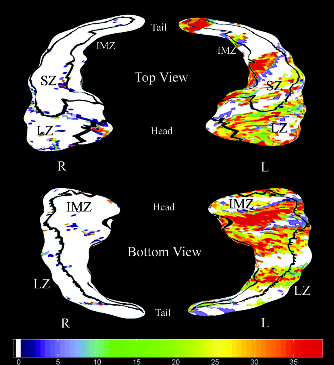
    Visualization of selected surface features by LOOCV experiment when the patch size is 50 × 100 in patients with AD compared with healthy control subjects. The first row shows the left and right hippocampi from the top view, whereas the second row shows them from the bottom view. Boundaries between the 3 zones of the hippocampal surface (ie, LZ, SZ, and IMZ) are drawn in black. LZ represents the lateral zone of the hippocampal surface, and approximates the CA1 subfield. SZ represents the superior zone approximating the combined CA2, CA3, and CA4 subfields and the gyrus dentatus, and IMZ represents the inferior-medial zone approximating the subiculum. All 3 zones are labeled. The white subregions represent no differences in the hippocampal surfaces between patients with AD and healthy control subjects. The significance of features is measured by the repeatability in the subsets. The more significant features are shaded on the purple-red hue scale shown in the third row. The purple represents lower significance, and the red represents higher significance.

Tables

  • Figures
    • View popup
    Table 1:

    Demographics and clinical findings

    VariableAD (n = 19)Controls (n = 20)P
    Sex, F/M9/1010/10>.95*
    Age, mean ± SD72.6 ± 6.970.7 ± 6.4.38†
    Education, mean ± SD11.3 ± 4.010.9 ± 4.5.76†
    MMSE score, mean ± SD18.9 ± 3.929.5 ± 0.9<.0001†
    • Note:—MMSE indicates Mini-Mental State Examination; AD, Alzheimer disease; F, female; M, male.

    • * The P value was obtained by Pearson χ2 2-tailed test, with continuity correction for n < 5.

    • † The P value was obtained by a 2-sample 2-tailed t test.

    • View popup
    Table 2:

    Cross-validation accuracies in the experiment

    Patch SizeSideLeave-1-Out Cross-Validation Accuracy, %*3-Fold Cross-Validation Accuracy*
    50 × 100Right84.683.4% (81.8%–84.9%)†
    Left94.984.5% (81.2%–87.9%)†
    25 × 50Right84.681.6% (79.8%–83.4%)†
    Left94.989.6% (87.1%–92.1%)†
    20 × 40Right84.682.6% (80.7%–84.6%)†
    Left94.989.3% (86.8%–91.9%)†
    • * Note:—All of the features appearing in subsets are selected to construct the classifier.

    • † The 95% confidence intervals of classification accuracy by 100 times cross-validation experiments.

PreviousNext
Back to top

In this issue

American Journal of Neuroradiology: 28 (7)
American Journal of Neuroradiology
Vol. 28, Issue 7
August 2007
  • Table of Contents
  • Index by author
Advertisement
Print
Download PDF
Email Article

Thank you for your interest in spreading the word on American Journal of Neuroradiology.

NOTE: We only request your email address so that the person you are recommending the page to knows that you wanted them to see it, and that it is not junk mail. We do not capture any email address.

Enter multiple addresses on separate lines or separate them with commas.
Hippocampal Shape Analysis of Alzheimer Disease Based on Machine Learning Methods
(Your Name) has sent you a message from American Journal of Neuroradiology
(Your Name) thought you would like to see the American Journal of Neuroradiology web site.
CAPTCHA
This question is for testing whether or not you are a human visitor and to prevent automated spam submissions.
Cite this article
S. Li, F. Shi, F. Pu, X. Li, T. Jiang, S. Xie, Y. Wang
Hippocampal Shape Analysis of Alzheimer Disease Based on Machine Learning Methods
American Journal of Neuroradiology Aug 2007, 28 (7) 1339-1345; DOI: 10.3174/ajnr.A0620

Citation Manager Formats

  • BibTeX
  • Bookends
  • EasyBib
  • EndNote (tagged)
  • EndNote 8 (xml)
  • Medlars
  • Mendeley
  • Papers
  • RefWorks Tagged
  • Ref Manager
  • RIS
  • Zotero
0 Responses
Respond to this article
Share
Bookmark this article
Hippocampal Shape Analysis of Alzheimer Disease Based on Machine Learning Methods
S. Li, F. Shi, F. Pu, X. Li, T. Jiang, S. Xie, Y. Wang
American Journal of Neuroradiology Aug 2007, 28 (7) 1339-1345; DOI: 10.3174/ajnr.A0620
del.icio.us logo Twitter logo Facebook logo Mendeley logo
  • Tweet Widget
  • Facebook Like
  • Google Plus One
Purchase

Jump to section

  • Article
    • Abstract
    • Materials and Methods
    • Results
    • Discussion
    • Conclusion
    • Acknowledgments
    • Footnotes
    • References
  • Figures & Data
  • Info & Metrics
  • Responses
  • References
  • PDF

Related Articles

  • No related articles found.
  • PubMed
  • Google Scholar

Cited By...

  • Identifying incident dementia by applying machine learning to a very large administrative claims dataset
  • Automated Segmentation of Hippocampal Subfields in Drug-Naive Patients with Alzheimer Disease
  • Crossref
  • Google Scholar

This article has not yet been cited by articles in journals that are participating in Crossref Cited-by Linking.

More in this TOC Section

  • White Matter Alterations in the Brains of Patients with Active, Remitted, and Cured Cushing Syndrome: A DTI Study
  • Qualitative and Quantitative Analysis of MR Imaging Findings in Patients with Middle Cerebral Artery Stroke Implanted with Mesenchymal Stem Cells
  • Fast Contrast-Enhanced 4D MRA and 4D Flow MRI Using Constrained Reconstruction (HYPRFlow): Potential Applications for Brain Arteriovenous Malformations
Show more Brain

Similar Articles

Advertisement

Indexed Content

  • Current Issue
  • Accepted Manuscripts
  • Article Preview
  • Past Issues
  • Editorials
  • Editor's Choice
  • Fellows' Journal Club
  • Letters to the Editor
  • Video Articles

Cases

  • Case Collection
  • Archive - Case of the Week
  • Archive - Case of the Month
  • Archive - Classic Case

More from AJNR

  • Trainee Corner
  • Imaging Protocols
  • MRI Safety Corner
  • Book Reviews

Multimedia

  • AJNR Podcasts
  • AJNR Scantastics

Resources

  • Turnaround Time
  • Submit a Manuscript
  • Submit a Video Article
  • Submit an eLetter to the Editor/Response
  • Manuscript Submission Guidelines
  • Statistical Tips
  • Fast Publishing of Accepted Manuscripts
  • Graphical Abstract Preparation
  • Imaging Protocol Submission
  • Evidence-Based Medicine Level Guide
  • Publishing Checklists
  • Author Policies
  • Become a Reviewer/Academy of Reviewers
  • News and Updates

About Us

  • About AJNR
  • Editorial Board
  • Editorial Board Alumni
  • Alerts
  • Permissions
  • Not an AJNR Subscriber? Join Now
  • Advertise with Us
  • Librarian Resources
  • Feedback
  • Terms and Conditions
  • AJNR Editorial Board Alumni

American Society of Neuroradiology

  • Not an ASNR Member? Join Now

© 2025 by the American Society of Neuroradiology All rights, including for text and data mining, AI training, and similar technologies, are reserved.
Print ISSN: 0195-6108 Online ISSN: 1936-959X

Powered by HighWire