Advertisement

Main menu

  • Home
  • Content
    • Current Issue
    • Accepted Manuscripts
    • Article Preview
    • Past Issue Archive
    • Video Articles
    • AJNR Case Collection
    • Case of the Week Archive
    • Case of the Month Archive
    • Classic Case Archive
  • Special Collections
    • AJNR Awards
    • Low-Field MRI
    • Alzheimer Disease
    • ASNR Foundation Special Collection
    • Photon-Counting CT
    • View All
  • Multimedia
    • AJNR Podcasts
    • AJNR SCANtastic
    • Trainee Corner
    • MRI Safety Corner
    • Imaging Protocols
  • For Authors
    • Submit a Manuscript
    • Submit a Video Article
    • Submit an eLetter to the Editor/Response
    • Manuscript Submission Guidelines
    • Statistical Tips
    • Fast Publishing of Accepted Manuscripts
    • Graphical Abstract Preparation
    • Imaging Protocol Submission
    • Author Policies
  • About Us
    • About AJNR
    • Editorial Board
    • Editorial Board Alumni
  • More
    • Become a Reviewer/Academy of Reviewers
    • Subscribers
    • Permissions
    • Alerts
    • Feedback
    • Advertisers
    • ASNR Home

User menu

  • Alerts
  • Log in

Search

  • Advanced search
American Journal of Neuroradiology

American Journal of Neuroradiology

ASHNR American Society of Functional Neuroradiology ASHNR American Society of Pediatric Neuroradiology ASSR
  • Alerts
  • Log in

Advanced Search

  • Home
  • Content
    • Current Issue
    • Accepted Manuscripts
    • Article Preview
    • Past Issue Archive
    • Video Articles
    • AJNR Case Collection
    • Case of the Week Archive
    • Case of the Month Archive
    • Classic Case Archive
  • Special Collections
    • AJNR Awards
    • Low-Field MRI
    • Alzheimer Disease
    • ASNR Foundation Special Collection
    • Photon-Counting CT
    • View All
  • Multimedia
    • AJNR Podcasts
    • AJNR SCANtastic
    • Trainee Corner
    • MRI Safety Corner
    • Imaging Protocols
  • For Authors
    • Submit a Manuscript
    • Submit a Video Article
    • Submit an eLetter to the Editor/Response
    • Manuscript Submission Guidelines
    • Statistical Tips
    • Fast Publishing of Accepted Manuscripts
    • Graphical Abstract Preparation
    • Imaging Protocol Submission
    • Author Policies
  • About Us
    • About AJNR
    • Editorial Board
    • Editorial Board Alumni
  • More
    • Become a Reviewer/Academy of Reviewers
    • Subscribers
    • Permissions
    • Alerts
    • Feedback
    • Advertisers
    • ASNR Home
  • Follow AJNR on Twitter
  • Visit AJNR on Facebook
  • Follow AJNR on Instagram
  • Join AJNR on LinkedIn
  • RSS Feeds

AJNR Awards, New Junior Editors, and more. Read the latest AJNR updates

OtherBRAIN

Assessment of Collateral Supply by Two-Coil Continuous Arterial Spin Labeling after Coil Occlusion of the Internal Carotid Artery

H.E. Möller, T. Mildner, C. Preul, C. Zimmer and D. Yves von Cramon
American Journal of Neuroradiology August 2007, 28 (7) 1304-1305; DOI: https://doi.org/10.3174/ajnr.A0552
H.E. Möller
  • Find this author on Google Scholar
  • Find this author on PubMed
  • Search for this author on this site
T. Mildner
  • Find this author on Google Scholar
  • Find this author on PubMed
  • Search for this author on this site
C. Preul
  • Find this author on Google Scholar
  • Find this author on PubMed
  • Search for this author on this site
C. Zimmer
  • Find this author on Google Scholar
  • Find this author on PubMed
  • Search for this author on this site
D. Yves von Cramon
  • Find this author on Google Scholar
  • Find this author on PubMed
  • Search for this author on this site
  • Article
  • Figures & Data
  • Info & Metrics
  • Responses
  • References
  • PDF
Loading

Abstract

SUMMARY: A patient undergoing coil occlusion of a left internal carotid artery aneurysm was investigated by continuous arterial spin labeling MR imaging to evaluate perfusion territory mapping. Labeling was restricted to the left- or right-sided carotid artery by use of a separate neck coil. Before embolization, perfusion contrast was largely restricted to the labeled hemisphere. After embolization, perfusion contrast was created symmetrically in both hemispheres on labeling the right side, verifying sufficient collateral supply.

In carotid artery occlusive disease, the degree of stenosis is a critical parameter in the potential development of stroke.1 Knowledge of the amount of compromise of cerebral blood flow (CBF), of the affected perfusion territories, and of the status of collaterals would additionally be desirable for assessing the hemodynamic status of the brain and predicting the outcome. Perfusion MR imaging is increasingly being used for quantifying CBF and assessing cerebrovascular function. A totally noninvasive technique is arterial spin labeling, which exploits magnetically labeled arterial water as an endogenous, diffusible tracer to quantify perfusion from 2 sets of images obtained with labeling and a control condition. Approaches for mapping the flow territories of major brain-supplying arteries have also been suggested.2–6 For this study, we used continuous arterial spin labeling (CASL) with a local transmit coil at the neck for selectively labeling the blood of either the right or left carotid artery and a separate head coil for imaging.2 The goal was to evaluate the potential of perfusion territory imaging for an assessment of the hemodynamic status of the brain in a patient undergoing coil occlusion of the internal carotid artery (ICA).

Case Report

In a male 28-year-old patient, a dissecting aneurysm of the left ICA had developed after an accident during childhood and caused a minor stroke. Carotid balloon occlusion test revealed that the patient would tolerate permanent ICA occlusion because of sufficient collateral supply to the left hemisphere. Multiple Guglielmi detachable coils (Boston Scientific, Natick, Mass) were then inserted for the occlusion of the distal ICA aneurysm through a microcatheter. Postembolization angiograms verified total occlusion of the left ICA.

After obtaining informed written consent, CASL was performed on a 3T MedSpec 30/100 scanner (Bruker, Ettlingen, Germany), 1 day before and 3 months after embolization. A 6-cm-diameter surface coil was centered in the region of the right carotid bifurcation, and radiofrequency irradiation (1.2 W) and a magnetic field gradient (2.5 mT/m) along the z-direction were applied during labeling periods of τ = 3.5 seconds. Estimates of the labeling efficiency yielded α = 85%.7 One-hundred repetitions (TR, 7 seconds) were acquired with and without application of CASL during odd and even repetitions, respectively. To account for the finite transit time from the labeling plane to the region of interest, a postlabeling delay of w = 1.5 seconds was introduced before image acquisition. Eight axial sections (thickness, 5 mm; intersection distance, 6 mm) were recorded sequentially from superior to inferior with spin-echo echo-planar imaging (TE, 50 ms; bandwidth, 100 kHz; matrix, 64 × 64; FOV, 192 mm) and a quadrature helmet coil.8 T1-weighted images were recorded at the identical section positions as anatomic reference. In the pre-embolization examination, the whole procedure was repeated after repositioning the label coil for application of CASL on the left side.

All of the perfusion maps were registered to a 3D dataset acquired in a separate session for a direct comparison of individual sections. Images measured during the CASL and control conditions were averaged separately to create a difference map. Estimates of CBF were obtained from the signal intensity change, S̄CASL − S̄ctrl, according to the following9: Math In this equation, a blood-brain partition coefficient λ is 0.9 mL/g, and a longitudinal relaxation time T1b is 1.3 seconds for cortical gray matter (GM). S̄CASL and S̄ctrl are averaged signal intensity amplitudes obtained with and without application of CASL, respectively, and T1app is the apparent longitudinal relaxation time.

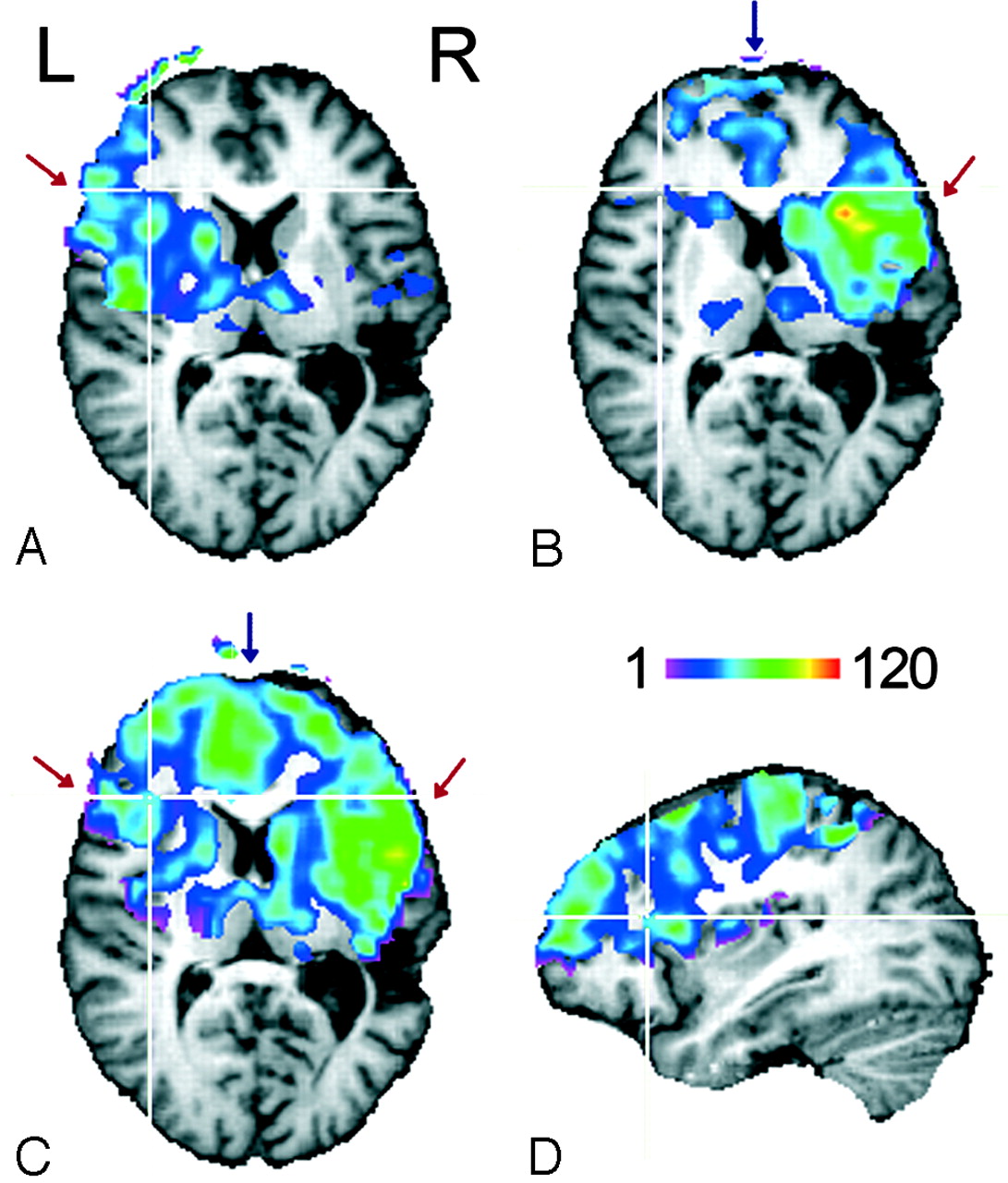
Quantitative perfusion maps obtained before left ICA occlusion are shown in Fig 1A, B. With left-sided CASL, perfusion-related signal intensity changes were found only in leptomeningeal branches of the left middle cerebral artery (MCA) flow territory (Fig 1B, red arrow), whereas with right-sided CASL, signal intensity changes were present in the flow territory of the right MCA (Fig 1A, red arrow) and also in the territories of leptomeningeal branches of both the right and left anterior cerebral artery (ACA) surrounding the interhemispheric fissure (Fig 1B, blue arrow). Figure 1C shows maps recorded with right-sided CASL after left ICA occlusion. Perfusion contrast was now created in both hemispheres in flow territories of the right and left ACA (Fig 1C, blue arrow), as well as that of the right and left MCA (Fig 1C, red arrow). CBF values averaged separately over the left and right hemisphere are summarized in the Table.

Fig 1.
  • Download figure
  • Open in new tab
  • Download powerpoint
Fig 1.

Perfusion territories of the (A) left and (B) right ICA recorded during the first examination and of the (C) right ICA recorded 3 months after coil occlusion of the left ICA. The color code indicates CBF values in milliliters per minute per 100 grams. Only voxels with a significant signal intensity change between label and no-label conditions (thresholded at P < .05) are displayed.

D, The coverage of the section package can be assessed from the sagittal section (left hemisphere).

View this table:
  • View inline
  • View popup

Gray Matter CBF values of the ICA flow territories

Discussion

The pre-embolization results demonstrate that the flow territories of the left and right ICA can be delineated selectively with the 2-coil CASL technique. With the current setup, coverage in deeper brain structures (eg, the inferior parietal lobe) was limited by the sensitive volume of the helmet coil (length, 13 cm) and the angulation of the section package (Fig 1D). Improvement is straightforward by use of an appropriately modified imaging coil.

The distribution of perfusion contrast over both hemispheres after left ICA occlusion, which was observed on unilateral, right-sided CASL, verifies the existence of sufficient collateral supply, presumably via the circle of Willis as the primary pathway. This finding indicating a high sensitivity of the CASL technique agrees well with the result from the preceding carotid balloon occlusion test. Other than such qualitative correspondence, quantitative assessment of the adequacy of collaterals is obtained from the data in the table demonstrating equal postembolization levels of GM perfusion in both hemispheres within the experimental accuracy. The averaged CBF of GM in the unaffected right hemisphere and its variation by 10% between successive examinations are similar to values observed in a previous scan/rescan CASL study in normal volunteers.10 The data recorded before embolization in the flow territory of the left ICA seem to indicate a CBF reduction of borderline significance (P = .09 obtained from a section-by-section comparison and paired-samples t test). Alternatively, a prolonged transit time, which may be attributed to the presence of the aneurysm, could also lead to artificially reduced CBF values. Recently, δ = 1930 ± 110 ms was measured for the time the arterial water needs to travel from the labeling plane at the neck to the tissue in the ICA flow territory.11 A quantitative analysis within the framework of the 2-compartment model (compare equation 1 from Wang et al12) indicates that a transit time increase by 200 ms leads to a reduction of the signal intensity change by 13%, which would explain the observed interhemispheric difference.

Conclusion

Two-coil CASL has a potential to assess the significance of carotid artery stenosis and compensating collateral flow. Because of the possibility to perform repeated studies, it may be suited for screening purposes and for monitoring perfusion changes after therapeutic interventions, which is not adequately achieved by more invasive methods. A 10% range of normal variation and potentially confounding transit time effects impose limitations for detecting subtle CBF changes and should be addressed in further investigations.

Footnotes

  • H.E.M. and T.M. contributed equally to this work.

    Paper previously presented at: Annual Scientific Meeting of the International Society for Magnetic Resonance in Medicine, May 6–12, 2006; Seattle, Wash.

References

  1. ↵
    Executive Committee for the Asymptomatic Carotid Atherosclerosis Study. Endarterectomy for asymptomatic carotid artery stenosis. JAMA 1995;273:1421–28
    CrossRefPubMed
  2. ↵
    Zaharchuk G, Ledden PJ, Kwong KK, et al. Multislice perfusion and perfusion territory imaging in humans with separate label and image coils. Magn Reson Med 1999;41:1093–98
    CrossRefPubMed
  3. Trampel R, Mildner T, Goerke U, et al. Continuous arterial spin labeling using a local magnetic field gradient coil. Magn Reson Med 2002;48:543–46
    CrossRefPubMed
  4. Davies NP, Jezzard P. Selective arterial spin labeling (SASL): perfusion territory mapping of selected feeding arteries tagged using two-dimensional radiofrequency pulses. Magn Reson Med 2003;49:1133–42
    CrossRefPubMed
  5. Werner R, Alfke K, Schaeffter T, et al. Brain perfusion territory imaging applying oblique-plane arterial spin labeling with a standard send/receive head coil. Magn Reson Med 2004;52:1443–47
    CrossRefPubMed
  6. ↵
    Hendrikse J, van der Grond J, Lu H, et al. Flow territory mapping of the cerebral arteries with regional perfusion MRI. Stroke 2004;35:882–87
    Abstract/FREE Full Text
  7. ↵
    Trampel R, Jochimsen TH, Mildner T, et al. Efficiency of flow-driven adiabatic spin inversion under realistic experimental conditions: a computer simulation. Magn Reson Med 2004;51:1187–93
    CrossRefPubMed
  8. ↵
    Driesel W, Merkle H, Hetzer S, et al. Reengineered helmet coil for human brain imaging. Concepts Magn Reson B (Magn Reson Eng)2005;27B:64–74
  9. ↵
    Mildner T, Trampel R, Möller HE, et al. Functional perfusion imaging using continuous arterial spin labeling with separate labeling and imaging coils at 3 T. Magn Reson Med 2003;49:791–95
    CrossRefPubMed
  10. ↵
    Floyd TF, Ratcliffe SJ, Wang J, et al. Precision of the CASL-perfusion MRI technique for the measurement of cerebral blood flow in whole brain and vascular territories. J Magn Reson Imaging 2003;18:649–55
    CrossRefPubMed
  11. ↵
    Mildner T, Möller HE, Driesel W, et al. Continuous arterial spin labeling at the human common carotid artery: the influence of transit times. NMR Biomed 2005;18:19–23
    CrossRefPubMed
  12. ↵
    Wang J, Alsop DC, Li L, et al. Comparison of quantitative perfusion imaging using arterial spin labeling at 1.5 and 4.0 tesla. Magn Reson Med 2002;48:242–54
    CrossRefPubMed
  • Received October 13, 2006.
  • Accepted after revision November 27, 2006.
  • Copyright © American Society of Neuroradiology
PreviousNext
Back to top

In this issue

American Journal of Neuroradiology: 28 (7)
American Journal of Neuroradiology
Vol. 28, Issue 7
August 2007
  • Table of Contents
  • Index by author
Advertisement
Print
Download PDF
Email Article

Thank you for your interest in spreading the word on American Journal of Neuroradiology.

NOTE: We only request your email address so that the person you are recommending the page to knows that you wanted them to see it, and that it is not junk mail. We do not capture any email address.

Enter multiple addresses on separate lines or separate them with commas.
Assessment of Collateral Supply by Two-Coil Continuous Arterial Spin Labeling after Coil Occlusion of the Internal Carotid Artery
(Your Name) has sent you a message from American Journal of Neuroradiology
(Your Name) thought you would like to see the American Journal of Neuroradiology web site.
CAPTCHA
This question is for testing whether or not you are a human visitor and to prevent automated spam submissions.
Cite this article
H.E. Möller, T. Mildner, C. Preul, C. Zimmer, D. Yves von Cramon
Assessment of Collateral Supply by Two-Coil Continuous Arterial Spin Labeling after Coil Occlusion of the Internal Carotid Artery
American Journal of Neuroradiology Aug 2007, 28 (7) 1304-1305; DOI: 10.3174/ajnr.A0552

Citation Manager Formats

  • BibTeX
  • Bookends
  • EasyBib
  • EndNote (tagged)
  • EndNote 8 (xml)
  • Medlars
  • Mendeley
  • Papers
  • RefWorks Tagged
  • Ref Manager
  • RIS
  • Zotero
0 Responses
Respond to this article
Share
Bookmark this article
Assessment of Collateral Supply by Two-Coil Continuous Arterial Spin Labeling after Coil Occlusion of the Internal Carotid Artery
H.E. Möller, T. Mildner, C. Preul, C. Zimmer, D. Yves von Cramon
American Journal of Neuroradiology Aug 2007, 28 (7) 1304-1305; DOI: 10.3174/ajnr.A0552
del.icio.us logo Twitter logo Facebook logo Mendeley logo
  • Tweet Widget
  • Facebook Like
  • Google Plus One
Purchase

Jump to section

  • Article
    • Abstract
    • Case Report
    • Discussion
    • Conclusion
    • Footnotes
    • References
  • Figures & Data
  • Info & Metrics
  • Responses
  • References
  • PDF

Related Articles

  • No related articles found.
  • PubMed
  • Google Scholar

Cited By...

  • MR Imaging of Individual Perfusion Reorganization Using Superselective Pseudocontinuous Arterial Spin-Labeling in Patients with Complex Extracranial Steno-Occlusive Disease
  • Crossref (4)
  • Google Scholar

This article has been cited by the following articles in journals that are participating in Crossref Cited-by Linking.

  • Arterial transit time effects in pulsed arterial spin labeling CBF mapping: Insight from a PET and MR study in normal human subjects
    Maolin Qiu, R. Paul Maguire, Jagriti Arora, Beata Planeta‐Wilson, David Weinzimmer, Jinghua Wang, Yuenan Wang, Hyeonjin Kim, Nallakkandi Rajeevan, Yiyun Huang, Richard E. Carson, R. Todd Constable
    Magnetic Resonance in Medicine 2010 63 2
  • MR Imaging of Individual Perfusion Reorganization Using Superselective Pseudocontinuous Arterial Spin-Labeling in Patients with Complex Extracranial Steno-Occlusive Disease
    V. Richter, M. Helle, M.J.P. van Osch, T. Lindner, A.S. Gersing, P. Tsantilas, H.-H. Eckstein, C. Preibisch, C. Zimmer
    American Journal of Neuroradiology 2017 38 4
  • Shielded dual‐loop resonator for arterial spin labeling at the neck
    Stefan Hetzer, Toralf Mildner, Wolfgang Driesel, Manfred Weder, Harald E. Möller
    Journal of Magnetic Resonance Imaging 2009 29 6
  • Influence of selecting EPI readout-encoding bandwidths on arterial spin labeling perfusion MRI
    Geon-Ho Jahng, Norbert Schuff
    Magnetic Resonance Materials in Physics, Biology and Medicine 2009 22 5

More in this TOC Section

  • Multimodal CT Provides Improved Performance for Lacunar Infarct Detection
  • Optimal MRI Sequence for Identifying Occlusion Location in Acute Stroke: Which Value of Time-Resolved Contrast-Enhanced MRA?
  • Evaluating the Effects of White Matter Multiple Sclerosis Lesions on the Volume Estimation of 6 Brain Tissue Segmentation Methods
Show more BRAIN

Similar Articles

Advertisement

Indexed Content

  • Current Issue
  • Accepted Manuscripts
  • Article Preview
  • Past Issues
  • Editorials
  • Editor's Choice
  • Fellows' Journal Club
  • Letters to the Editor
  • Video Articles

Cases

  • Case Collection
  • Archive - Case of the Week
  • Archive - Case of the Month
  • Archive - Classic Case

More from AJNR

  • Trainee Corner
  • Imaging Protocols
  • MRI Safety Corner

Multimedia

  • AJNR Podcasts
  • AJNR Scantastics

Resources

  • Turnaround Time
  • Submit a Manuscript
  • Submit a Video Article
  • Submit an eLetter to the Editor/Response
  • Manuscript Submission Guidelines
  • Statistical Tips
  • Fast Publishing of Accepted Manuscripts
  • Graphical Abstract Preparation
  • Imaging Protocol Submission
  • Evidence-Based Medicine Level Guide
  • Publishing Checklists
  • Author Policies
  • Become a Reviewer/Academy of Reviewers
  • News and Updates

About Us

  • About AJNR
  • Editorial Board
  • Editorial Board Alumni
  • Alerts
  • Permissions
  • Not an AJNR Subscriber? Join Now
  • Advertise with Us
  • Librarian Resources
  • Feedback
  • Terms and Conditions
  • AJNR Editorial Board Alumni

American Society of Neuroradiology

  • Not an ASNR Member? Join Now

© 2025 by the American Society of Neuroradiology All rights, including for text and data mining, AI training, and similar technologies, are reserved.
Print ISSN: 0195-6108 Online ISSN: 1936-959X

Powered by HighWire