Advertisement

Main menu

  • Home
  • Content
    • Current Issue
    • Accepted Manuscripts
    • Article Preview
    • Past Issue Archive
    • Video Articles
    • AJNR Case Collection
    • Case of the Week Archive
    • Case of the Month Archive
    • Classic Case Archive
  • Special Collections
    • AJNR Awards
    • Low-Field MRI
    • Alzheimer Disease
    • ASNR Foundation Special Collection
    • Photon-Counting CT
    • View All
  • Multimedia
    • AJNR Podcasts
    • AJNR SCANtastic
    • Trainee Corner
    • MRI Safety Corner
    • Imaging Protocols
  • For Authors
    • Submit a Manuscript
    • Submit a Video Article
    • Submit an eLetter to the Editor/Response
    • Manuscript Submission Guidelines
    • Statistical Tips
    • Fast Publishing of Accepted Manuscripts
    • Graphical Abstract Preparation
    • Imaging Protocol Submission
    • Author Policies
  • About Us
    • About AJNR
    • Editorial Board
    • Editorial Board Alumni
  • More
    • Become a Reviewer/Academy of Reviewers
    • Subscribers
    • Permissions
    • Alerts
    • Feedback
    • Advertisers
    • ASNR Home

User menu

  • Alerts
  • Log in

Search

  • Advanced search
American Journal of Neuroradiology

American Journal of Neuroradiology

ASHNR American Society of Functional Neuroradiology ASHNR American Society of Pediatric Neuroradiology ASSR
  • Alerts
  • Log in

Advanced Search

  • Home
  • Content
    • Current Issue
    • Accepted Manuscripts
    • Article Preview
    • Past Issue Archive
    • Video Articles
    • AJNR Case Collection
    • Case of the Week Archive
    • Case of the Month Archive
    • Classic Case Archive
  • Special Collections
    • AJNR Awards
    • Low-Field MRI
    • Alzheimer Disease
    • ASNR Foundation Special Collection
    • Photon-Counting CT
    • View All
  • Multimedia
    • AJNR Podcasts
    • AJNR SCANtastic
    • Trainee Corner
    • MRI Safety Corner
    • Imaging Protocols
  • For Authors
    • Submit a Manuscript
    • Submit a Video Article
    • Submit an eLetter to the Editor/Response
    • Manuscript Submission Guidelines
    • Statistical Tips
    • Fast Publishing of Accepted Manuscripts
    • Graphical Abstract Preparation
    • Imaging Protocol Submission
    • Author Policies
  • About Us
    • About AJNR
    • Editorial Board
    • Editorial Board Alumni
  • More
    • Become a Reviewer/Academy of Reviewers
    • Subscribers
    • Permissions
    • Alerts
    • Feedback
    • Advertisers
    • ASNR Home
  • Follow AJNR on Twitter
  • Visit AJNR on Facebook
  • Follow AJNR on Instagram
  • Join AJNR on LinkedIn
  • RSS Feeds

AJNR Awards, New Junior Editors, and more. Read the latest AJNR updates

Research ArticleBrain

Metabolic Findings on 3T 1H-MR Spectroscopy in Peritumoral Brain Edema

R. Ricci, A. Bacci, V. Tugnoli, S. Battaglia, M. Maffei, R. Agati and M. Leonardi
American Journal of Neuroradiology August 2007, 28 (7) 1287-1291; DOI: https://doi.org/10.3174/ajnr.A0564
R. Ricci
  • Find this author on Google Scholar
  • Find this author on PubMed
  • Search for this author on this site
A. Bacci
  • Find this author on Google Scholar
  • Find this author on PubMed
  • Search for this author on this site
V. Tugnoli
  • Find this author on Google Scholar
  • Find this author on PubMed
  • Search for this author on this site
S. Battaglia
  • Find this author on Google Scholar
  • Find this author on PubMed
  • Search for this author on this site
M. Maffei
  • Find this author on Google Scholar
  • Find this author on PubMed
  • Search for this author on this site
R. Agati
  • Find this author on Google Scholar
  • Find this author on PubMed
  • Search for this author on this site
M. Leonardi
  • Find this author on Google Scholar
  • Find this author on PubMed
  • Search for this author on this site
  • Article
  • Figures & Data
  • Info & Metrics
  • Responses
  • References
  • PDF
Loading

Article Figures & Data

Figures

  • Tables
  • Fig 1.
    • Download figure
    • Open in new tab
    • Download powerpoint
    Fig 1.

    A T2-weighted FSE axial image of a normal brain.

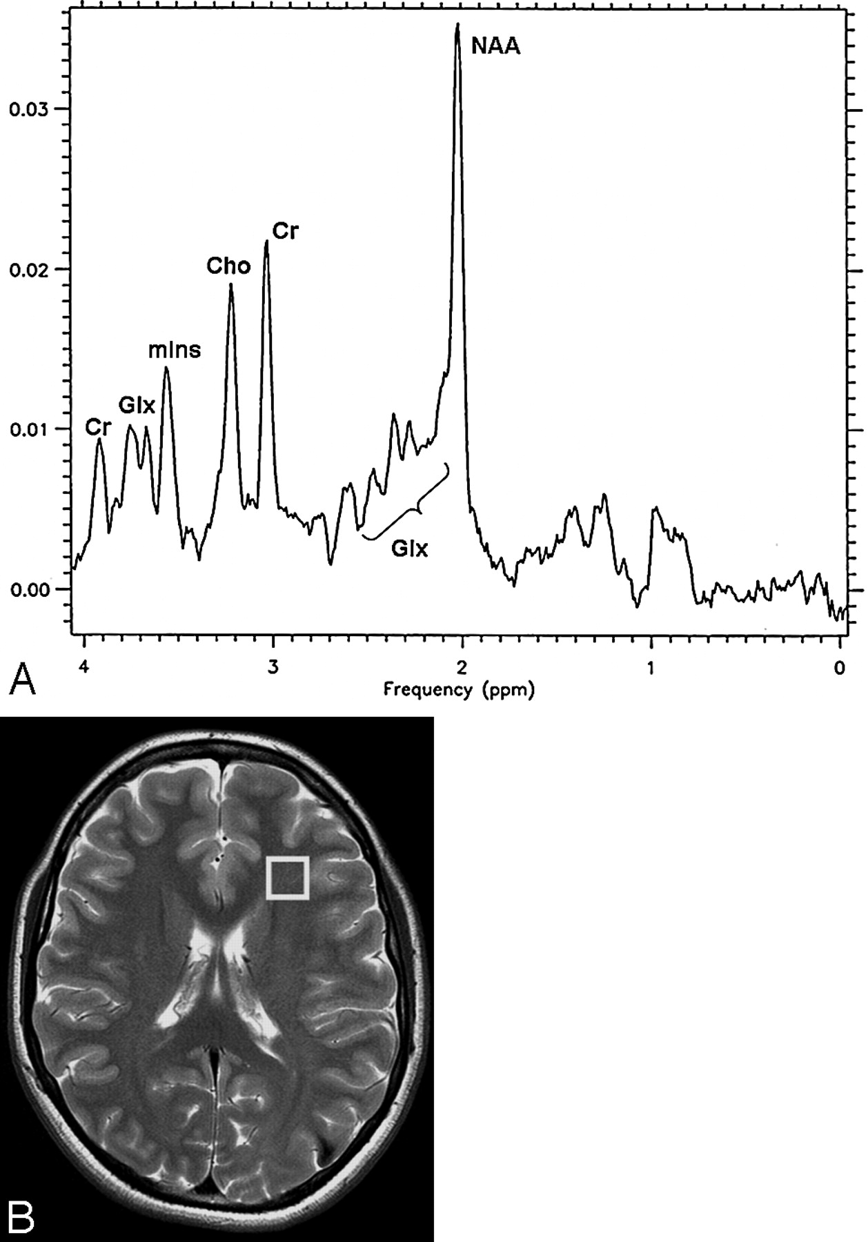
    A, Localized proton spectrum from the VOI (8 cm3) in the white matter of the left frontal region of 16 healthy volunteers, recorded with use of the PRESS sequence at 3T (TR, 2000 ms; TE, 35 ms). (NAA indicates N-acetylaspartate; Cho, choline-containing compounds; Cr, creatine and phosphocreatine; mIns, myo-inositol; Glx, glutamate plus glutamine.)

    B, Location of voxel used for localized proton spectra in healthy volunteers. A T2-weighted FSE axial image (TR, 4200 ms; TE, 93 ms; NEX, 2).

  • Fig 2.
    • Download figure
    • Open in new tab
    • Download powerpoint
    Fig 2.

    A glioblastoma in patient 3.

    A, Localized proton spectrum of the edema surrounding the large temporofrontal glioblastoma. The NAA/Cr, NAA/Cho, Cho/Cr, and mIns/Cr ratios are in the normal range. The Glx/Cr ratio is increased to 2.2 (normal value, 1.8 ± 0.2). Glc (3.78 and 3.44 ppm), Lac, Lip, Val, Leu, Ile, and Ala are detected.

    B, Axial FSE T2-weighted image (TR, 4200 ms; TE, 93 ms; NEX, 2) with the location of the voxel (6.4 cm3) in the edematous tissue.

    C, Axial SE T1-weighted image (TR, 560 ms; TE, 18 ms) after administration of contrast agent. At this level, the upper part of the tumor can be seen.

  • Fig 3.
    • Download figure
    • Open in new tab
    • Download powerpoint
    Fig 3.

    A meningioma in patient 12.

    A, Localized proton spectrum of the edema surrounding the meningioma. The NAA/Cho, Cho/Cr, and mIns/Cr ratios are in the normal range. NAA/Cr is decreased to 1.4 (normal value, 1.8 ± 0.3), and Glx/Cr is increased to 2.5 (normal value, 1.8 ± 0.2). Glc, Lac, Lip, Val, Leu, and Ile are detected.

    B, Axial FSE T2-weighted image (TR, 4200 ms; TE, 93 ms; NEX, 2) with the voxel (8 cm3) location in the edematous tissue.

    C, Axial SE T1-weighted image (TR, 560 ms; TE, 18 ms) after administration of contrast agent with strong enhancement of the tumor.

Tables

  • Figures
    • View popup
    Table 1:

    Ratios of the main metabolites detected in the cerebral edema of 13 patients*

    NAA/CrNAA/ChoCho/CrmIns/CrGlx/Cr
    Glioblastomas (pt no.)
        11.71.61.10.81.8
        20.9†0.9†10.61.6
        322.20.90.72.2†
        41†1†10.62.1†
        51.2†1.4†0.90.72.6†
        61.51.70.90.52
        71.51.90.80.52.8†
    Metastases (pt no.)
        81.81.710.42.8†
        91.92.30.80.43†
        101.61.4†1.10.42.7†
    Meningiomas (pt no.)
        111.81.90.90.43.2†
        121.91.810.73.7†
        131.4†1.80.80.42.5†
    • Note:—NAA indicates N-acetylaspartate; Cr, creatine and phosphocreatine; Cho, choline- containing compounds; Glx, glutamate plus glutamine; mIns, myo-inositol.

    • * Ratios of the main metabolites are detected by PRESS sequences (TR, 2000 ms; TE, 35 ms) in edema surrounding 7 glioblastomas, 3 metastases, and 3 meningiomas. These values were compared with those obtained in the white matter of the left frontal region with use of the same technique in 16 healthy volunteers between 25 and 67 years old (mean age, 40.7 ± 9 years). The normal values are NAA/Cr 1.8 ± 0.3; NAA/Cho 2 ± 0.4; Cho/Cr 0.9 ± 0.2; mIns/Cr 0.6 ± 0.2 and Glx/Cr 1.8 ± 0.2.

    • † Altered values.

    • View popup
    Table 2:

    Detection of metabolites normally not found in the human brain

    Glc 3.44 ppm*Lac*Lip*Other Metabolites*
    Glioblastomas (pt no.)
        1+++Val, Leu, Ile
        2++Val, Leu, Ile
        3++++Val, Leu, Ile, Ala
        4++Val, Leu, Ile
        5+++
        6+++Val, Leu, Ile
        7+Val, Leu, Ile
    Metastases (pt no.)
        8+++Val, Leu, Ile
        9++Val, Leu, Ile
        10++Val, Leu, Ile
    Meningiomas (pt no.)
        11++++Val, Leu, Ile
        12+++++Val, Leu, Ile
        13++++Val, Leu, Ile, Ala
    • Note:—Glc indicates glucose; Lac, lactate; Lip, lipids; Val, valine; Leu, leucine; Ile, isoleucine; and Ala, alanine.

    • * Peak resonances of Glc, Lac, and Lip detected and arbitrarily assigned to 1 of 3 grades, low (+), medium (++), or high (+++) on the basis of the ratio of the integral of the metabolite peak to the integral of unsuppressed water peak. For Val, Leu, Ile, and Ala, only their presence is indicated.

PreviousNext
Back to top

In this issue

American Journal of Neuroradiology: 28 (7)
American Journal of Neuroradiology
Vol. 28, Issue 7
August 2007
  • Table of Contents
  • Index by author
Advertisement
Print
Download PDF
Email Article

Thank you for your interest in spreading the word on American Journal of Neuroradiology.

NOTE: We only request your email address so that the person you are recommending the page to knows that you wanted them to see it, and that it is not junk mail. We do not capture any email address.

Enter multiple addresses on separate lines or separate them with commas.
Metabolic Findings on 3T 1H-MR Spectroscopy in Peritumoral Brain Edema
(Your Name) has sent you a message from American Journal of Neuroradiology
(Your Name) thought you would like to see the American Journal of Neuroradiology web site.
CAPTCHA
This question is for testing whether or not you are a human visitor and to prevent automated spam submissions.
Cite this article
R. Ricci, A. Bacci, V. Tugnoli, S. Battaglia, M. Maffei, R. Agati, M. Leonardi
Metabolic Findings on 3T 1H-MR Spectroscopy in Peritumoral Brain Edema
American Journal of Neuroradiology Aug 2007, 28 (7) 1287-1291; DOI: 10.3174/ajnr.A0564

Citation Manager Formats

  • BibTeX
  • Bookends
  • EasyBib
  • EndNote (tagged)
  • EndNote 8 (xml)
  • Medlars
  • Mendeley
  • Papers
  • RefWorks Tagged
  • Ref Manager
  • RIS
  • Zotero
0 Responses
Respond to this article
Share
Bookmark this article
Metabolic Findings on 3T 1H-MR Spectroscopy in Peritumoral Brain Edema
R. Ricci, A. Bacci, V. Tugnoli, S. Battaglia, M. Maffei, R. Agati, M. Leonardi
American Journal of Neuroradiology Aug 2007, 28 (7) 1287-1291; DOI: 10.3174/ajnr.A0564
del.icio.us logo Twitter logo Facebook logo Mendeley logo
  • Tweet Widget
  • Facebook Like
  • Google Plus One
Purchase

Jump to section

  • Article
    • Abstract
    • Patients and Methods
    • Results
    • Discussion
    • Conclusions
    • Acknowledgments
    • References
  • Figures & Data
  • Info & Metrics
  • Responses
  • References
  • PDF

Related Articles

  • No related articles found.
  • PubMed
  • Google Scholar

Cited By...

  • Application of 7T MRS to High-Grade Gliomas
  • Discrimination between Glioblastoma and Solitary Brain Metastasis: Comparison of Inflow-Based Vascular-Space-Occupancy and Dynamic Susceptibility Contrast MR Imaging
  • Crossref
  • Google Scholar

This article has not yet been cited by articles in journals that are participating in Crossref Cited-by Linking.

More in this TOC Section

  • White Matter Alterations in the Brains of Patients with Active, Remitted, and Cured Cushing Syndrome: A DTI Study
  • Qualitative and Quantitative Analysis of MR Imaging Findings in Patients with Middle Cerebral Artery Stroke Implanted with Mesenchymal Stem Cells
  • Fast Contrast-Enhanced 4D MRA and 4D Flow MRI Using Constrained Reconstruction (HYPRFlow): Potential Applications for Brain Arteriovenous Malformations
Show more Brain

Similar Articles

Advertisement

Indexed Content

  • Current Issue
  • Accepted Manuscripts
  • Article Preview
  • Past Issues
  • Editorials
  • Editor's Choice
  • Fellows' Journal Club
  • Letters to the Editor
  • Video Articles

Cases

  • Case Collection
  • Archive - Case of the Week
  • Archive - Case of the Month
  • Archive - Classic Case

More from AJNR

  • Trainee Corner
  • Imaging Protocols
  • MRI Safety Corner
  • Book Reviews

Multimedia

  • AJNR Podcasts
  • AJNR Scantastics

Resources

  • Turnaround Time
  • Submit a Manuscript
  • Submit a Video Article
  • Submit an eLetter to the Editor/Response
  • Manuscript Submission Guidelines
  • Statistical Tips
  • Fast Publishing of Accepted Manuscripts
  • Graphical Abstract Preparation
  • Imaging Protocol Submission
  • Evidence-Based Medicine Level Guide
  • Publishing Checklists
  • Author Policies
  • Become a Reviewer/Academy of Reviewers
  • News and Updates

About Us

  • About AJNR
  • Editorial Board
  • Editorial Board Alumni
  • Alerts
  • Permissions
  • Not an AJNR Subscriber? Join Now
  • Advertise with Us
  • Librarian Resources
  • Feedback
  • Terms and Conditions
  • AJNR Editorial Board Alumni

American Society of Neuroradiology

  • Not an ASNR Member? Join Now

© 2025 by the American Society of Neuroradiology All rights, including for text and data mining, AI training, and similar technologies, are reserved.
Print ISSN: 0195-6108 Online ISSN: 1936-959X

Powered by HighWire