Advertisement

Main menu

  • Home
  • Content
    • Current Issue
    • Accepted Manuscripts
    • Article Preview
    • Past Issue Archive
    • Video Articles
    • AJNR Case Collection
    • Case of the Week Archive
    • Case of the Month Archive
    • Classic Case Archive
  • Special Collections
    • AJNR Awards
    • Low-Field MRI
    • Alzheimer Disease
    • ASNR Foundation Special Collection
    • Photon-Counting CT
    • View All
  • Multimedia
    • AJNR Podcasts
    • AJNR SCANtastic
    • Trainee Corner
    • MRI Safety Corner
    • Imaging Protocols
  • For Authors
    • Submit a Manuscript
    • Submit a Video Article
    • Submit an eLetter to the Editor/Response
    • Manuscript Submission Guidelines
    • Statistical Tips
    • Fast Publishing of Accepted Manuscripts
    • Graphical Abstract Preparation
    • Imaging Protocol Submission
    • Author Policies
  • About Us
    • About AJNR
    • Editorial Board
    • Editorial Board Alumni
  • More
    • Become a Reviewer/Academy of Reviewers
    • Subscribers
    • Permissions
    • Alerts
    • Feedback
    • Advertisers
    • ASNR Home

User menu

  • Alerts
  • Log in

Search

  • Advanced search
American Journal of Neuroradiology

American Journal of Neuroradiology

ASHNR American Society of Functional Neuroradiology ASHNR American Society of Pediatric Neuroradiology ASSR
  • Alerts
  • Log in

Advanced Search

  • Home
  • Content
    • Current Issue
    • Accepted Manuscripts
    • Article Preview
    • Past Issue Archive
    • Video Articles
    • AJNR Case Collection
    • Case of the Week Archive
    • Case of the Month Archive
    • Classic Case Archive
  • Special Collections
    • AJNR Awards
    • Low-Field MRI
    • Alzheimer Disease
    • ASNR Foundation Special Collection
    • Photon-Counting CT
    • View All
  • Multimedia
    • AJNR Podcasts
    • AJNR SCANtastic
    • Trainee Corner
    • MRI Safety Corner
    • Imaging Protocols
  • For Authors
    • Submit a Manuscript
    • Submit a Video Article
    • Submit an eLetter to the Editor/Response
    • Manuscript Submission Guidelines
    • Statistical Tips
    • Fast Publishing of Accepted Manuscripts
    • Graphical Abstract Preparation
    • Imaging Protocol Submission
    • Author Policies
  • About Us
    • About AJNR
    • Editorial Board
    • Editorial Board Alumni
  • More
    • Become a Reviewer/Academy of Reviewers
    • Subscribers
    • Permissions
    • Alerts
    • Feedback
    • Advertisers
    • ASNR Home
  • Follow AJNR on Twitter
  • Visit AJNR on Facebook
  • Follow AJNR on Instagram
  • Join AJNR on LinkedIn
  • RSS Feeds

AJNR Awards, New Junior Editors, and more. Read the latest AJNR updates

Research ArticleSpine Imaging and Spine Image-Guided Interventions

Spinal Epidural Hemangiomas: Various Types of MR Imaging Features with Histopathologic Correlation

J.W. Lee, E.Y. Cho, S.H. Hong, H.W. Chung, J.H. Kim, K.-H. Chang, J.-Y. Choi, J.-S. Yeom and H.S. Kang
American Journal of Neuroradiology August 2007, 28 (7) 1242-1248; DOI: https://doi.org/10.3174/ajnr.A0563
J.W. Lee
  • Find this author on Google Scholar
  • Find this author on PubMed
  • Search for this author on this site
E.Y. Cho
  • Find this author on Google Scholar
  • Find this author on PubMed
  • Search for this author on this site
S.H. Hong
  • Find this author on Google Scholar
  • Find this author on PubMed
  • Search for this author on this site
H.W. Chung
  • Find this author on Google Scholar
  • Find this author on PubMed
  • Search for this author on this site
J.H. Kim
  • Find this author on Google Scholar
  • Find this author on PubMed
  • Search for this author on this site
K.-H. Chang
  • Find this author on Google Scholar
  • Find this author on PubMed
  • Search for this author on this site
J.-Y. Choi
  • Find this author on Google Scholar
  • Find this author on PubMed
  • Search for this author on this site
J.-S. Yeom
  • Find this author on Google Scholar
  • Find this author on PubMed
  • Search for this author on this site
H.S. Kang
  • Find this author on Google Scholar
  • Find this author on PubMed
  • Search for this author on this site
  • Article
  • Figures & Data
  • Supplemental
  • Info & Metrics
  • Responses
  • References
  • PDF
Loading

Article Figures & Data

Figures

  • Tables
  • Fig 1.
    • Download figure
    • Open in new tab
    • Download powerpoint
    Fig 1.

    Case 3, a cystlike mass with T1 hyperintensity (type A).

    A, The transverse T2-weighted spin-echo MR image demonstrates a well-defined mass at the anterior epidural space of L5. The mass shows bright high signal intensity, same as that of CSF. There is a rim of low signal intensity.

    B, On the transverse noncontrast T1-weighted image, the mass shows heterogeneous high signal intensity with a rim of low signal intensity. Signal intensity of the anterior portion of the mass is lower than that of the posterior portion.

    C, On the transverse fat-suppressed, postcontrast T1-weighted image, the anterior portion of the mass also shows strong enhancement.

    D, On the sagittal T2-weighted image, the mass is located at the anterior epidural space of the L5 spinal level. The mass shows bright high signal intensity with a rim of low signal intensity.

    E, On the sagittal noncontrast T1-weighted image, the mass (arrow) shows higher signal intensity than that of the intervertebral disk. We can see the rim of low signal intensity in the distal margin of the mass.

    F, On the sagittal fat-suppressed, postcontrast T1-weighted image, the mass shows strong heterogeneous enhancement.

    G, On microscopic examination, there is a vascular tumor composed of variable-sized, thick-walled, muscularized vessels, suggestive of an arteriovenous hemangioma. There is a focus of an organized hematoma with pigments of hemosiderin (H&E, × 100).
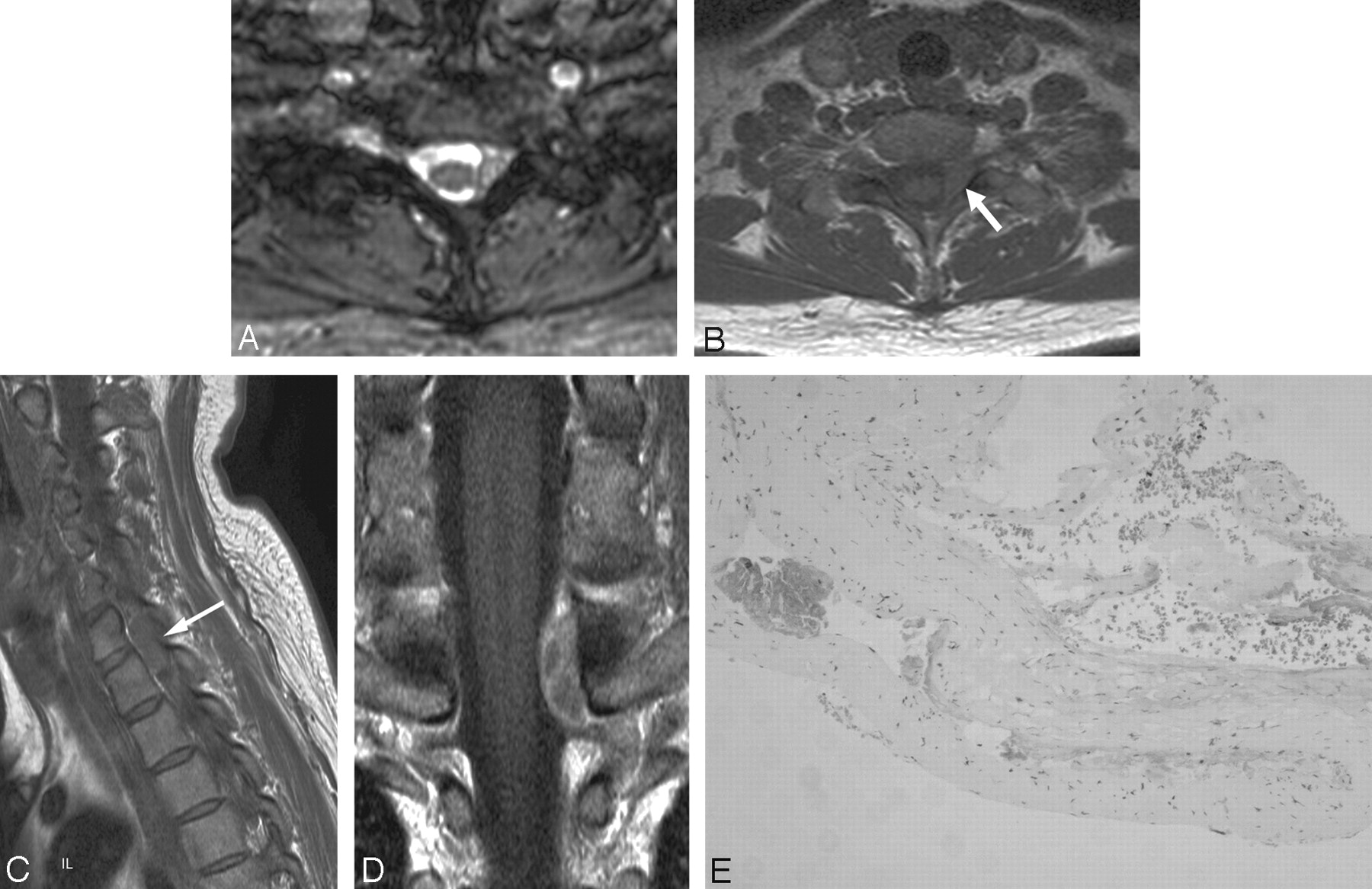
  • Fig 2.
    • Download figure
    • Open in new tab
    • Download powerpoint
    Fig 2.

    Case 2, a cystlike mass with T1 isointensity (type B).

    A, Transverse T2-weighted gradient-echo MR image demonstrates a well-defined mass with high signal intensity at the left posterior epidural space of C7-T1.

    B, On transverse noncontrast T1-weighted MR image, the mass (arrow) shows homogeneous isointensity.

    C, On sagittal noncontrast T1-weighted MR image, the signal intensity of the mass (arrow) is isointense to that of the intervertebral disk.

    D, On coronal postcontrast T1-weighted image, the mass shows peripheral thick enhancement with inner septumlike enhancement.

    E, Histologic section shows cystic dilated vessels with smooth muscle in the walls, suggestive of a venous hemangioma (H&E, × 100). There is no evidence of an organized hematoma.

  • Fig 3.
    • Download figure
    • Open in new tab
    • Download powerpoint
    Fig 3.

    Case 6, a solid hypervascular mass (type C).

    A, Sagittal T2-weighted spin-echo MR image demonstrates the mass at the posterior epidural space of T9-T11. The mass shows homogeneous high signal intensity. The spinal cord is severely compressed by the mass and shows high signal intensity, suggestive of compressive myelopathy.

    B, On noncontrast transverse T1-weighted spin-echo MR image, the mass shows heterogeneous isointensity with area of high signal intensity (arrow) at the posterior portion of the mass. The mass extends to the left neural foramen.

    C, On sagittal postcontrast T1-weighted image, the mass shows homogeneous strong enhancement. A dural tail sign is also seen (arrows).

    D, Photomicrograph reveals the formation of large cavernous vascular channels separated by a scant connective stroma. The spaces are lined by a flattened monolayer of endothelial cells (H&E, × 100).

  • Fig 4.
    • Download figure
    • Open in new tab
    • Download powerpoint
    Fig 4.

    Case 10, an epidural hematoma (type D).

    A, Sagittal T2-weighed spin-echo MR image demonstrates an ill-defined lesion with heterogeneous low and high signal intensity at the posterior epidural space of C5-C7.

    B, On sagittal noncontrast T1-weighed spin-echo MR image, there is high signal intensity in that lesion, suggestive of a hematoma.

    C, On sagittal postcontrast T1-weighed spin-echo MR image, the lesion shows heterogeneous enhancement.

    D, On histologic examination, most of the area is an organized thrombus. There are scattered small foci of large, dilated, blood-filled vessels lined by flattened endothelium, which represents a cavernous hemangioma (H&E, × 100).

Tables

  • Figures
    • View popup
    Table 1:

    Summary of MR imaging features of spinal epidural hemangiomas—location, level, and signal characteristics

    Case No.LocationLevelEpiduralT1WIT2WICEMR Type
    1L5-S11AntHighBright high with rimNAA
    2C7-T12PostIsoHigh with rimPeripheral strongB
    3L51AntHT highBright high with rimHT strongA
    4T11-T122PostIsoHM highHM strongC
    5T2-T44PostIsoHM bright highHM strongC
    6T9-T114PostIso with bright fociHT highHM strongC
    7C6-T24PostIsoHM highHM strongC
    8C5-T47BothHighHT high and lowHT strongD
    9T6–72PostIsoHM highHM strongC
    10C5-C74PostHigh with bright fociHT high and lowHT strongD
    11T3-T42PostIsoHM highHM strongC
    12L4–51AntIsoBright high with rimPeripheral strongB
    13L31AntIsoBright high with rimPeripheral strongB
    14C6-T15AntIsoHM highHM strongC
    • Note:—Level indicates craniocaudal extension described as the number of vertebrae where mass was present; Epidural, location of masses either anterior (Ant) or posterior (Post) epidural space; T1WI, T1-weighted MR image; T2WI, T2-weighted MR image; CE, contrast-enhanced MR image; A, cystlike mass with T1 hyperintensity; B, cystlike mass with T1 isointensity; C, solid hypervascular mass; D, epidural hematoma; Iso, isointense; HM, homogeneous signal intensity; HT, heterogeneous signal intensity; Peripheral, peripheral enhancement.

    • View popup
    Table 2:

    Summary of MR imaging features of spinal epidural hemangiomas—low signal rim, dural tail sign, and mass effect

    Case No.MR TypeContourLow Signal Rim on T1WILow Signal Rim on T2WILow Signal Rim on CEDural Tail SignCord CompNF WidenVertebral Body
    1ASmoothYesYesNANANoNoIntact
    2BLobularNoYesNoNoNoNoIntact
    3ALobularYesYesYesNoNoNoIntact
    4CSmoothYesYesNoYesNoNoIntact
    5CLobularNoNoNoNoNoYesIntact
    6CLobularNoNoNoYesYesYesIntact
    7CLobularYesYesNoYesYesYesErosion
    8DIll-definedNoNoNoYesYesYesIntact
    9CSmoothYesYesYesNoYesNoIntact
    10DIll-definedNoNoNoNoYesNoIntact
    11CLobularNoYesYesNoYesYesErosion
    12BLobularNoYesNoNoNoNoIntact
    13BSmoothNoNoNoNoNoNoIntact
    14CLobularYesYesNoNoYesYesErosion
    • Note:—T1WI indicates T1-weighted MR image; T2WI, T2-weighted MR image; CE, contrast-enhanced MR image; Cord comp, cord compression; NF widen, neural foraminal widening; Vertebral body, involvement of vertebral body.

    • View popup
    Table 3:

    MR imaging–pathologic correlation of spinal epidural hemangiomas

    Case No.MR TypeHistological FeatureHematomaLocationEpiduralLevelCC
    1AArteriovenousYesLumbosacralAnt1Radiculopathy
    2BVenousNoCervicothoracicPost2Radiculopathy
    3AArteriovenousYesLumbarAnt1Radiculopathy
    4CCavernousNoThoracicPost2Radiculopathy
    5CCavernousNoThoracicPost4Axial pain
    6CCavernousNoThoracicPost4Myelopathy
    7CCavernousNoCervicothoracicPost4Myelopathy
    8DCavernousYesCervicothoracicPost7Myelopathy
    9CCavernousNoThoracicPost2Myelopathy
    10DCavernousYesCervicalPost4Radiculopathy
    11CCavernousNoThoracicPost2Myelopathy
    12BVenousNoLumbarAnt1Radiculopathy
    13BVenousNoLumbarAnt1Radiculopathy
    14CCavernousNoCervicothoracicAnt5Myelopathy
    • Note:—A indicates cystlike mass with T1 hyperintensity; B, cystlike mass with T1 isointensity; C, solid hypervascular mass; D, epidural hematoma; Epidural, location of masses either anterior (Ant) or posterior (Post) epidural space; Level, craniocaudal extension described as the number of vertebrae where mass was present; CC, chief complaint.

PreviousNext
Back to top

In this issue

American Journal of Neuroradiology: 28 (7)
American Journal of Neuroradiology
Vol. 28, Issue 7
August 2007
  • Table of Contents
  • Index by author
Advertisement
Print
Download PDF
Email Article

Thank you for your interest in spreading the word on American Journal of Neuroradiology.

NOTE: We only request your email address so that the person you are recommending the page to knows that you wanted them to see it, and that it is not junk mail. We do not capture any email address.

Enter multiple addresses on separate lines or separate them with commas.
Spinal Epidural Hemangiomas: Various Types of MR Imaging Features with Histopathologic Correlation
(Your Name) has sent you a message from American Journal of Neuroradiology
(Your Name) thought you would like to see the American Journal of Neuroradiology web site.
CAPTCHA
This question is for testing whether or not you are a human visitor and to prevent automated spam submissions.
Cite this article
J.W. Lee, E.Y. Cho, S.H. Hong, H.W. Chung, J.H. Kim, K.-H. Chang, J.-Y. Choi, J.-S. Yeom, H.S. Kang
Spinal Epidural Hemangiomas: Various Types of MR Imaging Features with Histopathologic Correlation
American Journal of Neuroradiology Aug 2007, 28 (7) 1242-1248; DOI: 10.3174/ajnr.A0563

Citation Manager Formats

  • BibTeX
  • Bookends
  • EasyBib
  • EndNote (tagged)
  • EndNote 8 (xml)
  • Medlars
  • Mendeley
  • Papers
  • RefWorks Tagged
  • Ref Manager
  • RIS
  • Zotero
0 Responses
Respond to this article
Share
Bookmark this article
Spinal Epidural Hemangiomas: Various Types of MR Imaging Features with Histopathologic Correlation
J.W. Lee, E.Y. Cho, S.H. Hong, H.W. Chung, J.H. Kim, K.-H. Chang, J.-Y. Choi, J.-S. Yeom, H.S. Kang
American Journal of Neuroradiology Aug 2007, 28 (7) 1242-1248; DOI: 10.3174/ajnr.A0563
del.icio.us logo Twitter logo Facebook logo Mendeley logo
  • Tweet Widget
  • Facebook Like
  • Google Plus One
Purchase

Jump to section

  • Article
    • Abstract
    • Materials and Methods
    • Results
    • Discussion
    • Footnotes
    • References
  • Figures & Data
  • Supplemental
  • Info & Metrics
  • Responses
  • References
  • PDF

Related Articles

  • PubMed
  • Google Scholar

Cited By...

  • Spinal extradural cavernous haemangioma in an elderly man
  • Crossref (59)
  • Google Scholar

This article has been cited by the following articles in journals that are participating in Crossref Cited-by Linking.

  • Spinal Hematomas: What a Radiologist Needs to Know
    Jennifer L. Pierce, Joseph H. Donahue, Nicholas C. Nacey, Cody R. Quirk, Michael T. Perry, Nicholas Faulconer, Gene A. Falkowski, Michael D. Maldonado, Catherine A. Shaeffer, Francis H. Shen
    RadioGraphics 2018 38 5
  • Primary spinal epidural cavernous hemangioma: clinical features and surgical outcome in 14 cases
    Teng-yu Li, Yu-lun Xu, Jun Yang, James Wang, Gui-Huai Wang
    Journal of Neurosurgery: Spine 2015 22 1
  • Haemangioma, an uncommon cause of an extradural or intradural extramedullary mass: case series with radiological pathological correlation
    S. H. McEvoy, M. Farrell, F. Brett, S. Looby
    Insights into Imaging 2016 7 1
  • Posterior epidural migration of herniated lumbar disc fragment: a literature review
    Alaa Eldin Elsharkawy, Anne Hagemann, Peter Douglas Klassen
    Neurosurgical Review 2019 42 4
  • Case Study of a Spinal Epidural Capillary Hemangioma: A 4-Year Postoperative Follow-Up
    Arsen Seferi, Ridvan Alimehmeti, Gentian Vyshka, Teona Bushati, Mentor Petrela
    Global Spine Journal 2014 4 1
  • Posterior epidural intervertebral disc migration and sequestration: A systematic review
    Paolo Palmisciano, Kishore Balasubramanian, Gianluca Scalia, Navraj S. Sagoo, Ali S. Haider, Othman Bin Alamer, Vishal Chavda, Bipin Chaurasia, Harsh Deora, Maurizio Passanisi, Valerio Da Ros, Giuseppe R. Giammalva, Rosario Maugeri, Domenico G. Iacopino, Salvatore Cicero, Salah G. Aoun, Giuseppe E. Umana
    Journal of Clinical Neuroscience 2022 98
  • Clinical and radiological presentation of spinal epidural haemangiomas: clinical series in a tertiary care centre during a 10-year period
    Mario Mühmer, Richard Bostelmann, Sevgi Sarikaya-Seiwert, Marcel Schneiderhan, Hans-Jakob Steiger, Jan Frederick Cornelius
    European Spine Journal 2014 23 2
  • Pure Spinal Epidural Cavernous Hemangioma with Intralesional Hemorrhage: A Rare Cause of Thoracic Myelopathy
    Donghwan Jang, Choonghyo Kim, Seung Jin Lee, Young-Joon Ryu, Jiha Kim
    Korean Journal of Spine 2014 11 2
  • Posterior migration of lumbar disc herniation - imaging dilemma due to contrast contraindication: a case report
    Fabrício Guimarães Gonçalves, Prasad Baladev Hanagandi, Carlos Ignacio Torres, Raquel DelCarpio-O'Donovan
    Radiologia Brasileira 2012 45 3
  • Clinical features and long-term surgical outcomes of pure spinal epidural cavernous hemangioma—report of 23 cases
    Liang Zhang, Guangyu Qiao, Aijia Shang, Xinguang Yu
    Acta Neurochirurgica 2020 162 11

More in this TOC Section

  • Cone Beam CT Myelography
  • Post-Procedural Brachial Neuritis Features
  • Diagnostic Value of Brain WMH in SIH
Show more Spine Imaging and Spine Image-Guided Interventions

Similar Articles

Advertisement

Indexed Content

  • Current Issue
  • Accepted Manuscripts
  • Article Preview
  • Past Issues
  • Editorials
  • Editor's Choice
  • Fellows' Journal Club
  • Letters to the Editor
  • Video Articles

Cases

  • Case Collection
  • Archive - Case of the Week
  • Archive - Case of the Month
  • Archive - Classic Case

More from AJNR

  • Trainee Corner
  • Imaging Protocols
  • MRI Safety Corner
  • Book Reviews

Multimedia

  • AJNR Podcasts
  • AJNR Scantastics

Resources

  • Turnaround Time
  • Submit a Manuscript
  • Submit a Video Article
  • Submit an eLetter to the Editor/Response
  • Manuscript Submission Guidelines
  • Statistical Tips
  • Fast Publishing of Accepted Manuscripts
  • Graphical Abstract Preparation
  • Imaging Protocol Submission
  • Evidence-Based Medicine Level Guide
  • Publishing Checklists
  • Author Policies
  • Become a Reviewer/Academy of Reviewers
  • News and Updates

About Us

  • About AJNR
  • Editorial Board
  • Editorial Board Alumni
  • Alerts
  • Permissions
  • Not an AJNR Subscriber? Join Now
  • Advertise with Us
  • Librarian Resources
  • Feedback
  • Terms and Conditions
  • AJNR Editorial Board Alumni

American Society of Neuroradiology

  • Not an ASNR Member? Join Now

© 2025 by the American Society of Neuroradiology All rights, including for text and data mining, AI training, and similar technologies, are reserved.
Print ISSN: 0195-6108 Online ISSN: 1936-959X

Powered by HighWire