
Article Figures & Data
Figures
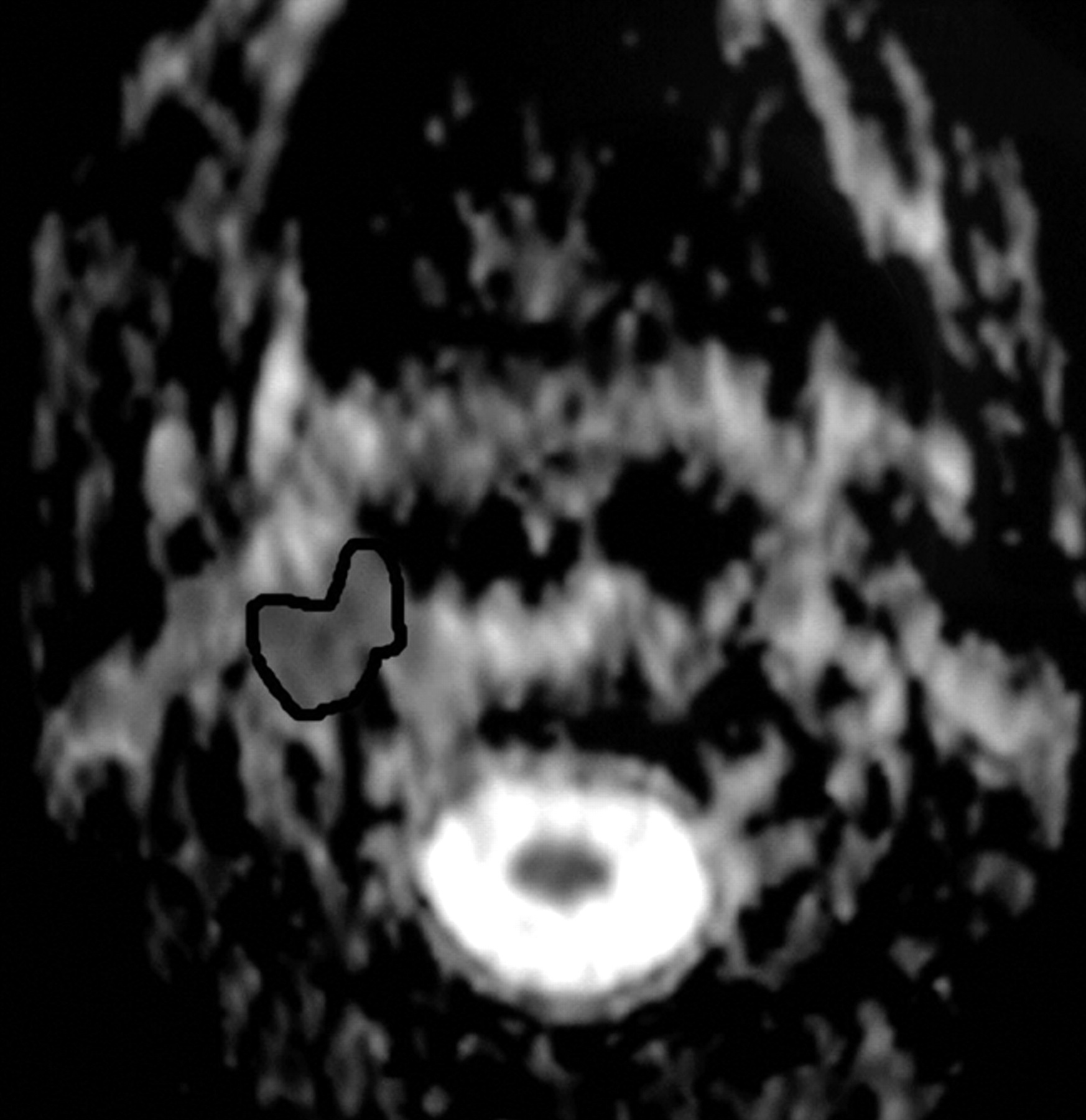
- Fig 1.
ADC map demonstrating the location of ROI in a patient with a recurrent nasopharyngeal mass.
- Fig 2.
Recurrent oncocytic carcinoma of the right parotid gland.
A, Axial T2WI, shown as ill defined, is seen at the site of the right parotid region after surgical resection and irradiation.
B, Axial postcontrast T1WI shows the inhomogenous pattern of enhancement. Recurrence could not be excluded.
C, ADC map shows low signal intensity at the site of the lesion with a mean ADC value of 1.07 ± 0.18 × 10⁻³ mm2/s. Biopsy revealed recurrent tumor.
- Fig 3.
Recurrent squamous cell carcinoma of the nasal cavity.
A, Axial postcontrast T1-weighted MR image shows that enhancing lesion is seen in the right side of the nasal cavity. Recurrent tumor cannot be differentiated from postradiation changes.
B, ADC map shows hypointensity within the lesion with a low ADC value (1.17 ± 0.17 × 10⁻³ mm2/s).
- Fig 4.
Recurrent squamous cell carcinoma of the oropharynx with metastatic cervical lymph node.
A, Axial T2WI shows an ill-defined irregular mass of inhomogeneous signal intensity involving the right side of the oropharynx. An enlarged cervical lymph node (arrow) with inhomogeneous high signal intensity is also noted at the right side of the neck.
B, ADC map shows low signal intensity of both the lesion and the lymph node with a mean ADC value of 1.20 ± 0.22 × 10⁻³ mm2/s and 1.05 ± 0.20 × 10⁻³ mm2/s, respectively, suggestive of tumor recurrence with metastatic lymph nodes. This was proved by biopsy.
- Fig 5.
Bone marrow infiltration.
A, Axial T2-weighted MR image shows an ill-defined inhomogeneous signal intensity involving the right greater wing of the sphenoid bone.
B, ADC map shows low signal intensity at this region with a mean ADC value of 0.84 ± 0.30 × 10⁻³ mm2/s, suggestive of tumor infiltration, which was proved on bone marrow biopsy.
- Fig 6.
Posttreatment changes after surgery and radiation therapy.
A, Axial postcontrast T1WI shows an ill-defined, mildly enhancing mass at the region of the ethmoidal sinuses. Recurrence was suspected.
B, ADC map shows high signal intensity at the site of the lesion denoting posttreatment fibrous tissue, which was proved by biopsy. The mean ADC value at the site of the posttreatment changes was 1.89 ± 0.19 × 10⁻³ mm2/s.
- Fig 7.
Posttreatment changes after surgery and radiation therapy.
A, Axial postcontrast T1WI shows a small ill-defined enhancing lesion at the site of the right parotid gland after surgical resection and irradiation. Recurrence could not be differentiated from posttreatment changes.
B, ADC map shows low signal intensity at the site of the lesion with a mean ADC value of 1.07 ± 0.18 × 10⁻³ mm2/s. Biopsy revealed only attenuated fibrous tissue with no tumor cells.
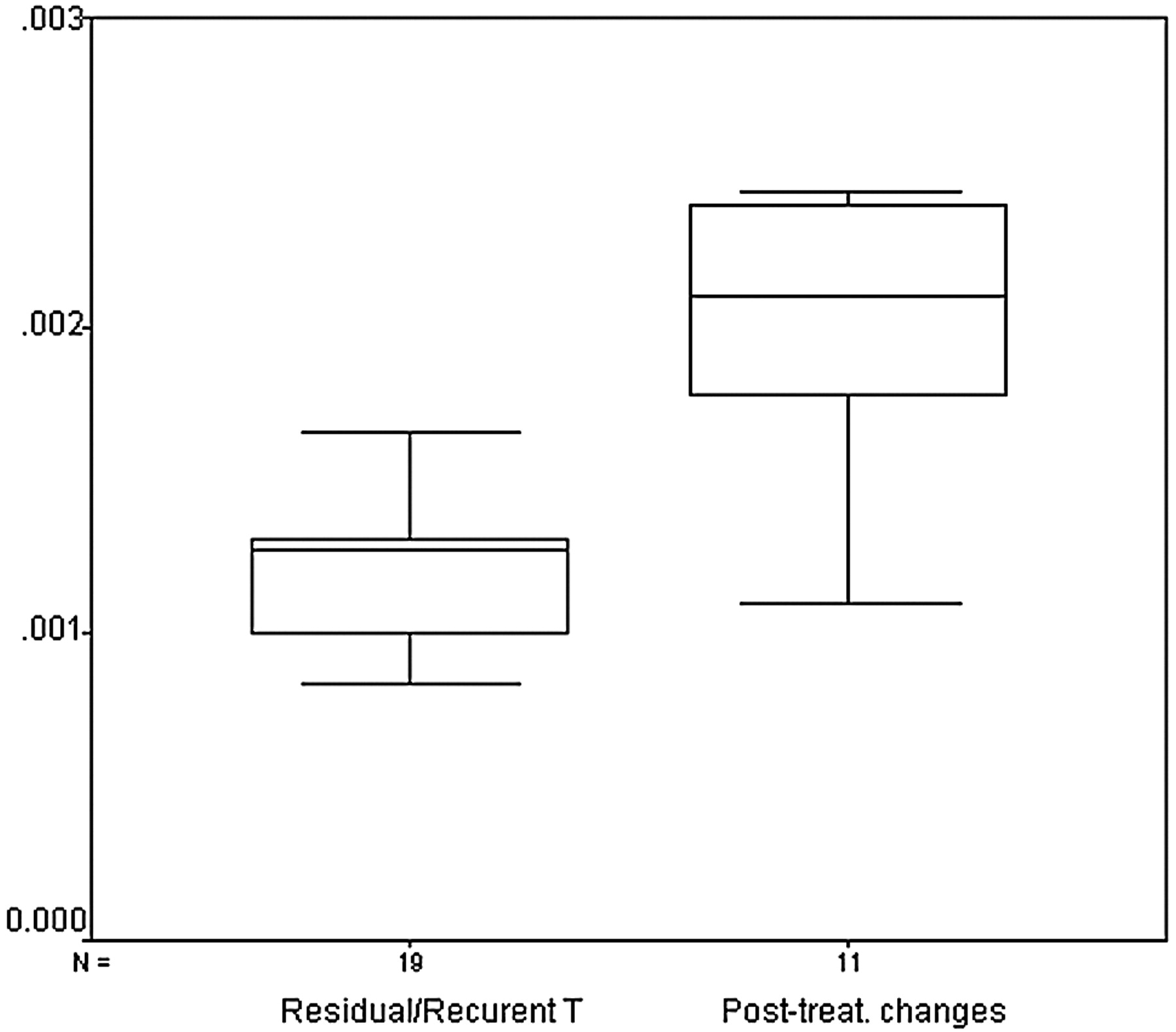
- Fig 8.
Box and whisker plot compares the mean ADCs of residual or recurrent tumors and posttreatment changes. The horizontal line is the median (50th percentile) of the measured values, the top and bottom of the box represent the 25th and 75th percentiles, respectively, and whiskers indicate the range from the largest to smallest observed data points. Note that despite the overlap between the ADC values of both groups, the ADCs of posttreatment changes are significantly higher than that of residual or recurrent tumors.


Tables
Patient Characteristic No. of Patients Location Nasopharynx 8 Oropharynx 4 Larynx 3 Paranasal sinuses and nasal cavity 7 Salivary glands 6 Skull base 1 Cheek 1 Method of treatment Surgery 3 Radiotherapy 12 Surgery and radiotherapy 15 Variable Range of ADC Value Mean ADC Value Residual or recurrent tumor (0.83–1.65) × 10−3 mm2/s 1.17 ± 0.33 × 10⁻³ mm2/s Posttreatment changes (1.10–2.44) × 10−3 mm2/s 2.07 ± 0.25 × 10⁻³ mm2/s Note:—ADC indicates apparent diffusion coefficient.
Threshold of ADC Value (×10−3 mm2/s) Sensitivity % (n/N) Specificity % (n/N) Accuracy % Positive Predictive Value % (n/N) Negative Predictive Value % (n/N) ≤1.00 37 (7/19) 100 (11/11) 60 100 (7/7) 47 (11/23) ≤1.30 84 (16/19) 90 (10/11) 87 94 (16/17) 76 (10/13) ≤1.50 89 (16/19) 73 (9/11) 83 85 (17/20) 80 (8/10) ≤2.00 100 (19/19) 45 (5/11) 80 76 (19/25) 100 (5/5) ≤2.40 100 (19/19) 9 (1/11) 66 65 (19/29) 100 (1/1) Note:—ADC indicates apparent diffusion coefficient.
In this issue


Jump to section
Related Articles
Cited By...
- MRI Posttreatment Surveillance for Head and Neck Squamous Cell Carcinoma: Proposed MR NI-RADS Criteria
- MRI with DWI for the Detection of Posttreatment Head and Neck Squamous Cell Carcinoma: Why Morphologic MRI Criteria Matter
- Diffusion-Weighted Imaging of the Head and Neck: Influence of Fat-Suppression Technique and Multishot 2D Navigated Interleaved Acquisitions
- Independent Poor Prognostic Factors for True Progression after Radiation Therapy and Concomitant Temozolomide in Patients with Glioblastoma: Subependymal Enhancement and Low ADC Value
- Diffusivity Measurements Differentiate Benign from Malignant Lesions in Patients with Peripheral Neuropathy or Plexopathy
- Differentiation of Recurrent Tumor and Posttreatment Changes in Head and Neck Squamous Cell Carcinoma: Application of High b-Value Diffusion-Weighted Imaging
- Biologic Imaging of Head and Neck Cancer: The Present and the Future
- Non-Gaussian Analysis of Diffusion-Weighted MR Imaging in Head and Neck Squamous Cell Carcinoma: A Feasibility Study
- Diffusion-Weighted Magnetic Resonance Imaging for Predicting and Detecting Early Response to Chemoradiation Therapy of Squamous Cell Carcinomas of the Head and Neck