
Article Figures & Data
Figures
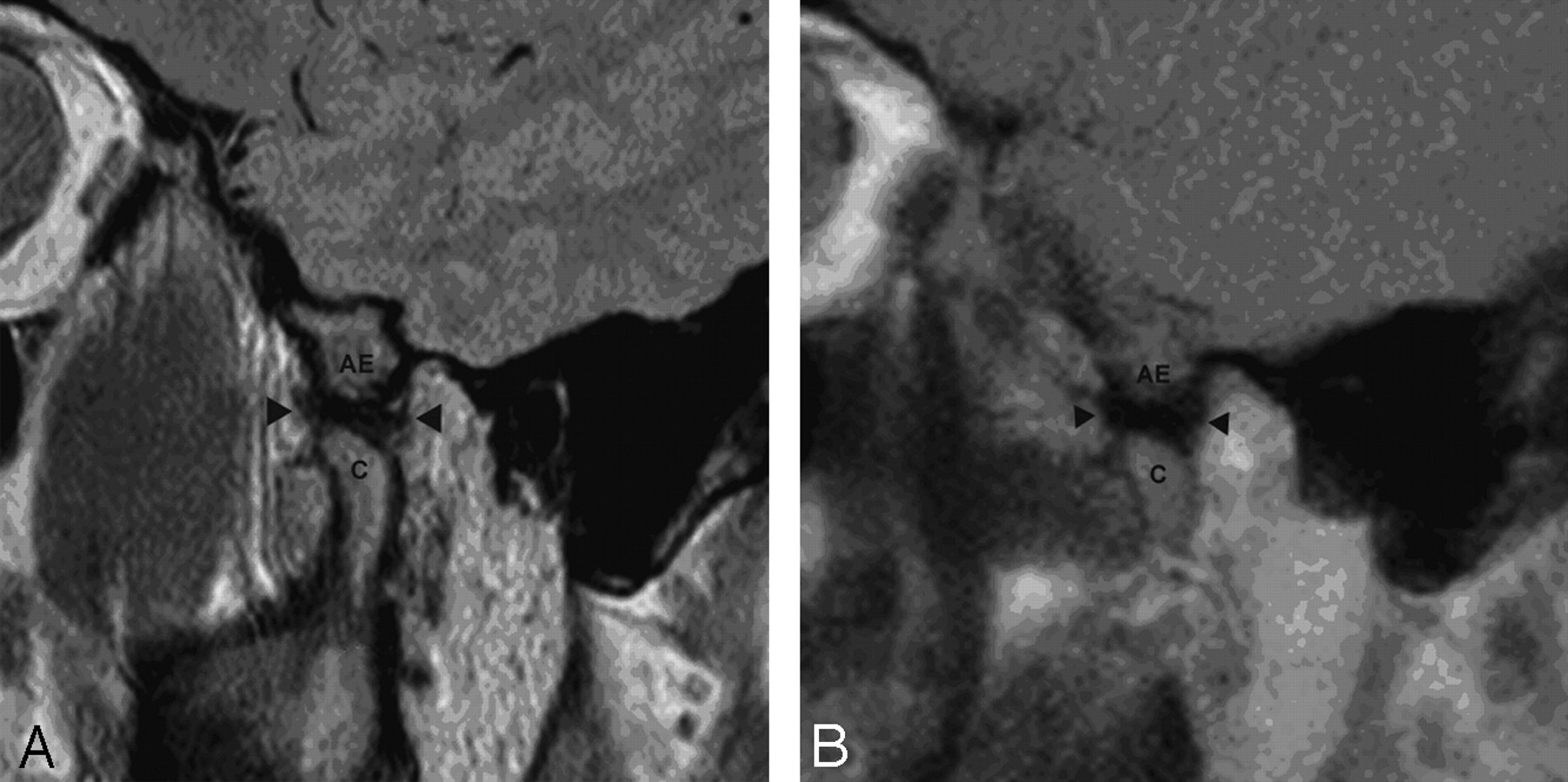
- Fig 1.
A, Static sagittal oblique proton-attenuation image of the TMJ in the open-mouthed position. B, Single frame from sagittal HASTE imaging series of the same temporomandibular joint. In both studies, the articular eminence (AE), condyle (C), and articular disk (arrowheads) are demonstrated.
- Fig 2.
Selected images from dynamic HASTE series of imaging, from full closure to maximal opening of the TMJ.
- Fig 3.
A, Static sagittal oblique proton-attenuation image of the TMJ. B, Single frame from a sagittal HASTE imaging series of the same TMJ. Reduced susceptibility artifact (asterisks on both images) is noted on the HASTE imaging.
- Fig 4.
Single frame from a dynamic sagittal HASTE series demonstrates poor delineation of the posterior band of the articular disk.
- Fig 5.
A, Closed- and open-mouthed static sagittal oblique proton-attenuation images and (B) selected frames from dynamic sagittal HASTE image series, acquired from the same TMJ. Anteriorly displaced disk material (arrowheads) is more clearly identified on the dynamic series, with reduction of material seen by the 3rd image.


Tables
Results from reader assessments of dynamic and static examinations
Examination Type Dynamic Static % of cases with motion artifact Reader 1 0 8.8 Reader 2 20.6 11.8 Reader 3 0 38.2 Overall (P = .02) 6.9 19.6 % of cases with limited range of motion Reader 1 23.5 29.4 Reader 2 8.8 26.5 Reader 3 20.6 35.3 Overall (P = .02) 17.7 30.4 Consensus (χ2 = 0.85, P > .25) 14.7 23.5 % of cases of dislocations rated Reader 1 17.7 38.2 Reader 2 2.9 20.6 Reader 3 47.1 35.3 Overall (P = .02) 22.6 31.4 Consensus (χ2 = 0.06, P > .5) 38.2 35.3 % of cases with high confidence ratings (>3) Reader 1 64.7 61.8 Reader 2 76.5 55.9 Reader 3 72.6 50.0 Overall (P = .06) 72.6 55.9 Mean and SD of confidence scores Reader 1 3.85 ± 1.0 3.91 ± 1.1 Reader 2 4.24 ± 1.1 3.74 ± 1.1 Reader 3 4.24 ± 0.9 3.56 ± 1.1 Overall (P = .02) 4.11 ± 1.0 3.74 ± 1.1


