
Article Figures & Data
Figures
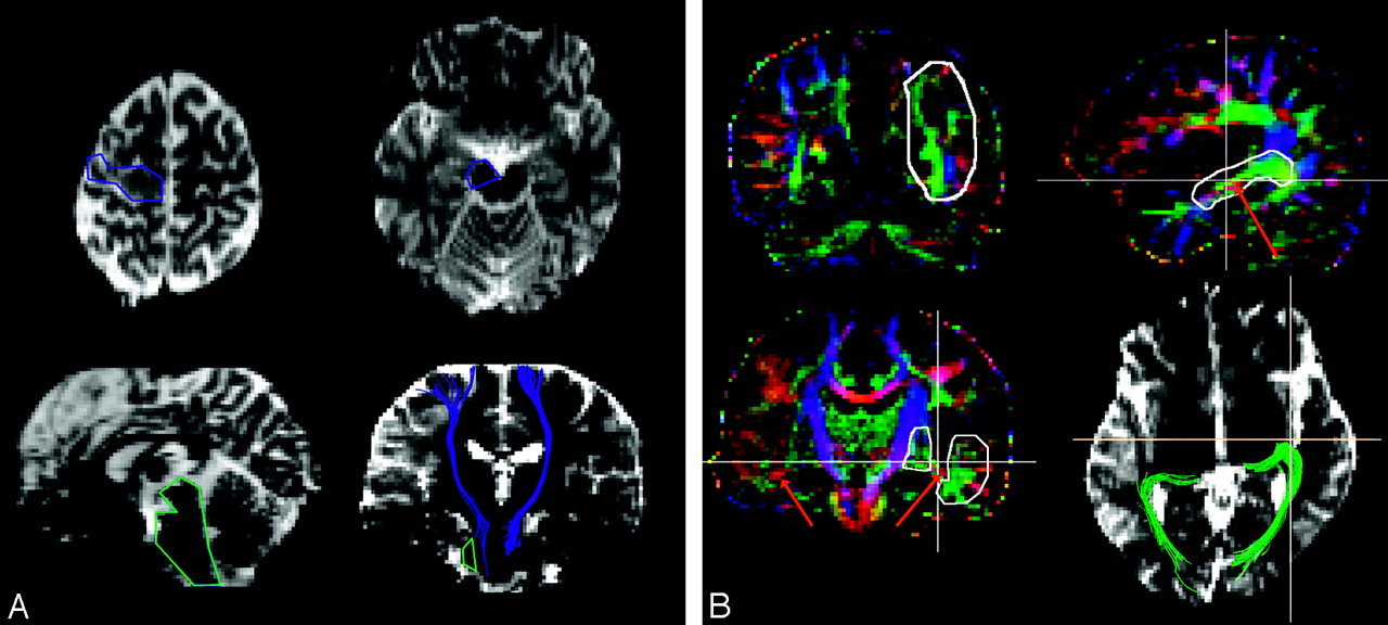
- Fig 1.
A, ROI segmentation for corticospinal tract tractography. Polygonal ROIs are placed on transverse b = 0 images (TR/TE, 7000 ms/79 ms). The first “OR” ROI (blue) is placed at either side of the cerebral peduncle on the plane where the characteristic Ω shape of the central sulcus is at the center of cerebral hemisphere (top left). The second “AND” ROI (blue) is placed at the ipsilateral precentral gyrus (top right). Left and right ROI segmentations were separately performed. “NOT” ROIs (green) are placed on midline structures connecting right and left corticospinal tracts on sagittal reconstructed image (bottom left) and fibers projecting into ipsilateral cerebellar peduncle on coronal reconstructed image (bottom right). Examples of bilateral corticospinal tract overlaid on coronal reconstructed image are shown (bottom right).
B, ROI segmentation for optic radiation tractography. The 4 kinds of ROIs (white polygon) are placed on coronal or sagittal color-coded maps. Cross lines indicate the orthogonal planes. The first “OR” ROI is placed at either side of the occipital lobe, including the calcarine cortex on the coronal plane through the anterior edge of the occipital-parietal sulcus (top left). The second “AND” ROI is placed at the ipsilateral temporal stem, including the Meyer loop on the sagittal plane (top right). Temporal stem is identified as green, and the Meyer loop is identified as a small red area inside the temporal stem (red arrow). The third and fourth “NOT” ROIs are placed on the same coronal plane through the dorsal end of the Sylvian fissure (bottom left). Bilateral Meyer loops are indicated as red arrows. Occipital-frontal connections medial to the Meyer loop and fibers projecting to the temporal horn passing through lateral to the Meyer loop are removed using the “NOT” operation. Examples of bilateral optic radiation overlaid on transverse b = 0 images (TR/TE, 7000 ms/79 ms) are shown (bottom right).
- Fig 2.
A, Corticospinal tract tractography. Patients were classified into 1 of 3 groups based on the distance between AVM nidus and tract and the presence of motor weakness. Three groups (A–C) are assigned from top row to bottom row. Left column displays transverse b = 0 images (TR/TE, 7000 ms/79 ms). Blue areas represent the voxel where fiber tracts penetrate the image. Middle column displays transverse maximum intensity projection (MIP) images of MR angiography. Red arrows indicate AVM nidus. Right column displays 3D reconstruction of fibers and MR angiography superimposed on b = 0 images. 3D reconstructions of AVM nidus (red) and hemorrhage (yellow) segmented from MR angiography were displayed by using shaded surface rendering. Patients in groups A and B are subjects without hemorrhage, and the group C patient is a subject with hemorrhage in this figure. In this group C patient, left corticospinal tract veers laterally, and projection fibers from the medial precentral gyrus are not visualized (green arrow).
B, Optic radiation tractography. Patients were classified into 1 of 3 groups based on the distance between AVM nidus and tract and the presence of visual field defect. Optic radiation tractography is represented in green, and imaging methods are the same as explained in A. The group B patient is the same as the group A patient presented in A. In this group C patient, left optic radiation was disrupted around the occipital pole.
- Fig 3.
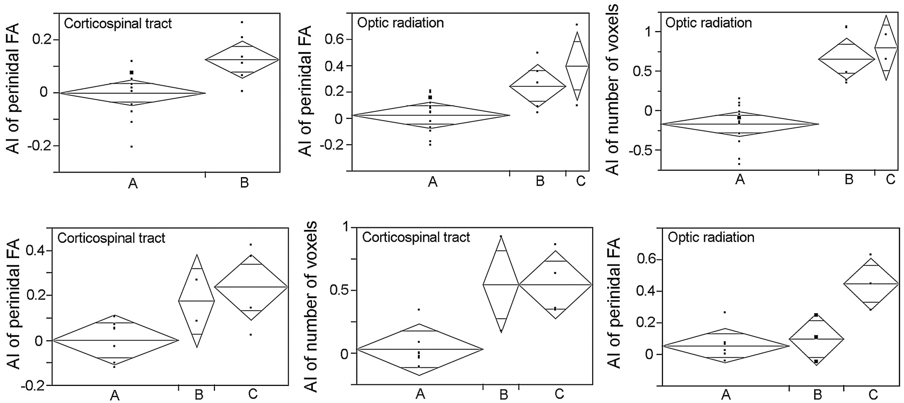
Results of statistical analysis with significant differences are demonstrated. Top row displays patients without intracranial hemorrhage, and bottom row displays patients with intracranial hemorrhage. Gray rhomboids indicate mean and its 95% confident intervals. Horizontal axes represent groups A–C, and vertical axes represent AIs. In patients without intracranial hemorrhage, AI of perinidal FA along corticospinal tract (left, P = .006) and optic radiation (middle, P = .01) and AI of optic radiation volume (right, P < .0001) differed significantly between groups. In patients with hemorrhage, AIs of perinidal FA (P = .04), tract volume (P = .01) of corticospinal tract, and AI of perinidal FA along optic radiation (P = .004) differed significantly between groups.


Tables
Patient No. Sex Age at MR Imaging (years) Location of Nidus Hemorrhage Perifocal Hyperintensity Neurologic Symptoms Spetzler-Martin Grade 1 F 29 Right occipital No No No 2 2 M 31 Right frontal No No No 1 3 F 27 Left occipital No Yes Right homonymous hemianopsia 3 4 M 64 Right occipital No No No 2 5 M 49 Left frontal No Yes No 2 6 M 72 Left parietal No No No 1 7 M 25 Left occipital No No No 2 8 M 45 Right frontal No Yes No 3 9 M 28 Left frontal No No No 3 10 M 49 Left putamen No No No 2 11 M 25 Right temporal No Yes No 2 12 M 28 Right occipital No No Left lower quadranopsia 4 13 F 5 Left parietal No No No 1 14 M 59 Right frontal No Yes No 1 15 F 28 Right parietal No No No 2 16 M 45 Left cingulum No No No 2 17 F 33 Right cingulum No No No 4 18 F 29 Left frontal No No No 2 19 M 28 Right insula No No No 4 20 F 17 Left cerebellum No Yes Cerebellar ataxia 3 21 F 20 Left frontal No No No 3 22 F 45 Right occipital Yes No No 3 23 F 29 Left occipital Yes No No 2 24 M 22 Right cerebellum Yes No Cerebellar ataxia 4 25 F 15 Right occipital Yes No No 3 26 F 56 Right cerebellum Yes Yes Cerebellar ataxia, right trigeminal palsy 1 27 M 45 Right parietal Yes Yes Recent memory disturbance 3 28 F 21 Right occipital Yes Yes Left upper quadranopsia 2 29 F 27 Right parietal Yes Yes Left hemianopsia 2 30 F 6 Right frontal Yes Yes Left hemiparesis (MMT4/5) 2 31 M 38 Right parietal Yes No Left hemiparesis (MMT 4/5) 2 32 M 50 Left frontal Yes Yes Motor aphasia 5 33 F 23 Right frontal Yes Yes Left hemiparesis (MMT 4/5) 2 34 M 48 Left parietal Yes Yes Right hemiparesis (MMT 3/5), right spatial neglect 3 Note:—M indicates male; F, female; MMT, manual muscle testing. Patients 1–21 were without intracranial hemorrhage, and patients 22–34 displayed hemorrhage.
- Table 2:
Tractographic appearances and neurologic symptoms for corticospinal tract and optic radiation close to nidus
Variable Group B Group C Nonhemorrhagic Hemorrhagic Nonhemorrhagic Hemorrhagic Corticospinal tract No change 6 0 0 2 Compression 1 2 0 1 Disruption 0 0 0 1 Optic radiation No change 1 1 1 1 Compression 2 1 0 2 Penetration 0 1 0 0 Disruption 2 0 1 0
In this issue


Jump to section
Related Articles
Cited By...
- Classification of brain arteriovenous malformations located in motor-related areas based on location and anterior choroidal artery feeding
- Individual variations of the human corticospinal tract and its hand-related motor fibers using diffusion MRI tractography
- Principles and Limitations of Computational Algorithms in Clinical Diffusion Tensor MR Tractography
- Identifying the human optic radiation using diffusion imaging and fiber tractography