
Article Figures & Data
Figures
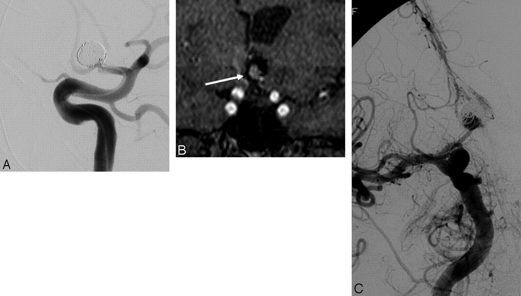
- Fig 1.
A, Internal carotid angiogram demonstrates near-complete coil embolization of a large anterior communicating artery aneurysm. Follow-up short TE MRA was performed with MPR and MIP postprocessing. B, An image from the coronal MPR demonstrates a large recurrence (arrow) within the central aspect of the coil mass. A black signal-intensity void is distributed about the periphery of this central recanalization, indicative of the displaced and compacted coil mass that is now draped around the periphery of the recanalized pocket of central flow. Signal-intensity voids running though the central area of flow-related enhancement correspond to individual coil strands that bridge the recanalization. C, Correlative follow-up right internal carotid angiogram in a projection performed to replicate the coronal MPR confirms the large central recanalization with the coil mass displaced into the periphery of the fundus and a few individual coil strands bridging the recurrent aneurysm.
- Fig 2.
A, Conventional left internal carotid angiogram demonstrates a tiny 1- to 2-mm residual neck at the base of a coiled anterior communicating artery aneurysm. B, Correlative MIP projection from a TOF-MRA accurately demonstrates this tiny residual.
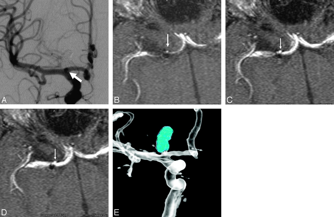
- Fig 3.
A, Conventional right internal carotid angiogram depicts a coil mass within a right carotid terminus aneurysm. A tiny (2-mm) residual aneurysm (arrow) is evident only as a double attenuation overlapping the parent vessel. On the other A-plane angiographic views, this small residual projected over the coil mass and was obscured. On the lateral projections, the residual was obscured by overlying anterior cerebral artery and middle cerebral artery branches. B–D, Correlative TOF-MRA source images demonstrate that the tiny residual arises from the anterior aspect of the aneurysm neck and projects superiorly along the anterior aspect of the coil mass (arrows). In retrospect, it can be appreciated just how this small residual would be obscured by either the coil mass or adjacent vessels on the most angiographic projections. E, An image from a rotational angiogram with the coil mass attenuation mapped to a blue color provides additional evidence of the tiny residual at the base of the coil mass, projecting superiorly from the anterior aneurysm neck.
- Fig 4.
A, Conventional angiogram demonstrates a small right posterior communicating artery aneurysm after coil embolization. The unsubtracted image (left) depicts a tail of coil projecting anteriorly (arrow). The subtracted image (right) demonstrates that this coil tail projects toward the parent artery from which the aneurysm arises. A small amount of filling between the interstices of the coil mass is also noted along the anterior aspect of the aneurysm neck. B, Correlative TOF-MRA source image demonstrates the coil mass as a signal-intensity void. A linear signal-intensity void (arrow) projects anteriorly into the parent posterior communicating artery corresponding to the coil tail visualized on the native image. The small pocket of residual filling is also appreciated along the lateral aspect of the coil mass. Reading the MRA without the posttreatment angiographic correlation could lead to confusion with respect to the interpretation of these structures in the region of the aneurysm neck. A correlation of the 2 techniques provides a detailed and complimentary understanding of the anatomy in this region.
- Fig 5.
A, Axial source image from a contrast-enhanced MRA unambiguously demonstrates a tiny (<2-mm) residual superior hypophyseal-region aneurysm (arrow). B, Correlative left internal carotid conventional angiogram shows that this tiny pocket of residual filling (arrow) is surrounded by strands of coil.
- Fig 6.
Axial source images from a contrast-enhanced MR image obtained immediately after treatment (left image) demonstrate no enhancement about the posterior aspect of a large basilar aneurysm coil mass (arrows). Follow-up imaging performed 6 weeks later (right image) demonstrates the interval development of a thin rim of enhancement marginating the posterior aspect of the coil mass (arrows). This pattern of enhancement is not representative of recanalization but more likely of mural enhancement as a sequela of the in-growth of granulation tissue about the periphery of the coil mass.
- Fig 7.
CE-MRA image following coil embolization of a large right ICA posterior wall aneurysm. Arrows depict several scattered pixels of hyperintensity distributed within the signal-intensity void of the coil mass. Although commonly observed, the etiology and significance of these tiny foci of hyperintensity within the coil mass remain unclear.


Tables
MRA protocol
Routine MRA Coiled Aneurysm Ultrashort TE TOF MRA Contrast-Enhanced MRA Technique 6-Slab MOTSA 3-Slab MOTSA 3D SPGR single volume Flow comp ON OFF OFF BW 15 kHz 32 kHz 32 kHz Minimal TE 3.7 ms ∼2 ms ∼2 ms TR 33 ms 33 ms 14 ms Flip angle 20° 20° 30° Section thickness 1.4 mm 1.4 mm 1.4 mm F × P 320 × 192 384 × 244 512 × 256 Note:—MOTSA indicates multiple overlapping thin-slab acquisition; TR, repetition time; SPGR, spoiled gradient-recalled-echo; Flow comp, flow compensation; BW, bandwidth; F, frequency; P, phase.
In this issue


Jump to section
- Article
- Abstract
- Neurovascular Imaging Following Coil Embolization
- Available Noninvasive Imaging Techniques and Their Utility in the Assessment of Coiled Aneurysms
- MRA Techniques for the Evaluation of Coiled Aneurysms: Efficacy
- MRA Techniques for the Evaluation of Coiled Aneurysms: Limitations and Technical Points
- Contrast-Versus-Noncontrast MRA
- MRA Techniques for the Evaluation of Coiled Aneurysms: Recommended Follow-Up Protocol
- Conclusions
- References
- Figures & Data
- Info & Metrics
- Responses
- References
Related Articles
Cited By...
- Noninvasive Angiographic Results of Clipped or Coiled Intracranial Aneurysms: An Inter- and Intraobserver Reliability Study
- Long-Term Rupture Risk in Patients with Unruptured Intracranial Aneurysms Treated with Endovascular Therapy: A Systematic Review and Meta-Analysis
- Usefulness of Silent MR Angiography for Intracranial Aneurysms Treated with a Flow-Diverter Device
- Visualization of Aneurysmal Neck and Dome after Coiling with 3D Multifusion Imaging of Silent MRA and FSE-MR Cisternography
- Vessel Wall Enhancement in Treated Unruptured Aneurysms
- Anatomical results, rebleeding and factors that affect the degree of occlusion in ruptured cerebral aneurysms after endovascular therapy
- Contrast-Enhanced Time-Resolved MRA for Follow-Up of Intracranial Aneurysms Treated with the Pipeline Embolization Device
- MRA Versus DSA for Follow-Up of Coiled Intracranial Aneurysms: A Meta-Analysis
- In Vitro and In Vivo Imaging Characteristics Assessment of Polymeric Coils Compared with Standard Platinum Coils for the Treatment of Intracranial Aneurysms
- Temporal Evolution of Susceptibility Artifacts from Coiled Aneurysms on MR Angiography: An In Vivo Canine Study
- Matrix2 Coils in Embolization of Intracranial Aneurysms: 1-Year Outcome and Comparison with Bare Platinum Coil Group in a Single Institution
- A Prospective Trial of 3T and 1.5T Time-of-Flight and Contrast-Enhanced MR Angiography in the Follow-Up of Coiled Intracranial Aneurysms
- MR Angiographic Follow-Up of Intracranial Aneurysms Treated with Detachable Coils: Evaluation of a Blood-Pool Contrast Medium
- Advances in Interventional Neuroradiology 2007