Advertisement

Main menu

  • Home
  • Content
    • Current Issue
    • Accepted Manuscripts
    • Article Preview
    • Past Issue Archive
    • Video Articles
    • AJNR Case Collection
    • Case of the Week Archive
    • Case of the Month Archive
    • Classic Case Archive
  • Special Collections
    • AJNR Awards
    • Low-Field MRI
    • Alzheimer Disease
    • ASNR Foundation Special Collection
    • Photon-Counting CT
    • View All
  • Multimedia
    • AJNR Podcasts
    • AJNR SCANtastic
    • Trainee Corner
    • MRI Safety Corner
    • Imaging Protocols
  • For Authors
    • Submit a Manuscript
    • Submit a Video Article
    • Submit an eLetter to the Editor/Response
    • Manuscript Submission Guidelines
    • Statistical Tips
    • Fast Publishing of Accepted Manuscripts
    • Graphical Abstract Preparation
    • Imaging Protocol Submission
    • Author Policies
  • About Us
    • About AJNR
    • Editorial Board
    • Editorial Board Alumni
  • More
    • Become a Reviewer/Academy of Reviewers
    • Subscribers
    • Permissions
    • Alerts
    • Feedback
    • Advertisers
    • ASNR Home

User menu

  • Alerts
  • Log in

Search

  • Advanced search
American Journal of Neuroradiology

American Journal of Neuroradiology

ASHNR American Society of Functional Neuroradiology ASHNR American Society of Pediatric Neuroradiology ASSR
  • Alerts
  • Log in

Advanced Search

  • Home
  • Content
    • Current Issue
    • Accepted Manuscripts
    • Article Preview
    • Past Issue Archive
    • Video Articles
    • AJNR Case Collection
    • Case of the Week Archive
    • Case of the Month Archive
    • Classic Case Archive
  • Special Collections
    • AJNR Awards
    • Low-Field MRI
    • Alzheimer Disease
    • ASNR Foundation Special Collection
    • Photon-Counting CT
    • View All
  • Multimedia
    • AJNR Podcasts
    • AJNR SCANtastic
    • Trainee Corner
    • MRI Safety Corner
    • Imaging Protocols
  • For Authors
    • Submit a Manuscript
    • Submit a Video Article
    • Submit an eLetter to the Editor/Response
    • Manuscript Submission Guidelines
    • Statistical Tips
    • Fast Publishing of Accepted Manuscripts
    • Graphical Abstract Preparation
    • Imaging Protocol Submission
    • Author Policies
  • About Us
    • About AJNR
    • Editorial Board
    • Editorial Board Alumni
  • More
    • Become a Reviewer/Academy of Reviewers
    • Subscribers
    • Permissions
    • Alerts
    • Feedback
    • Advertisers
    • ASNR Home
  • Follow AJNR on Twitter
  • Visit AJNR on Facebook
  • Follow AJNR on Instagram
  • Join AJNR on LinkedIn
  • RSS Feeds

AJNR Awards, New Junior Editors, and more. Read the latest AJNR updates

Research ArticleBRAIN

Hyperintense Putaminal Rim at 3T Reflects Fewer Ferritin Deposits in the Lateral Marginal Area of the Putamen

S. Fujii, E. Matsusue, T. Kinoshita, S. Sugihara, E. Ohama and T. Ogawa
American Journal of Neuroradiology April 2007, 28 (4) 777-781;
S. Fujii
  • Find this author on Google Scholar
  • Find this author on PubMed
  • Search for this author on this site
E. Matsusue
  • Find this author on Google Scholar
  • Find this author on PubMed
  • Search for this author on this site
T. Kinoshita
  • Find this author on Google Scholar
  • Find this author on PubMed
  • Search for this author on this site
S. Sugihara
  • Find this author on Google Scholar
  • Find this author on PubMed
  • Search for this author on this site
E. Ohama
  • Find this author on Google Scholar
  • Find this author on PubMed
  • Search for this author on this site
T. Ogawa
  • Find this author on Google Scholar
  • Find this author on PubMed
  • Search for this author on this site
  • Article
  • Figures & Data
  • Info & Metrics
  • Responses
  • References
  • PDF
Loading

Article Figures & Data

Figures

  • Fig 1.
    • Download figure
    • Open in new tab
    • Download powerpoint
    Fig 1.

    Classification of HPR on axial T2-weighted FSE images. A, Absent (15 years old). B, Vague (29 years old). C, Present (54 years old). D, Vague with marked and diffuse putaminal hypointenisty (77 years old).

  • Fig 2.
    • Download figure
    • Open in new tab
    • Download powerpoint
    Fig 2.

    Frequency of HPR in 8 age groups. HPR becomes gradually prominent after 20 years of age and tends to become vague or absent after 70 years of age.

  • Fig 3.
    • Download figure
    • Open in new tab
    • Download powerpoint
    Fig 3.

    Comparison of postmortem MR images and histologic findings in a 12-year-old subject. Postmortem axial (A) and coronal (B) T2-weighted images. A section corresponding to B stained with Klüver-Barrera (C), Berlin blue (D), and ferritin immunohistochemistry (E). Postmortem MR images (A and B) show absent HPR. With Klüver-Barrera staining (C), myelinated fibers are small in number in the lateral marginal area of the putamen (dotted lines) in comparison with the remainder. With Berlin blue staining, hemosiderin is scattered in the inner portion of the putamen but is scarce in the outer portion (D). With ferritin immunohistochemistry, ferritin deposits are mild in the inner portion but are scarce in the lateral marginal area of the putamen (dotted lines, E).

  • Fig 4.
    • Download figure
    • Open in new tab
    • Download powerpoint
    Fig 4.

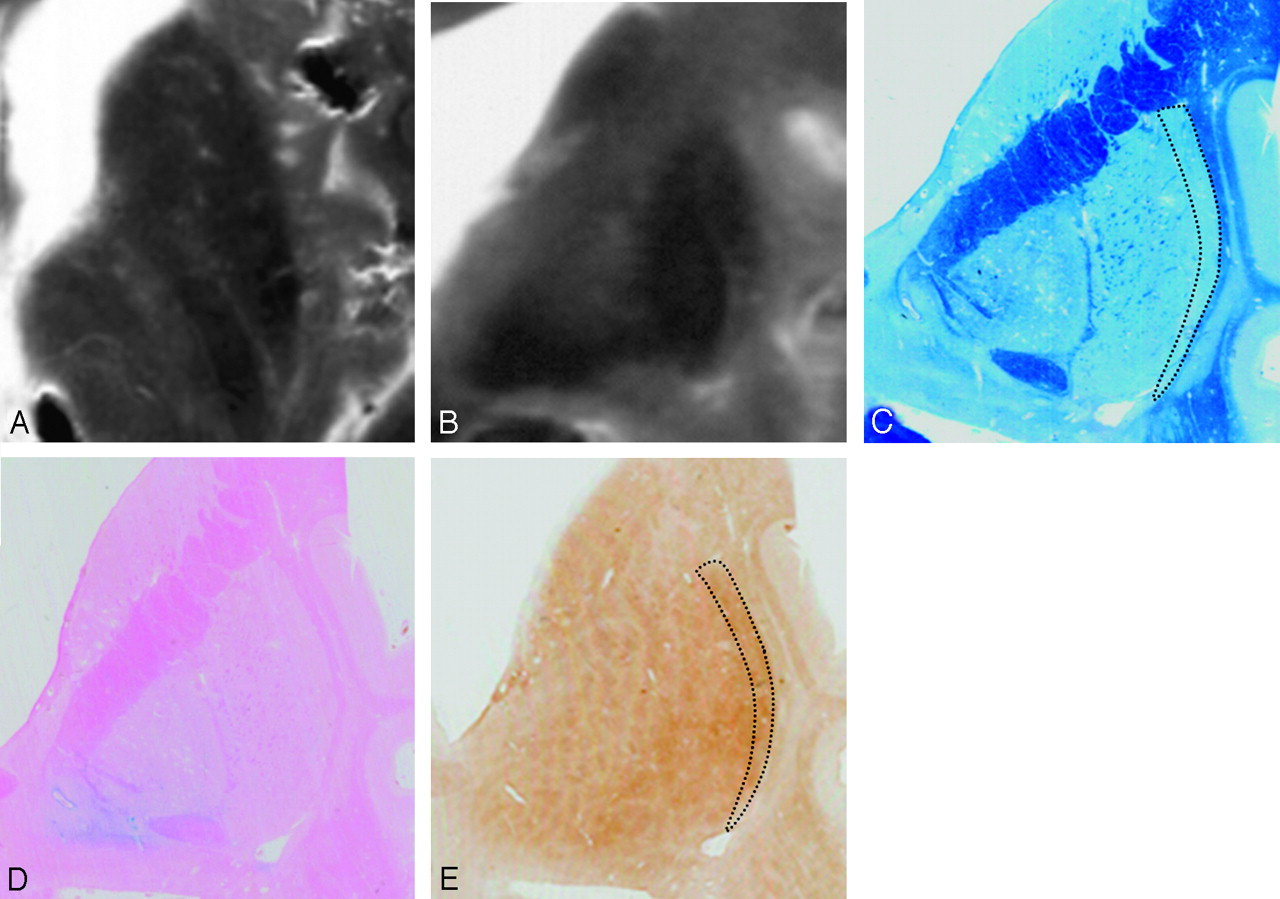
    Comparison of postmortem MR images and histologic findings in a 63-year-old subject. Postmortem axial (A) and coronal (B) T2-weighted images. Sections corresponding to B are stained with Klüver-Barrera (C), Berlin blue (D), and ferritin immunohistochemistry (E). Postmortem MR images (A and B) show present HPR. With Klüver-Barrera staining (C), HPR on a T2-weighed FSE image corresponds to the lateral marginal area of the putamen (dotted lines), where myelinated fibers are small in number in comparison with the remainder of the putamen. With Berlin blue staining (D), hemosiderin-stained blue is scattered in the inner portion of the putamen but is scarce in the lateral marginal area. With ferritin immunohistochemistry (E), ferritin deposits are mild in the lateral marginal area of the putamen (dotted lines), corresponding to HPR on the postmortem MR images (B). By contrast, the deposits are increased in the remainder of the putamen corresponding to the area of decreased signal intensity on the postmortem MR images (B).

  • Fig 5.
    • Download figure
    • Open in new tab
    • Download powerpoint
    Fig 5.

    Comparison of postmortem MR images and histologic findings in an 83-year-old subject. Postmortem axial (A) and coronal (B) T2-weighted images. A section corresponding to B stained with Klüver-Barrera (C), Berlin blue (D), and ferritin immunohistochemistry (E). A and B, Postmortem MR images show diffuse putaminal hypointensity, and HPR is not detectable. With Klüver-Barrera staining (C), myelinated fibers are few in the lateral marginal area of the putamen (dotted lines). With Berlin blue staining (D), hemosiderin is scattered in the inner portion of the putamen but is scarce in the outer portion. Ferritin immunohistochemistry reveals diffuse ferritin deposits in the putamen, including the lateral marginal area (E).

PreviousNext
Back to top

In this issue

American Journal of Neuroradiology: 28 (4)
American Journal of Neuroradiology
Vol. 28, Issue 4
April 2007
  • Table of Contents
  • Index by author
Advertisement
Print
Download PDF
Email Article

Thank you for your interest in spreading the word on American Journal of Neuroradiology.

NOTE: We only request your email address so that the person you are recommending the page to knows that you wanted them to see it, and that it is not junk mail. We do not capture any email address.

Enter multiple addresses on separate lines or separate them with commas.
Hyperintense Putaminal Rim at 3T Reflects Fewer Ferritin Deposits in the Lateral Marginal Area of the Putamen
(Your Name) has sent you a message from American Journal of Neuroradiology
(Your Name) thought you would like to see the American Journal of Neuroradiology web site.
CAPTCHA
This question is for testing whether or not you are a human visitor and to prevent automated spam submissions.
Cite this article
S. Fujii, E. Matsusue, T. Kinoshita, S. Sugihara, E. Ohama, T. Ogawa
Hyperintense Putaminal Rim at 3T Reflects Fewer Ferritin Deposits in the Lateral Marginal Area of the Putamen
American Journal of Neuroradiology Apr 2007, 28 (4) 777-781;

Citation Manager Formats

  • BibTeX
  • Bookends
  • EasyBib
  • EndNote (tagged)
  • EndNote 8 (xml)
  • Medlars
  • Mendeley
  • Papers
  • RefWorks Tagged
  • Ref Manager
  • RIS
  • Zotero
0 Responses
Respond to this article
Share
Bookmark this article
Hyperintense Putaminal Rim at 3T Reflects Fewer Ferritin Deposits in the Lateral Marginal Area of the Putamen
S. Fujii, E. Matsusue, T. Kinoshita, S. Sugihara, E. Ohama, T. Ogawa
American Journal of Neuroradiology Apr 2007, 28 (4) 777-781;
del.icio.us logo Twitter logo Facebook logo Mendeley logo
  • Tweet Widget
  • Facebook Like
  • Google Plus One

Jump to section

  • Article
    • Abstract
    • Materials and Methods
    • Results
    • Discussion
    • Conclusion
    • Acknowledgments
    • References
  • Figures & Data
  • Info & Metrics
  • Responses
  • References
  • PDF

Related Articles

  • No related articles found.
  • PubMed
  • Google Scholar

Cited By...

  • A new MRI rating scale for progressive supranuclear palsy and multiple system atrophy: validity and reliability
  • Crossref
  • Google Scholar

This article has not yet been cited by articles in journals that are participating in Crossref Cited-by Linking.

More in this TOC Section

  • Enhanced Axonal Metabolism during Early Natalizumab Treatment in Relapsing-Remitting Multiple Sclerosis
  • SWI or T2*: Which MRI Sequence to Use in the Detection of Cerebral Microbleeds? The Karolinska Imaging Dementia Study
  • Progression of Microstructural Damage in Spinocerebellar Ataxia Type 2: A Longitudinal DTI Study
Show more BRAIN

Similar Articles

Advertisement

Indexed Content

  • Current Issue
  • Accepted Manuscripts
  • Article Preview
  • Past Issues
  • Editorials
  • Editor's Choice
  • Fellows' Journal Club
  • Letters to the Editor
  • Video Articles

Cases

  • Case Collection
  • Archive - Case of the Week
  • Archive - Case of the Month
  • Archive - Classic Case

More from AJNR

  • Trainee Corner
  • Imaging Protocols
  • MRI Safety Corner
  • Book Reviews

Multimedia

  • AJNR Podcasts
  • AJNR Scantastics

Resources

  • Turnaround Time
  • Submit a Manuscript
  • Submit a Video Article
  • Submit an eLetter to the Editor/Response
  • Manuscript Submission Guidelines
  • Statistical Tips
  • Fast Publishing of Accepted Manuscripts
  • Graphical Abstract Preparation
  • Imaging Protocol Submission
  • Evidence-Based Medicine Level Guide
  • Publishing Checklists
  • Author Policies
  • Become a Reviewer/Academy of Reviewers
  • News and Updates

About Us

  • About AJNR
  • Editorial Board
  • Editorial Board Alumni
  • Alerts
  • Permissions
  • Not an AJNR Subscriber? Join Now
  • Advertise with Us
  • Librarian Resources
  • Feedback
  • Terms and Conditions
  • AJNR Editorial Board Alumni

American Society of Neuroradiology

  • Not an ASNR Member? Join Now

© 2025 by the American Society of Neuroradiology All rights, including for text and data mining, AI training, and similar technologies, are reserved.
Print ISSN: 0195-6108 Online ISSN: 1936-959X

Powered by HighWire