Advertisement

Main menu

  • Home
  • Content
    • Current Issue
    • Accepted Manuscripts
    • Article Preview
    • Past Issue Archive
    • Video Articles
    • AJNR Case Collection
    • Case of the Week Archive
    • Case of the Month Archive
    • Classic Case Archive
  • Special Collections
    • AJNR Awards
    • Low-Field MRI
    • Alzheimer Disease
    • ASNR Foundation Special Collection
    • Photon-Counting CT
    • View All
  • Multimedia
    • AJNR Podcasts
    • AJNR SCANtastic
    • Trainee Corner
    • MRI Safety Corner
    • Imaging Protocols
  • For Authors
    • Submit a Manuscript
    • Submit a Video Article
    • Submit an eLetter to the Editor/Response
    • Manuscript Submission Guidelines
    • Statistical Tips
    • Fast Publishing of Accepted Manuscripts
    • Graphical Abstract Preparation
    • Imaging Protocol Submission
    • Author Policies
  • About Us
    • About AJNR
    • Editorial Board
    • Editorial Board Alumni
  • More
    • Become a Reviewer/Academy of Reviewers
    • Subscribers
    • Permissions
    • Alerts
    • Feedback
    • Advertisers
    • ASNR Home

User menu

  • Alerts
  • Log in

Search

  • Advanced search
American Journal of Neuroradiology

American Journal of Neuroradiology

ASHNR American Society of Functional Neuroradiology ASHNR American Society of Pediatric Neuroradiology ASSR
  • Alerts
  • Log in

Advanced Search

  • Home
  • Content
    • Current Issue
    • Accepted Manuscripts
    • Article Preview
    • Past Issue Archive
    • Video Articles
    • AJNR Case Collection
    • Case of the Week Archive
    • Case of the Month Archive
    • Classic Case Archive
  • Special Collections
    • AJNR Awards
    • Low-Field MRI
    • Alzheimer Disease
    • ASNR Foundation Special Collection
    • Photon-Counting CT
    • View All
  • Multimedia
    • AJNR Podcasts
    • AJNR SCANtastic
    • Trainee Corner
    • MRI Safety Corner
    • Imaging Protocols
  • For Authors
    • Submit a Manuscript
    • Submit a Video Article
    • Submit an eLetter to the Editor/Response
    • Manuscript Submission Guidelines
    • Statistical Tips
    • Fast Publishing of Accepted Manuscripts
    • Graphical Abstract Preparation
    • Imaging Protocol Submission
    • Author Policies
  • About Us
    • About AJNR
    • Editorial Board
    • Editorial Board Alumni
  • More
    • Become a Reviewer/Academy of Reviewers
    • Subscribers
    • Permissions
    • Alerts
    • Feedback
    • Advertisers
    • ASNR Home
  • Follow AJNR on Twitter
  • Visit AJNR on Facebook
  • Follow AJNR on Instagram
  • Join AJNR on LinkedIn
  • RSS Feeds

AJNR Awards, New Junior Editors, and more. Read the latest AJNR updates

Research ArticleBRAIN

CT Angiography and Perfusion CT in Cerebral Vasospasm after Subarachnoid Hemorrhage

S. Binaghi, M.L. Colleoni, P. Maeder, A. Uské, L. Regli, A. R. Dehdashti, P. Schnyder and R. Meuli
American Journal of Neuroradiology April 2007, 28 (4) 750-758;
S. Binaghi
  • Find this author on Google Scholar
  • Find this author on PubMed
  • Search for this author on this site
M.L. Colleoni
  • Find this author on Google Scholar
  • Find this author on PubMed
  • Search for this author on this site
P. Maeder
  • Find this author on Google Scholar
  • Find this author on PubMed
  • Search for this author on this site
A. Uské
  • Find this author on Google Scholar
  • Find this author on PubMed
  • Search for this author on this site
L. Regli
  • Find this author on Google Scholar
  • Find this author on PubMed
  • Search for this author on this site
A. R. Dehdashti
  • Find this author on Google Scholar
  • Find this author on PubMed
  • Search for this author on this site
P. Schnyder
  • Find this author on Google Scholar
  • Find this author on PubMed
  • Search for this author on this site
R. Meuli
  • Find this author on Google Scholar
  • Find this author on PubMed
  • Search for this author on this site
  • Article
  • Figures & Data
  • Info & Metrics
  • Responses
  • References
  • PDF
Loading

Article Figures & Data

Figures

  • Tables
  • Fig 1.
    • Download figure
    • Open in new tab
    • Download powerpoint
    Fig 1.

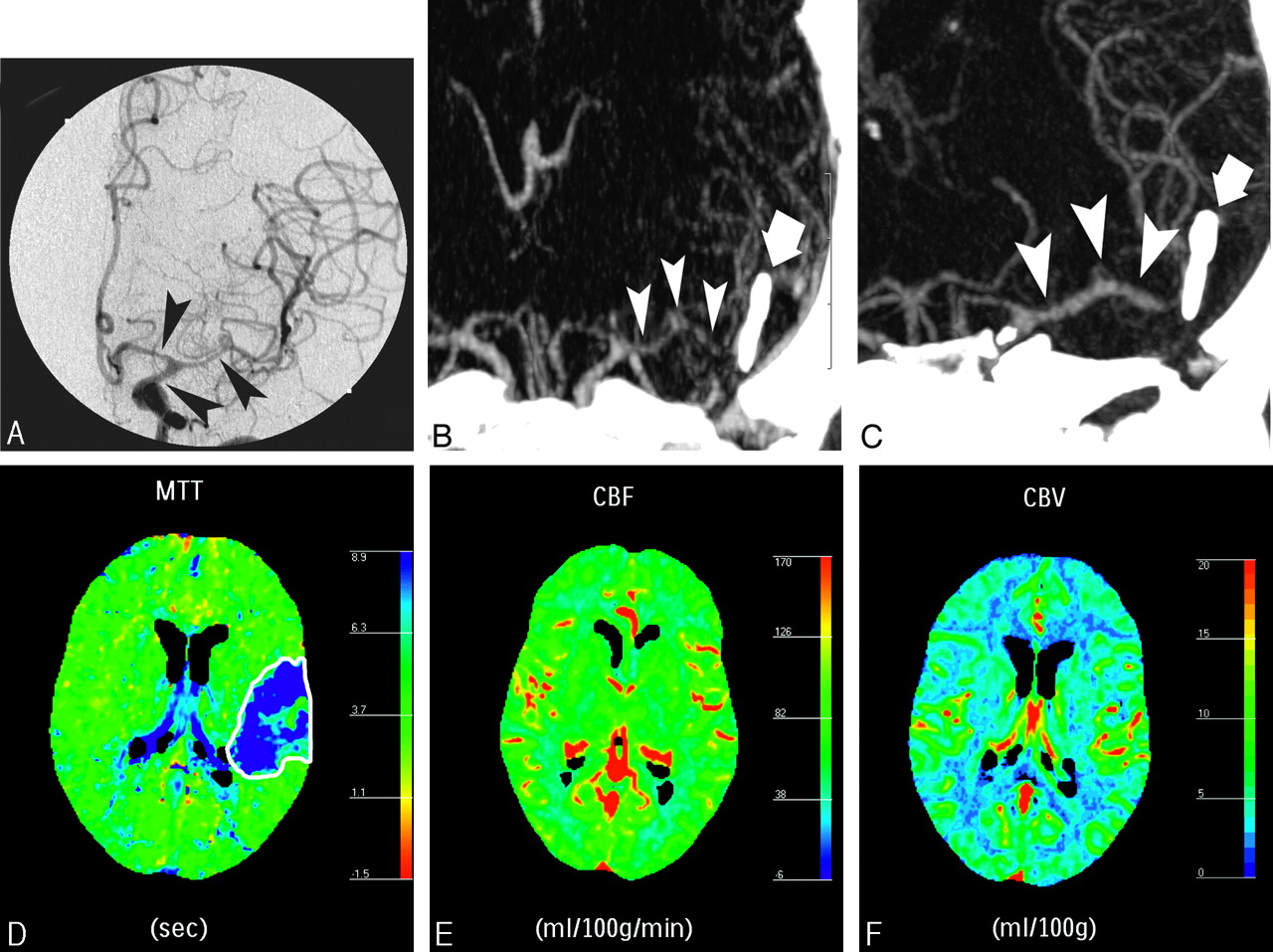
    A 44-year-old woman presenting with weakness of the right arm and leg, clinically attributed to cerebrovascular vasospasm 6 days after SAH related to a ruptured saccular aneurysm of the left MCA bifurcation, which was clipped (bold arrow). DSA showed moderate vasospasm on the distal carotid segment and severe vasospasm on the A1 segment of the left ACA and the M1 and proximal M2 segments of the left MCA (A, black arrowheads). Maximum intensity projection (MIP) MSCTA image before (B) and after (C) intra-arterial infusion of nimodipine showing resolution of the vasospasm (white arrowheads), and followed by the resolution of the symptoms. At pretreatment PCT, MTT was increased in the left MCA territory (D), CBF was normal (E), and a slight increase in CBV (F) was observed, representing vasospasm related auto regulation mechanisms.

  • Fig 2.
    • Download figure
    • Open in new tab
    • Download powerpoint
    Fig 2.

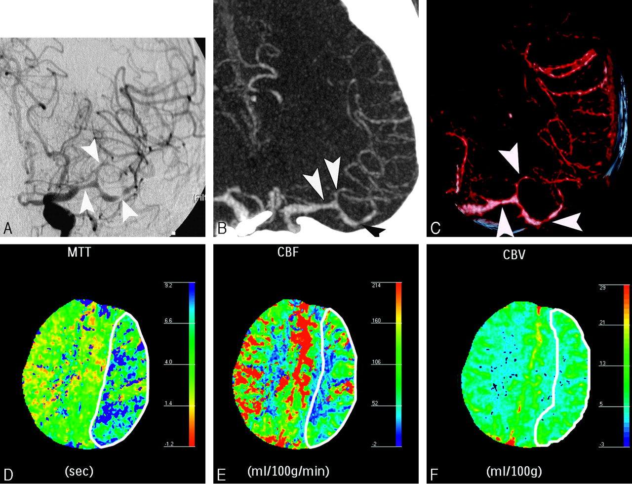
    A 44-year-old man with right-sided hemiparesis attributed to cerebrovascular vasospasm occurring 9 days after SAH consecutive to a ruptured aneurysm of the anterior communicating artery, treated by surgical clipping. DSA (A) showed moderate vasospasm on M1 segment of left MCA and focal severe vasospasm on the M2 segment (arrowheads). These findings were confirmed by MIP MSCTA reconstruction (B) and volume-rendered MSCTA reconstruction (C). Perfusion CT performed during the same CT session revealed an increase in MTT (D) and a decrease in rCBF (E), with slight increase of rCBV (F). This pattern of perfusion alterations corresponds to a reversible ischemic lesion consecutive to vasospasm. The patient was then treated by a local intra-arterial nimodipine infusion and a balloon angioplasty of the left M1 segment.

  • Fig 3.
    • Download figure
    • Open in new tab
    • Download powerpoint
    Fig 3.

    A 52-year-old man with symptoms of cerebrovascular vasospasm 5 days after SAH. He was treated with a surgical clipping of a ruptured aneurysm of the anterior communicating artery (AcomA). A first postoperative angiogram showed no abnormalities. Actual DSA (A) showed an absence of the distal segments of the right AcomA (black arrowheads), interpreted as secondary to a very tight vasospasm. Posteroanterior (B) MIP reconstructions of the AcomA at MSCTA confirmed the lack of enhancement of the right AcomA. A nonenhanced cerebral CT (not shown) disclosed a vague hypoattenuation in the territory of the right AcomA. Perfusion CT results confirmed an irreversible ischemic lesion in the territory of the right AcomA, characterized by an increased MTT (C), a decreased rCBF (D), and a decreased rCBV (E). Thus, no specific endovascular treatment of the right AcomA was undertaken.

Tables

  • Figures
    • View popup
    Table 1:

    Sensitivity, specificity (95% confidence limits in parentheses), positive predictive value, negative predictive value, and accuracy of MSCTA (compared with DSA) in the depiction of intracranial vasospasms, depending on anatomic location

    Location of VasospasmSensitivity (%)Specificity (%)Positive Predictive Value (%)Negative Predictive Value (%)Accuracy (%)
    Distal carotid artery45 (16–74)1001008385
    A1 segment87.5 (71–100)1001009295
    M1 segment10096 (88–100)9310097
    A2 segment100100100100100
    M2 segment100100100100100
    Basilar artery100100100100100
    Total87.7 (79–96)99.2 (98–100)98.394.195.4
    • Note:—MSCTA indicates multisection CT angiography; DSA, digital substraction angiography. Posterior cerebral artery location was not reported because no vasospasm was found on this segment.

    • View popup
    Table 2:

    Sensitivity, specificity (95% confidence limits in parentheses), positive predictive value, negative predictive value, and accuracy of MSCTA in the characterization of intracranial vasospasms at the level of the distal internal carotid artery and the intracranial cerebral arteries

    Vasospasm GradeTrue-PositiveFalse-PositiveFalse-NegativeTrue-NegativeSensitivity (%)Specificity (%)Accuracy (%)Positive Predictive Value (%)Negative Predictive Value (%)
    Mild-Moderate
        Distal ICA3063433.3 (64–3)1008610085
        Intracranial cerebral arteries336518386.8 (76–98)96.8 (94–99)95.284.697.3
    Severe
        Distal ICA20034100100100100100
        Intracranial cerebral arteries131418376.5 (57–96)99.5 (98–100)97.592.997.9
    • Note:—MSCTA indicates multisection CT angiography; ICA, internal carotid artery.

    • View popup
    Table 3:

    Sensitivity, specificity, accuracy, positive predictive value, and negative predictive value of perfusion CT in the characterization of intracranial vasospasms

    Vasospasm GradeSensitivity (%)Specificity (%)Accuracy (%)Positive Predictive Value (%)Negative Predictive Value (%)
    Mild-Moderate2010038.510027.3
    Severe9010092.310075
PreviousNext
Back to top

In this issue

American Journal of Neuroradiology: 28 (4)
American Journal of Neuroradiology
Vol. 28, Issue 4
April 2007
  • Table of Contents
  • Index by author
Advertisement
Print
Download PDF
Email Article

Thank you for your interest in spreading the word on American Journal of Neuroradiology.

NOTE: We only request your email address so that the person you are recommending the page to knows that you wanted them to see it, and that it is not junk mail. We do not capture any email address.

Enter multiple addresses on separate lines or separate them with commas.
CT Angiography and Perfusion CT in Cerebral Vasospasm after Subarachnoid Hemorrhage
(Your Name) has sent you a message from American Journal of Neuroradiology
(Your Name) thought you would like to see the American Journal of Neuroradiology web site.
CAPTCHA
This question is for testing whether or not you are a human visitor and to prevent automated spam submissions.
Cite this article
S. Binaghi, M.L. Colleoni, P. Maeder, A. Uské, L. Regli, A. R. Dehdashti, P. Schnyder, R. Meuli
CT Angiography and Perfusion CT in Cerebral Vasospasm after Subarachnoid Hemorrhage
American Journal of Neuroradiology Apr 2007, 28 (4) 750-758;

Citation Manager Formats

  • BibTeX
  • Bookends
  • EasyBib
  • EndNote (tagged)
  • EndNote 8 (xml)
  • Medlars
  • Mendeley
  • Papers
  • RefWorks Tagged
  • Ref Manager
  • RIS
  • Zotero
0 Responses
Respond to this article
Share
Bookmark this article
CT Angiography and Perfusion CT in Cerebral Vasospasm after Subarachnoid Hemorrhage
S. Binaghi, M.L. Colleoni, P. Maeder, A. Uské, L. Regli, A. R. Dehdashti, P. Schnyder, R. Meuli
American Journal of Neuroradiology Apr 2007, 28 (4) 750-758;
del.icio.us logo Twitter logo Facebook logo Mendeley logo
  • Tweet Widget
  • Facebook Like
  • Google Plus One

Jump to section

  • Article
    • Abstract
    • Materials and Methods
    • Results
    • Discussion
    • Acknowledgments
    • References
  • Figures & Data
  • Info & Metrics
  • Responses
  • References
  • PDF

Related Articles

  • No related articles found.
  • PubMed
  • Google Scholar

Cited By...

  • CTP for the Screening of Vasospasm and Delayed Cerebral Ischemia in Aneurysmal SAH: A Systematic Review and Meta-analysis
  • CTA Supplemented by CTP Increases Interrater Reliability and Endovascular Treatment Use in Patients with Aneurysmal SAH
  • Reliability of the Diagnosis of Cerebral Vasospasm Using Catheter Cerebral Angiography: A Systematic Review and Inter- and Intraobserver Study
  • Reliability of CT Angiography in Cerebral Vasospasm: A Systematic Review of the Literature and an Inter- and Intraobserver Study
  • Bedside cerebral microdialysis monitoring of delayed cerebral hypoperfusion in comatose patients with poor grade aneurysmal subarachnoid haemorrhage
  • Location, Infarct Load, and 3-Month Outcomes of Delayed Cerebral Infarction After Aneurysmal Subarachnoid Hemorrhage
  • Appropriate Use of CT Perfusion following Aneurysmal Subarachnoid Hemorrhage: A Bayesian Analysis Approach
  • Does the Location of the Arterial Input Function Affect Quantitative CTP in Patients with Vasospasm?
  • Invasive interventional management of post-hemorrhagic cerebral vasospasm in patients with aneurysmal subarachnoid hemorrhage
  • Using Quantitative CT Perfusion for Evaluation of Delayed Cerebral Ischemia Following Aneurysmal Subarachnoid Hemorrhage
  • Diagnostic Accuracy of CT Angiography and CT Perfusion for Cerebral Vasospasm: A Meta-Analysis
  • Diagnosing Delayed Cerebral Ischemia With Different CT Modalities in Patients With Subarachnoid Hemorrhage With Clinical Deterioration
  • Crossref
  • Google Scholar

This article has not yet been cited by articles in journals that are participating in Crossref Cited-by Linking.

More in this TOC Section

  • White Matter Alterations in the Brains of Patients with Active, Remitted, and Cured Cushing Syndrome: A DTI Study
  • Qualitative and Quantitative Analysis of MR Imaging Findings in Patients with Middle Cerebral Artery Stroke Implanted with Mesenchymal Stem Cells
  • Fast Contrast-Enhanced 4D MRA and 4D Flow MRI Using Constrained Reconstruction (HYPRFlow): Potential Applications for Brain Arteriovenous Malformations
Show more BRAIN

Similar Articles

Advertisement

Indexed Content

  • Current Issue
  • Accepted Manuscripts
  • Article Preview
  • Past Issues
  • Editorials
  • Editor's Choice
  • Fellows' Journal Club
  • Letters to the Editor
  • Video Articles

Cases

  • Case Collection
  • Archive - Case of the Week
  • Archive - Case of the Month
  • Archive - Classic Case

More from AJNR

  • Trainee Corner
  • Imaging Protocols
  • MRI Safety Corner
  • Book Reviews

Multimedia

  • AJNR Podcasts
  • AJNR Scantastics

Resources

  • Turnaround Time
  • Submit a Manuscript
  • Submit a Video Article
  • Submit an eLetter to the Editor/Response
  • Manuscript Submission Guidelines
  • Statistical Tips
  • Fast Publishing of Accepted Manuscripts
  • Graphical Abstract Preparation
  • Imaging Protocol Submission
  • Evidence-Based Medicine Level Guide
  • Publishing Checklists
  • Author Policies
  • Become a Reviewer/Academy of Reviewers
  • News and Updates

About Us

  • About AJNR
  • Editorial Board
  • Editorial Board Alumni
  • Alerts
  • Permissions
  • Not an AJNR Subscriber? Join Now
  • Advertise with Us
  • Librarian Resources
  • Feedback
  • Terms and Conditions
  • AJNR Editorial Board Alumni

American Society of Neuroradiology

  • Not an ASNR Member? Join Now

© 2025 by the American Society of Neuroradiology All rights, including for text and data mining, AI training, and similar technologies, are reserved.
Print ISSN: 0195-6108 Online ISSN: 1936-959X

Powered by HighWire