Advertisement

Main menu

  • Home
  • Content
    • Current Issue
    • Accepted Manuscripts
    • Article Preview
    • Past Issue Archive
    • Video Articles
    • AJNR Case Collection
    • Case of the Week Archive
    • Case of the Month Archive
    • Classic Case Archive
  • Special Collections
    • AJNR Awards
    • Low-Field MRI
    • Alzheimer Disease
    • ASNR Foundation Special Collection
    • Photon-Counting CT
    • View All
  • Multimedia
    • AJNR Podcasts
    • AJNR SCANtastic
    • Trainee Corner
    • MRI Safety Corner
    • Imaging Protocols
  • For Authors
    • Submit a Manuscript
    • Submit a Video Article
    • Submit an eLetter to the Editor/Response
    • Manuscript Submission Guidelines
    • Statistical Tips
    • Fast Publishing of Accepted Manuscripts
    • Graphical Abstract Preparation
    • Imaging Protocol Submission
    • Author Policies
  • About Us
    • About AJNR
    • Editorial Board
    • Editorial Board Alumni
  • More
    • Become a Reviewer/Academy of Reviewers
    • Subscribers
    • Permissions
    • Alerts
    • Feedback
    • Advertisers
    • ASNR Home

User menu

  • Alerts
  • Log in

Search

  • Advanced search
American Journal of Neuroradiology

American Journal of Neuroradiology

ASHNR American Society of Functional Neuroradiology ASHNR American Society of Pediatric Neuroradiology ASSR
  • Alerts
  • Log in

Advanced Search

  • Home
  • Content
    • Current Issue
    • Accepted Manuscripts
    • Article Preview
    • Past Issue Archive
    • Video Articles
    • AJNR Case Collection
    • Case of the Week Archive
    • Case of the Month Archive
    • Classic Case Archive
  • Special Collections
    • AJNR Awards
    • Low-Field MRI
    • Alzheimer Disease
    • ASNR Foundation Special Collection
    • Photon-Counting CT
    • View All
  • Multimedia
    • AJNR Podcasts
    • AJNR SCANtastic
    • Trainee Corner
    • MRI Safety Corner
    • Imaging Protocols
  • For Authors
    • Submit a Manuscript
    • Submit a Video Article
    • Submit an eLetter to the Editor/Response
    • Manuscript Submission Guidelines
    • Statistical Tips
    • Fast Publishing of Accepted Manuscripts
    • Graphical Abstract Preparation
    • Imaging Protocol Submission
    • Author Policies
  • About Us
    • About AJNR
    • Editorial Board
    • Editorial Board Alumni
  • More
    • Become a Reviewer/Academy of Reviewers
    • Subscribers
    • Permissions
    • Alerts
    • Feedback
    • Advertisers
    • ASNR Home
  • Follow AJNR on Twitter
  • Visit AJNR on Facebook
  • Follow AJNR on Instagram
  • Join AJNR on LinkedIn
  • RSS Feeds

AJNR Awards, New Junior Editors, and more. Read the latest AJNR updates

Research ArticleSpine Imaging and Spine Image-Guided Interventions

Anterior Spinal Artery as a Collateral Channel in Cases of Bilateral Vertebral Arterial Steno-Occlusive Diseases

H.-S. Kang, M.H. Han, S.H. Kim, O.-K. Kwon, H.G. Roh and Y.C. Ko
American Journal of Neuroradiology February 2007, 28 (2) 222-225;
H.-S. Kang
  • Find this author on Google Scholar
  • Find this author on PubMed
  • Search for this author on this site
M.H. Han
  • Find this author on Google Scholar
  • Find this author on PubMed
  • Search for this author on this site
S.H. Kim
  • Find this author on Google Scholar
  • Find this author on PubMed
  • Search for this author on this site
O.-K. Kwon
  • Find this author on Google Scholar
  • Find this author on PubMed
  • Search for this author on this site
H.G. Roh
  • Find this author on Google Scholar
  • Find this author on PubMed
  • Search for this author on this site
Y.C. Ko
  • Find this author on Google Scholar
  • Find this author on PubMed
  • Search for this author on this site
  • Article
  • Figures & Data
  • Info & Metrics
  • Responses
  • References
  • PDF
Loading

Article Figures & Data

Figures

  • Tables
  • Fig 1.
    • Download figure
    • Open in new tab
    • Download powerpoint
    Fig 1.

    Left vertebral angiograms in anteroposterior (A and C) and lateral (B and D) views show the occluded vertebral artery (VA) and retrograde upward flow through the anterior spinal artery (ASA) at early arterial (A and B) and later (C and D) phases in patient 1. The ASA is connected to the right distal VA and supplies the right anterior and posterior inferior cerebellar arteries.

  • Fig 2.
    • Download figure
    • Open in new tab
    • Download powerpoint
    Fig 2.

    Left vertebral angiograms in anteroposterior (AP) (A) and lateral (B) views show the anterior spinal artery (ASA; arrow) filled in retrograde direction as well as the occluded VA in patient 3, though it is not clear that the ASA is providing any significant collateral support to the posterior circulation. Left common carotid angiograms in AP (C) and lateral (D) views reveal contrast filling of the basilar artery from the posterior communicating artery. Diffuse severe arteriosclerotic narrowing and dilation of cerebral arteries is also noted.
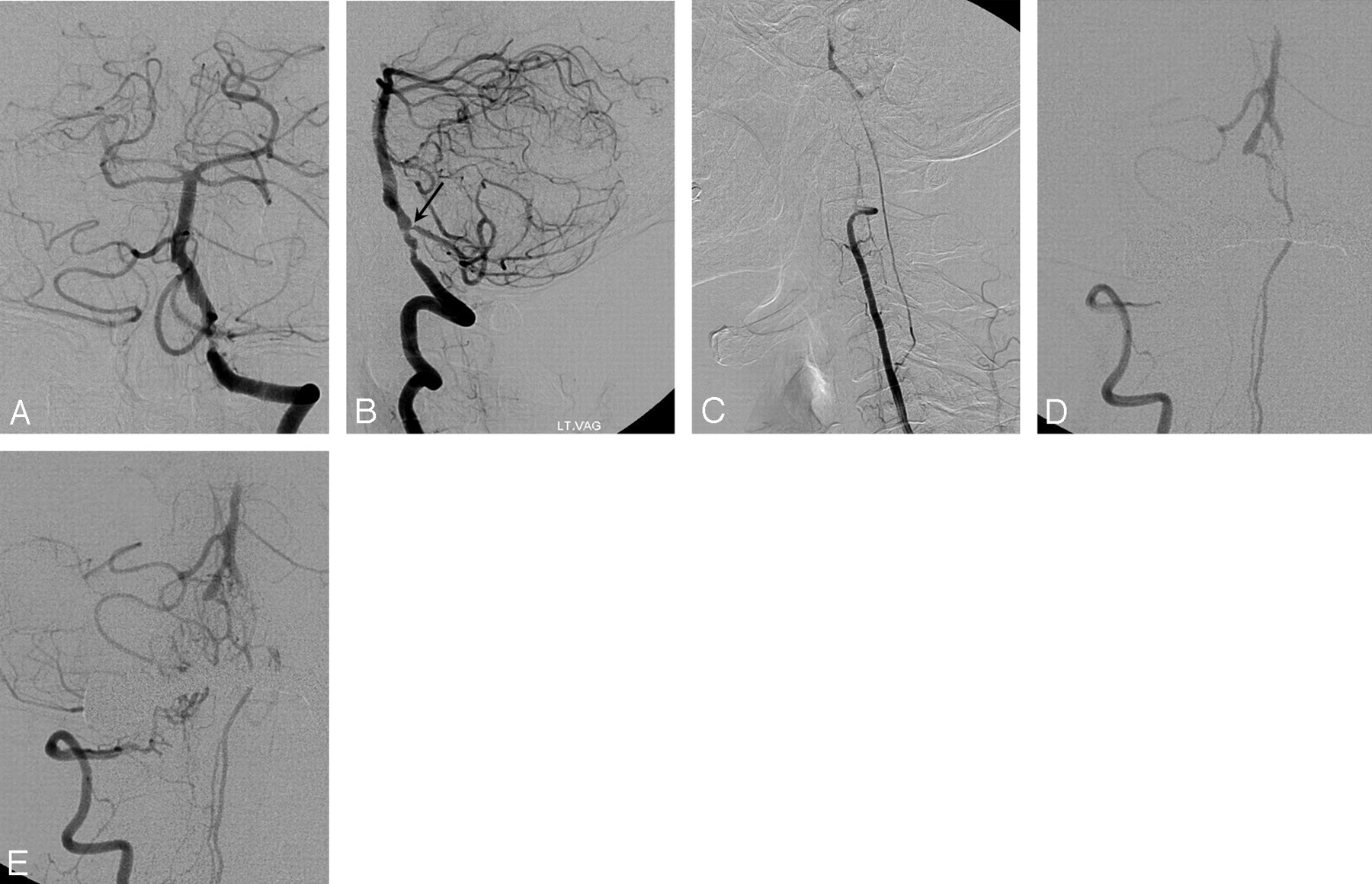
  • Fig 3.
    • Download figure
    • Open in new tab
    • Download powerpoint
    Fig 3.

    Right vertebral angiograms in lateral views (A and B) demonstrate the occluded vertebral artery (VA) and retrograde flow through the anterior spinal artery (ASA) and perfusion to pontine perforators after filling of the basilar artery at later phase (B, arrow) in patient 2. Angiograms in right anterior oblique view at early arterial (C) and later (D) phases reveals reversal (normalization) of blood flow through the ASA after successful stent-assisted recanalization of the right VA.

  • Fig 4.
    • Download figure
    • Open in new tab
    • Download powerpoint
    Fig 4.

    Irregular stenosis of the left vertebral artery (VA) is shown on the left vertebral angiograms in anteroposterior (AP) (A) and lateral (B) views in patient 5. Severe stenosis of the left posterior inferior cerebral artery (PICA) orifice is also noted (arrow). Right vertebral angiogram in the lateral view shows retrograde flow through the anterior spinal artery (ASA) (C). There is minimal perfusion to right distal VA. Follow-up angiography after 9 months shows increased perfusion through the ASA at early arterial (D) and later (E) phases. Both distal VAs are connected to the ASA, and perfusion to the common trunk of the right anterior interior cerebellar artery (AICA) and PICA and left AICA is maintained by this collateral route. The left VA was completely occluded (not shown).

Tables

  • Figures
    • View popup
    Table 1:

    Clinical characteristics and MR imaging features of the patients

    No/Gender/AgePresentationAcute Ischemic Lesions on MRIExtent of Parenchymal LesionsInterventional ProceduremRS at Discharge
    1/F/62Dizziness, gait disturbanceL pons, both CHFocalIA thrombolysis w/ stent-assisted angioplasty3
    2/M/64R hemiparesis, R facial palsyR midbrain, L CH, R occipital lobeFocalIA thrombolysis w/ stent-assisted angioplasty2
    3/F/75L hemiplegia, L facial palsy, dysphagiaR pons, both CHFocalNA4
    4/F/63Dizziness, gait ataxiaR CHFocalStent-assisted angioplasty1
    5/M/40Dizziness, gait ataxiaL CHTerritorialNA2
    6/M/81Quadriparesis, aphagiaL midbrain, L pons, L CHTerritorialIA thrombolysis w/ stent-assisted angioplasty4
    • Note:—MRI indicates MR imaging; mRS, modified Rankin scale; CH, cerebellar hemisphere; IA, intra-arterial; R, right; L, left; NA, not attempted.

    • View popup
    Table 2:

    Angiographic characteristics of the patients

    NoRight VALeft VACourse of ASAPerfusion via ASACollateral Supply from PcomAVBJ PatencyComment
    1OcclusionOcclusionL VA → R VAR AICA & PICABoth SCAs filling from L PcomAYesReversed flow of ASA disappeared after recanalization; reappearance of ASA
    2OcclusionSevere stenosisR VA → L VAPart of BANilYesBA filling from L VA; reversal of ASA flow direction after recanalization
    3OcclusionOcclusionL VA → R VANot profuseBA trunk filling from L PcomANo
    4Severe stenosisOcclusionR & L VA → L VANot profuseBoth SCAs filling from R PcomAYesReversal of ASA flow direction after recanalization
    5OcclusionOcclusionR VA → both VAsR AICA & PICA; L AICANilYesIncreased perfusion through ASA after occlusion of L VA
    6OcclusionSevere stenosisR VA → R VANot profuseNilYesReversal of ASA flow direction after recanalization
    • Note:—VA, vertebral artery; ASA, anterior spinal artery; PcomA, posterior communicating artery; VBJ, vertebrobasilar junction; L, left; R, right; AICA, anterior inferior cerebellar artery; PICA, posterior inferior cerebellar artery; SCA, superior cerebellar artery; BA, basilar artery.

PreviousNext
Back to top

In this issue

American Journal of Neuroradiology: 28 (2)
American Journal of Neuroradiology
Vol. 28, Issue 2
February 2007
  • Table of Contents
  • Index by author
Advertisement
Print
Download PDF
Email Article

Thank you for your interest in spreading the word on American Journal of Neuroradiology.

NOTE: We only request your email address so that the person you are recommending the page to knows that you wanted them to see it, and that it is not junk mail. We do not capture any email address.

Enter multiple addresses on separate lines or separate them with commas.
Anterior Spinal Artery as a Collateral Channel in Cases of Bilateral Vertebral Arterial Steno-Occlusive Diseases
(Your Name) has sent you a message from American Journal of Neuroradiology
(Your Name) thought you would like to see the American Journal of Neuroradiology web site.
CAPTCHA
This question is for testing whether or not you are a human visitor and to prevent automated spam submissions.
Cite this article
H.-S. Kang, M.H. Han, S.H. Kim, O.-K. Kwon, H.G. Roh, Y.C. Ko
Anterior Spinal Artery as a Collateral Channel in Cases of Bilateral Vertebral Arterial Steno-Occlusive Diseases
American Journal of Neuroradiology Feb 2007, 28 (2) 222-225;

Citation Manager Formats

  • BibTeX
  • Bookends
  • EasyBib
  • EndNote (tagged)
  • EndNote 8 (xml)
  • Medlars
  • Mendeley
  • Papers
  • RefWorks Tagged
  • Ref Manager
  • RIS
  • Zotero
0 Responses
Respond to this article
Share
Bookmark this article
Anterior Spinal Artery as a Collateral Channel in Cases of Bilateral Vertebral Arterial Steno-Occlusive Diseases
H.-S. Kang, M.H. Han, S.H. Kim, O.-K. Kwon, H.G. Roh, Y.C. Ko
American Journal of Neuroradiology Feb 2007, 28 (2) 222-225;
del.icio.us logo Twitter logo Facebook logo Mendeley logo
  • Tweet Widget
  • Facebook Like
  • Google Plus One

Jump to section

  • Article
    • Abstract
    • Materials and Methods
    • Results
    • Discussion
    • Conclusion
    • References
  • Figures & Data
  • Info & Metrics
  • Responses
  • References
  • PDF

Related Articles

  • ERRATUM
  • PubMed
  • Google Scholar

Cited By...

  • Time window for recanalization in basilar artery occlusion: Speculative synthesis
  • Imaging Characteristics of Cerebrovascular Arteriopathy and Stroke in Hutchinson-Gilford Progeria Syndrome
  • Crossref
  • Google Scholar

This article has not yet been cited by articles in journals that are participating in Crossref Cited-by Linking.

More in this TOC Section

  • Management Outcomes For VO Spine Biopsy
  • Cone Beam CT Myelography
  • Post-Procedural Brachial Neuritis Features
Show more SPINE IMAGING AND SPINE IMAGE-GUIDED INTERVENTIONS

Similar Articles

Advertisement

Indexed Content

  • Current Issue
  • Accepted Manuscripts
  • Article Preview
  • Past Issues
  • Editorials
  • Editor's Choice
  • Fellows' Journal Club
  • Letters to the Editor
  • Video Articles

Cases

  • Case Collection
  • Archive - Case of the Week
  • Archive - Case of the Month
  • Archive - Classic Case

More from AJNR

  • Trainee Corner
  • Imaging Protocols
  • MRI Safety Corner
  • Book Reviews

Multimedia

  • AJNR Podcasts
  • AJNR Scantastics

Resources

  • Turnaround Time
  • Submit a Manuscript
  • Submit a Video Article
  • Submit an eLetter to the Editor/Response
  • Manuscript Submission Guidelines
  • Statistical Tips
  • Fast Publishing of Accepted Manuscripts
  • Graphical Abstract Preparation
  • Imaging Protocol Submission
  • Evidence-Based Medicine Level Guide
  • Publishing Checklists
  • Author Policies
  • Become a Reviewer/Academy of Reviewers
  • News and Updates

About Us

  • About AJNR
  • Editorial Board
  • Editorial Board Alumni
  • Alerts
  • Permissions
  • Not an AJNR Subscriber? Join Now
  • Advertise with Us
  • Librarian Resources
  • Feedback
  • Terms and Conditions
  • AJNR Editorial Board Alumni

American Society of Neuroradiology

  • Not an ASNR Member? Join Now

© 2025 by the American Society of Neuroradiology All rights, including for text and data mining, AI training, and similar technologies, are reserved.
Print ISSN: 0195-6108 Online ISSN: 1936-959X

Powered by HighWire