Advertisement

Main menu

  • Home
  • Content
    • Current Issue
    • Accepted Manuscripts
    • Article Preview
    • Past Issue Archive
    • Video Articles
    • AJNR Case Collection
    • Case of the Week Archive
    • Case of the Month Archive
    • Classic Case Archive
  • Special Collections
    • AJNR Awards
    • Low-Field MRI
    • Alzheimer Disease
    • ASNR Foundation Special Collection
    • Photon-Counting CT
    • View All
  • Multimedia
    • AJNR Podcasts
    • AJNR SCANtastic
    • Trainee Corner
    • MRI Safety Corner
    • Imaging Protocols
  • For Authors
    • Submit a Manuscript
    • Submit a Video Article
    • Submit an eLetter to the Editor/Response
    • Manuscript Submission Guidelines
    • Statistical Tips
    • Fast Publishing of Accepted Manuscripts
    • Graphical Abstract Preparation
    • Imaging Protocol Submission
    • Author Policies
  • About Us
    • About AJNR
    • Editorial Board
    • Editorial Board Alumni
  • More
    • Become a Reviewer/Academy of Reviewers
    • Subscribers
    • Permissions
    • Alerts
    • Feedback
    • Advertisers
    • ASNR Home

User menu

  • Alerts
  • Log in

Search

  • Advanced search
American Journal of Neuroradiology

American Journal of Neuroradiology

ASHNR American Society of Functional Neuroradiology ASHNR American Society of Pediatric Neuroradiology ASSR
  • Alerts
  • Log in

Advanced Search

  • Home
  • Content
    • Current Issue
    • Accepted Manuscripts
    • Article Preview
    • Past Issue Archive
    • Video Articles
    • AJNR Case Collection
    • Case of the Week Archive
    • Case of the Month Archive
    • Classic Case Archive
  • Special Collections
    • AJNR Awards
    • Low-Field MRI
    • Alzheimer Disease
    • ASNR Foundation Special Collection
    • Photon-Counting CT
    • View All
  • Multimedia
    • AJNR Podcasts
    • AJNR SCANtastic
    • Trainee Corner
    • MRI Safety Corner
    • Imaging Protocols
  • For Authors
    • Submit a Manuscript
    • Submit a Video Article
    • Submit an eLetter to the Editor/Response
    • Manuscript Submission Guidelines
    • Statistical Tips
    • Fast Publishing of Accepted Manuscripts
    • Graphical Abstract Preparation
    • Imaging Protocol Submission
    • Author Policies
  • About Us
    • About AJNR
    • Editorial Board
    • Editorial Board Alumni
  • More
    • Become a Reviewer/Academy of Reviewers
    • Subscribers
    • Permissions
    • Alerts
    • Feedback
    • Advertisers
    • ASNR Home
  • Follow AJNR on Twitter
  • Visit AJNR on Facebook
  • Follow AJNR on Instagram
  • Join AJNR on LinkedIn
  • RSS Feeds

AJNR Awards, New Junior Editors, and more. Read the latest AJNR updates

Research ArticleBRAIN

Assessment of Hemorrhage in Pituitary Macroadenoma by T2*-Weighted Gradient-Echo MR Imaging

M. Tosaka, N. Sato, J. Hirato, H. Fujimaki, R. Yamaguchi, H. Kohga, K. Hashimoto, M. Yamada, M. Mori, N. Saito and Y. Yoshimoto
American Journal of Neuroradiology November 2007, 28 (10) 2023-2029; DOI: https://doi.org/10.3174/ajnr.A0692
M. Tosaka
  • Find this author on Google Scholar
  • Find this author on PubMed
  • Search for this author on this site
N. Sato
  • Find this author on Google Scholar
  • Find this author on PubMed
  • Search for this author on this site
J. Hirato
  • Find this author on Google Scholar
  • Find this author on PubMed
  • Search for this author on this site
H. Fujimaki
  • Find this author on Google Scholar
  • Find this author on PubMed
  • Search for this author on this site
R. Yamaguchi
  • Find this author on Google Scholar
  • Find this author on PubMed
  • Search for this author on this site
H. Kohga
  • Find this author on Google Scholar
  • Find this author on PubMed
  • Search for this author on this site
K. Hashimoto
  • Find this author on Google Scholar
  • Find this author on PubMed
  • Search for this author on this site
M. Yamada
  • Find this author on Google Scholar
  • Find this author on PubMed
  • Search for this author on this site
M. Mori
  • Find this author on Google Scholar
  • Find this author on PubMed
  • Search for this author on this site
N. Saito
  • Find this author on Google Scholar
  • Find this author on PubMed
  • Search for this author on this site
Y. Yoshimoto
  • Find this author on Google Scholar
  • Find this author on PubMed
  • Search for this author on this site
  • Article
  • Figures & Data
  • Info & Metrics
  • Responses
  • References
  • PDF
Loading

Article Figures & Data

Figures

  • Tables
  • Fig 1.
    • Download figure
    • Open in new tab
    • Download powerpoint
    Fig 1.

    Patient 1. A 65-year-old woman presented with classic pituitary apoplexy manifesting as a sudden onset of severe headache, nausea, vomiting, and visual disturbance. Also, she became drowsy because of severe dehydration and hyponatremia. A, Sagittal T1-weighted MR image showing low- to isointense masses in the nonenhanced pituitary lesion with diffuse high signal intensity. B, Sagittal T2-weighted MR image showing dark masses in the mixed high signal intensity area. C, Coronal T2*-weighted gradient-echo image showing dark masses indicating hematoma (white arrows). D, Photomicrograph showing hemorrhage and a small amount of chromophobic adenoma (H&E, original magnification ×40).

  • Fig 2.
    • Download figure
    • Open in new tab
    • Download powerpoint
    Fig 2.

    Patient 2. A 30-year-old man presented with subacute hematoma manifesting as right temporal hemianopia and progressive right visual disturbance. He occasionally had a mild headache but no sudden and eventful headaches. His serum prolactin level was 533.2 ng/mL. A, Coronal T1-weighted MR image showing a large pituitary lesion containing a large area of high signal intensity. B, Coronal T2-weighted MR image showing a large area of low signal intensity with a marked low signal intensity rim. C, Coronal T2*-weighted GE image showing low intensity hematomas with a clear dark rim (white arrow), and a dark mass in the solid portion of the tumor (white arrowhead). D, Photomicrograph showing hemorrhage in chromophobic adenoma (H&E, original magnification ×40).
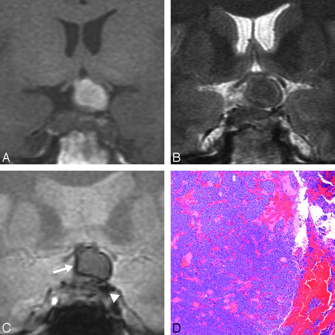
  • Fig 3.
    • Download figure
    • Open in new tab
    • Download powerpoint
    Fig 3.

    Patient 3. A 43-year-old man had typical cysts in a nonfunctioning adenoma. A, Coronal T1-weighted MR image showing a large pituitary lesion. B, Coronal T2-weighted MR image showing the equivocal hypointense rim of a cyst (white arrowheads). C, Coronal T2*-weighted GE image showing the faint hypointense rim of a cyst (white arrows). D, Photomicrograph showing hemorrhage in chromophobic adenoma (H&E, original magnification ×40).
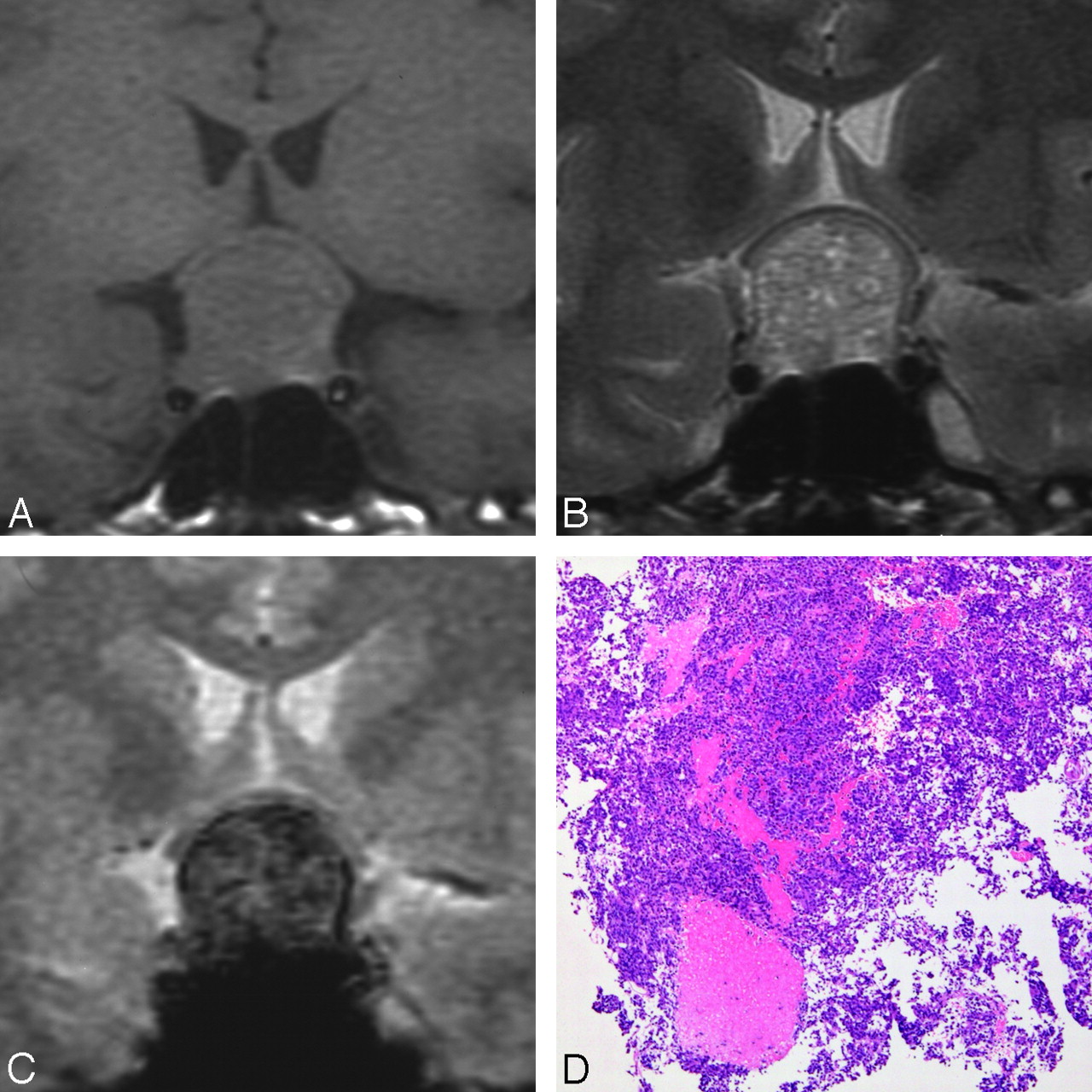
  • Fig 4.
    • Download figure
    • Open in new tab
    • Download powerpoint
    Fig 4.

    Patient 4. A 39-year-old woman had a solid nonfunctioning pituitary adenoma without cyst or hematoma. She had no past or present headache. A, Coronal T1-weighted MR image showing the isointense tumor. B, Coronal T2-weighted MR image showing the mixed high signal intensity pituitary lesion. C, Coronal T2*-weighted GE image showing diffuse dark appearance of the adenoma. D, Photomicrograph showing hemorrhage in chromophobic adenoma (H&E stain, original magnification ×40).

Tables

  • Figures
  • Intratumoral dark lesions on T2*-weighted GE MR imaging and other hemorrhagic findings

    Signs Relating to Intratumoral HemorrhageIntratumoral Dark Lesions on T2*-Weighted GE MR ImagingP Value
    YesNo
    Intratumoral high intensity on T1-weighted FSE MR imaging<.02*
            Yes71
            No512
    Intratumoral dark lesions on T2-weighted FSE MR imaging<.01*
            Yes81
            No412
    Intraoperative findings of intratumoral hemorrhage<.001*
            Yes†100
            No213
    Histologic findings of intratumoral hemorrhage<.001*
            Yes‡124
            No09
    • Note:—GE indicates gradient-echo; FSE, fast spin-echo.

    • * Statistically significant.

    • † Cysts including xanthochromic fluid, liquefied hematoma, and coagulation.

    • ‡ Positive histologic hemorrhagic finding (++ or +).

PreviousNext
Back to top

In this issue

American Journal of Neuroradiology: 28 (10)
American Journal of Neuroradiology
Vol. 28, Issue 10
November 2007
  • Table of Contents
  • Index by author
Advertisement
Print
Download PDF
Email Article

Thank you for your interest in spreading the word on American Journal of Neuroradiology.

NOTE: We only request your email address so that the person you are recommending the page to knows that you wanted them to see it, and that it is not junk mail. We do not capture any email address.

Enter multiple addresses on separate lines or separate them with commas.
Assessment of Hemorrhage in Pituitary Macroadenoma by T2*-Weighted Gradient-Echo MR Imaging
(Your Name) has sent you a message from American Journal of Neuroradiology
(Your Name) thought you would like to see the American Journal of Neuroradiology web site.
CAPTCHA
This question is for testing whether or not you are a human visitor and to prevent automated spam submissions.
Cite this article
M. Tosaka, N. Sato, J. Hirato, H. Fujimaki, R. Yamaguchi, H. Kohga, K. Hashimoto, M. Yamada, M. Mori, N. Saito, Y. Yoshimoto
Assessment of Hemorrhage in Pituitary Macroadenoma by T2*-Weighted Gradient-Echo MR Imaging
American Journal of Neuroradiology Nov 2007, 28 (10) 2023-2029; DOI: 10.3174/ajnr.A0692

Citation Manager Formats

  • BibTeX
  • Bookends
  • EasyBib
  • EndNote (tagged)
  • EndNote 8 (xml)
  • Medlars
  • Mendeley
  • Papers
  • RefWorks Tagged
  • Ref Manager
  • RIS
  • Zotero
0 Responses
Respond to this article
Share
Bookmark this article
Assessment of Hemorrhage in Pituitary Macroadenoma by T2*-Weighted Gradient-Echo MR Imaging
M. Tosaka, N. Sato, J. Hirato, H. Fujimaki, R. Yamaguchi, H. Kohga, K. Hashimoto, M. Yamada, M. Mori, N. Saito, Y. Yoshimoto
American Journal of Neuroradiology Nov 2007, 28 (10) 2023-2029; DOI: 10.3174/ajnr.A0692
del.icio.us logo Twitter logo Facebook logo Mendeley logo
  • Tweet Widget
  • Facebook Like
  • Google Plus One
Purchase

Jump to section

  • Article
    • Abstract
    • Materials and Methods
    • Results
    • Discussion
    • Conclusion
    • Footnotes
    • References
  • Figures & Data
  • Info & Metrics
  • Responses
  • References
  • PDF

Related Articles

  • No related articles found.
  • PubMed
  • Google Scholar

Cited By...

  • Clinical and imaging features of suspected pituitary apoplexy in a domestic rat
  • Differentiation between Cystic Pituitary Adenomas and Rathke Cleft Cysts: A Diagnostic Model Using MRI
  • Acute headache and visual field defect
  • Crossref (58)
  • Google Scholar

This article has been cited by the following articles in journals that are participating in Crossref Cited-by Linking.

  • Principles, Techniques, and Applications of T2*-based MR Imaging and Its Special Applications
    Govind B. Chavhan, Paul S. Babyn, Bejoy Thomas, Manohar M. Shroff, E. Mark Haacke
    RadioGraphics 2009 29 5
  • Pituitary Apoplexy
    Claire Briet, Sylvie Salenave, Jean-François Bonneville, Edward R. Laws, Philippe Chanson
    Endocrine Reviews 2015 36 6
  • Differentiation between Cystic Pituitary Adenomas and Rathke Cleft Cysts: A Diagnostic Model Using MRI
    M. Park, S.-K. Lee, J. Choi, S.-H. Kim, S.H. Kim, N.-Y. Shin, J. Kim, S.S. Ahn
    American Journal of Neuroradiology 2015 36 10
  • Pituitary apoplexy: evaluation, management, and prognosis
    Sally Murad-Kejbou, Eric Eggenberger
    Current Opinion in Ophthalmology 2009 20 6
  • Pituitary apoplexy: an update on clinical and imaging features
    Alessandro Boellis, Alberto di Napoli, Andrea Romano, Alessandro Bozzao
    Insights into Imaging 2014 5 6
  • Italian Association of Clinical Endocrinologists (AME) and International Chapter of Clinical Endocrinology (ICCE). Position statement for clinical practice: prolactin-secreting tumors
    Renato Cozzi, Maria Rosaria Ambrosio, Roberto Attanasio, Claudia Battista, Alessandro Bozzao, Marco Caputo, Enrica Ciccarelli, Laura De Marinis, Ernesto De Menis, Marco Faustini Fustini, Franco Grimaldi, Andrea Lania, Giovanni Lasio, Francesco Logoluso, Marco Losa, Pietro Maffei, Davide Milani, Maurizio Poggi, Michele Zini, Laurence Katznelson, Anton Luger, Catalina Poiana
    European Journal of Endocrinology 2022 186 3
  • Pituitary Apoplexy
    Claire Briet, Sylvie Salenave, Philippe Chanson
    Endocrinology and Metabolism Clinics of North America 2015 44 1
  • Evaluation of the Sellar and Parasellar Regions
    Brian M. Chin, Richard R. Orlandi, Richard H. Wiggins
    Magnetic Resonance Imaging Clinics of North America 2012 20 3
  • Magnetic resonance imaging in the management of prolactinomas; a review of the evidence
    Elena V. Varlamov, José Miguel Hinojosa-Amaya, Maria Fleseriu
    Pituitary 2020 23 1
  • The aetiologies of the unilateral oculomotor nerve palsy: a review of the literature
    Hafiz Khuram Raza, Hao Chen, Thitsavanh Chansysouphanthong, Guiyun Cui
    Somatosensory & Motor Research 2018 35 3-4

More in this TOC Section

  • Multimodal CT Provides Improved Performance for Lacunar Infarct Detection
  • Optimal MRI Sequence for Identifying Occlusion Location in Acute Stroke: Which Value of Time-Resolved Contrast-Enhanced MRA?
  • Evaluating the Effects of White Matter Multiple Sclerosis Lesions on the Volume Estimation of 6 Brain Tissue Segmentation Methods
Show more BRAIN

Similar Articles

Advertisement

Indexed Content

  • Current Issue
  • Accepted Manuscripts
  • Article Preview
  • Past Issues
  • Editorials
  • Editor's Choice
  • Fellows' Journal Club
  • Letters to the Editor
  • Video Articles

Cases

  • Case Collection
  • Archive - Case of the Week
  • Archive - Case of the Month
  • Archive - Classic Case

More from AJNR

  • Trainee Corner
  • Imaging Protocols
  • MRI Safety Corner
  • Book Reviews

Multimedia

  • AJNR Podcasts
  • AJNR Scantastics

Resources

  • Turnaround Time
  • Submit a Manuscript
  • Submit a Video Article
  • Submit an eLetter to the Editor/Response
  • Manuscript Submission Guidelines
  • Statistical Tips
  • Fast Publishing of Accepted Manuscripts
  • Graphical Abstract Preparation
  • Imaging Protocol Submission
  • Evidence-Based Medicine Level Guide
  • Publishing Checklists
  • Author Policies
  • Become a Reviewer/Academy of Reviewers
  • News and Updates

About Us

  • About AJNR
  • Editorial Board
  • Editorial Board Alumni
  • Alerts
  • Permissions
  • Not an AJNR Subscriber? Join Now
  • Advertise with Us
  • Librarian Resources
  • Feedback
  • Terms and Conditions
  • AJNR Editorial Board Alumni

American Society of Neuroradiology

  • Not an ASNR Member? Join Now

© 2025 by the American Society of Neuroradiology All rights, including for text and data mining, AI training, and similar technologies, are reserved.
Print ISSN: 0195-6108 Online ISSN: 1936-959X

Powered by HighWire