
Article Figures & Data
Figures
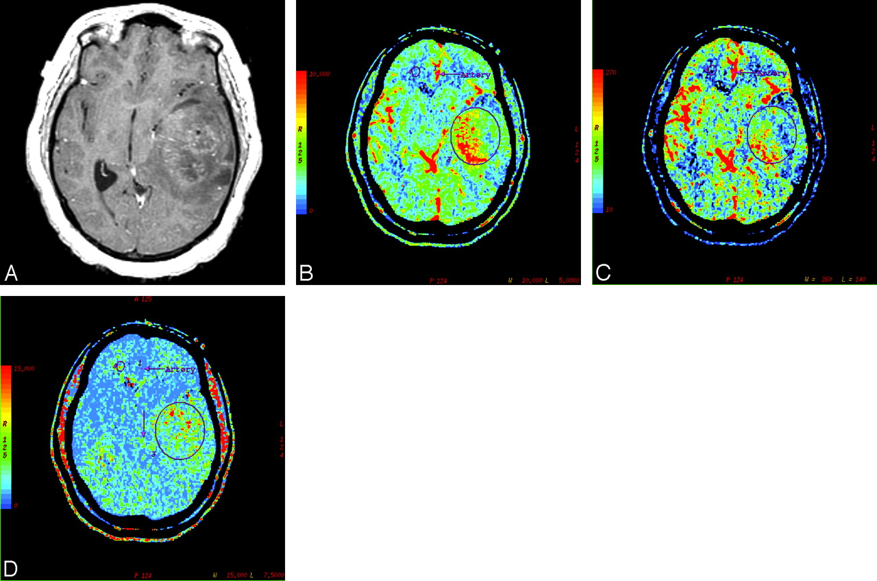
- Fig 1.
A, Postcontrast T1-weighted axial image in a 46-year-old man with WHO grade IV glioma showing a markedly enhancing bifrontal necrotic tumor with involvement of the genu of the corpus callosum and surrounding perilesional white matter edema. B, PCT CBV map showing elevated blood volume (nCBV = 4.3). C, CBF map showing elevated blood flow (nCBF = 6.27). D, MTT map showing decreased MTT (nMTT = 0.63) within the tumor.
- Fig 2.
WHO grade III glioma in a 39-year-old woman. A, Postcontrast T1-weighted axial image showing a large heterogeneously enhancing left temporal mass lesion with significant mass effect. B, Corresponding CBV map showing elevated blood volume (nCBV = 2.61). C, CBF map showing increased blood flow (nCBF = 1.65). D, MTT map showing decreased MTT (nMTT = 1.68) within the tumor. Also note the mismatch between areas of increased blood volume/blood flow on the CBV/CBF maps and the areas of gadolinium enhancement.
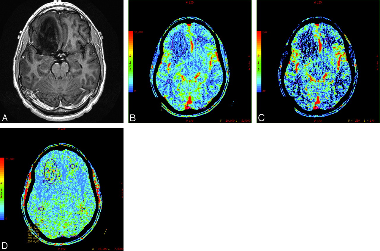
- Fig 3.
A 34-year-old man with WHO grade II glioma. A, Postcontrast T1-weighted axial image showing a nonenhancing mass in the right frontal lobe. B, CBV map showing low blood volume (nCBV = 0.94). C, CBF map showing decreased blood flow (nCBF = 1.26). D, MTT map showing increased transit time (nMTT = 1.07) within the tumor.
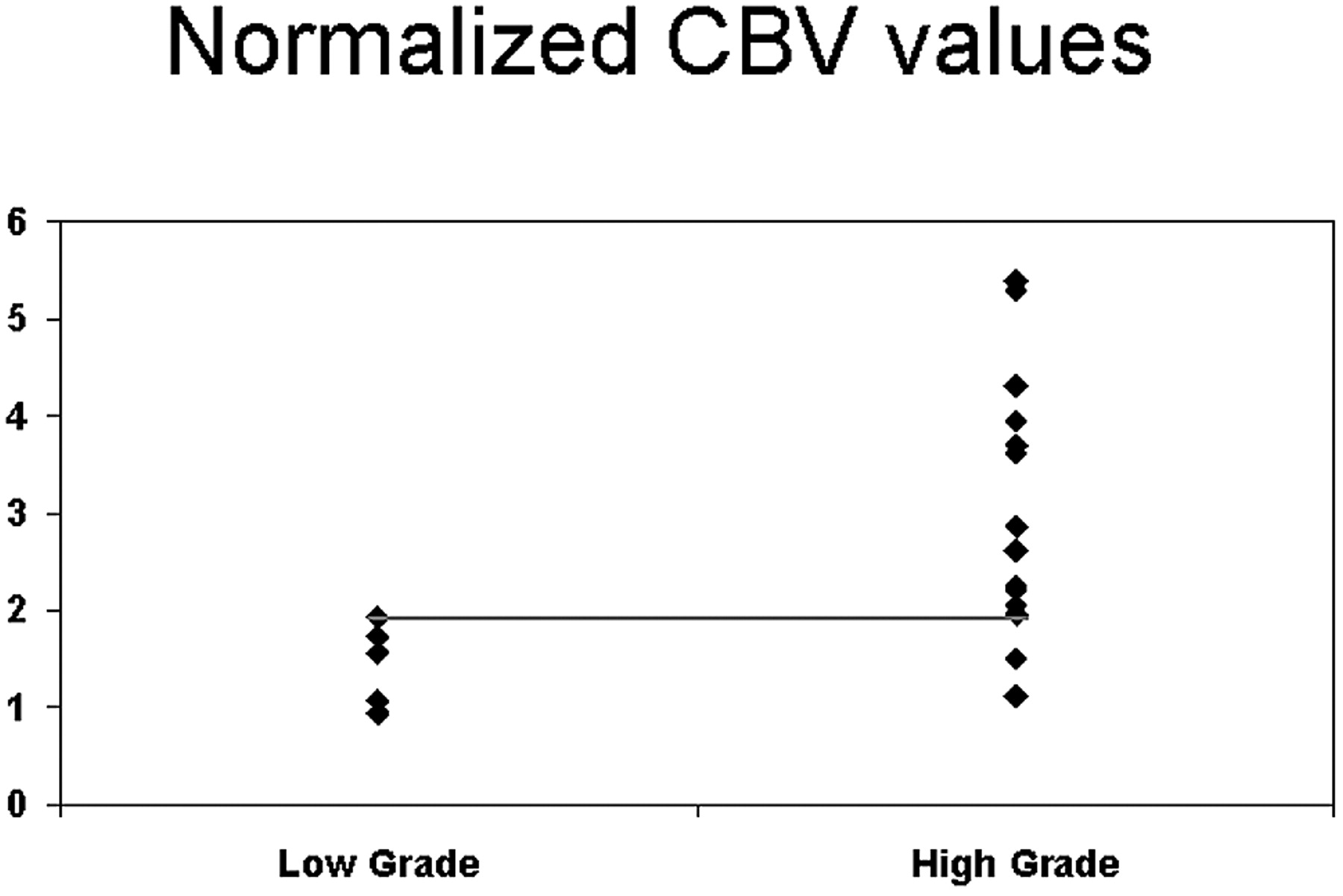
- Fig 4.
Scatter plot for nCBV versus grade of tumor showing the threshold to differentiate low- from high-grade tumors.
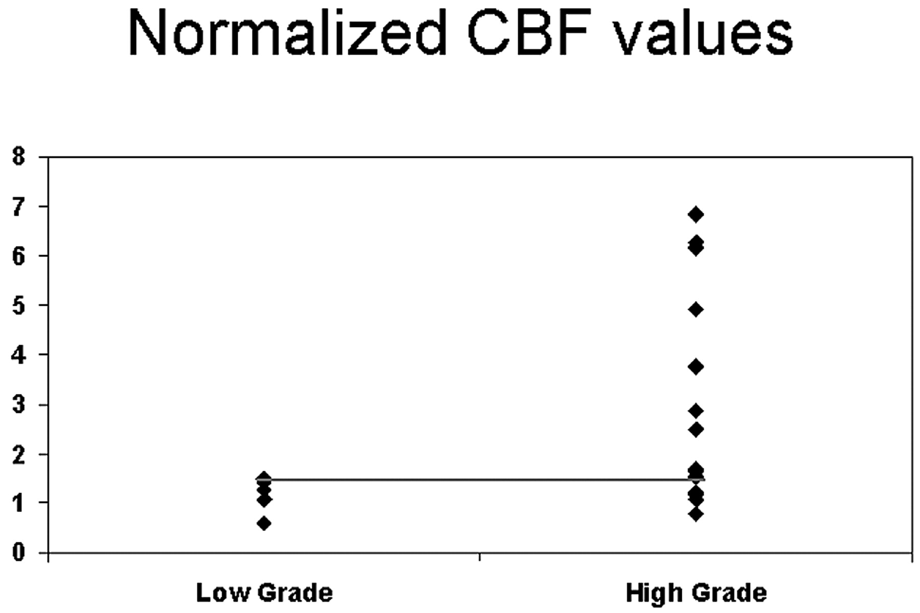
- Fig 5.
Scatter plot for nCBF versus grade of tumor showing the threshold to differentiate low- from high-grade tumors.
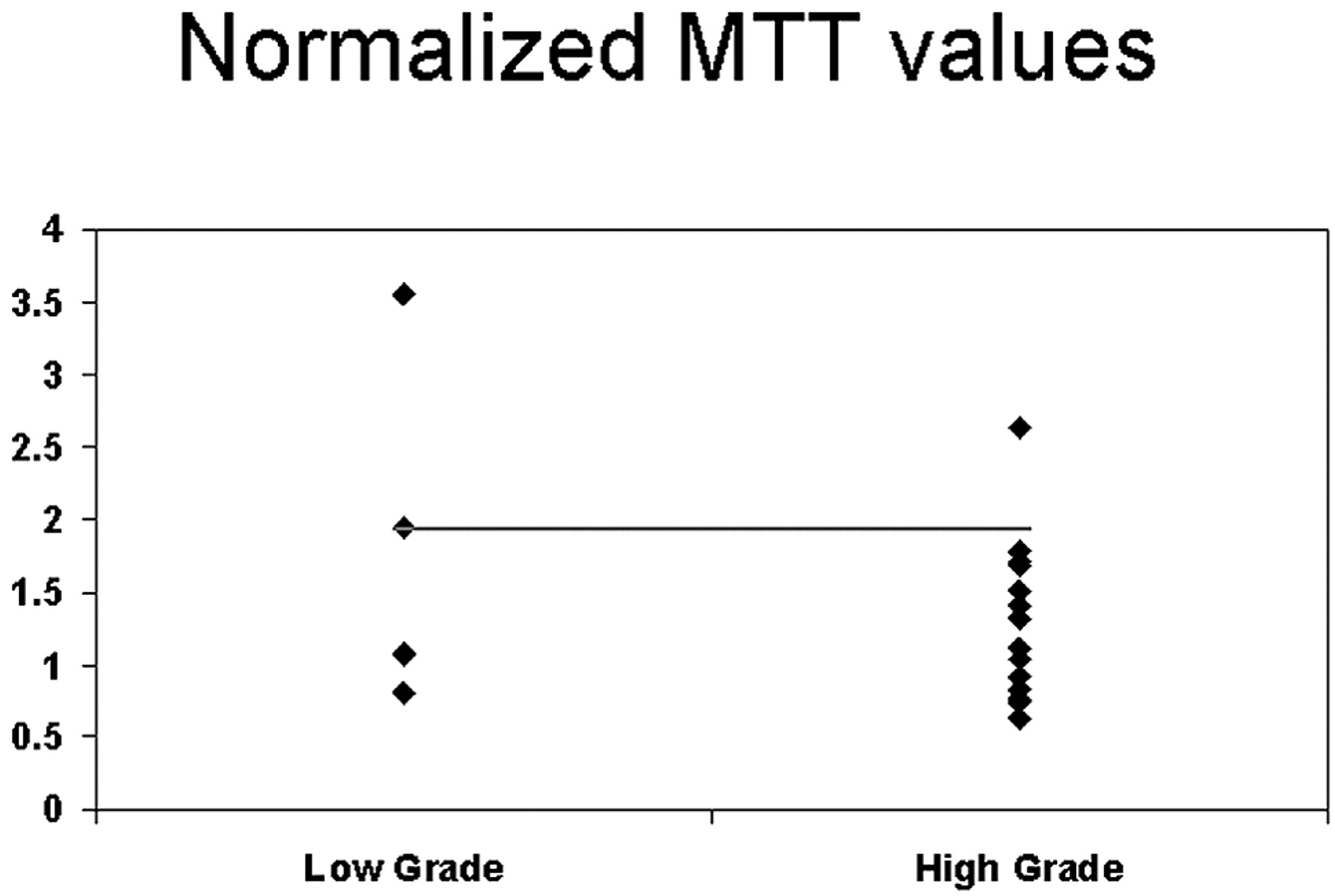
- Fig 6.
Scatter plot for nMTT versus grade of tumor showing the threshold to differentiate low- from high-grade tumors.


Tables
No. Age/Sex Contrast Enhance-ment Hemor-rhage Necro-sis Vasogenic Edema Mass Effect Signal Heterogeneity Involvement of Corpus Callosum Crossing Midline MR Grade of Tumor WHO Grade of Tumor nCBV nCBF nMTT 1 20/M HE – – + + HT + + HG I 1.92 0.58 3.55 2 27/F NCE – – + + HO – – LG II 1.07 1.48 0.83 3 34/M NCE – CD + ++ HT + + LG II 0.94 1.26 1.07 4 56/F HE – + ++ ++ HT – – HG II 1.56 1.07 1.94 5 34/F NCE – – – + HO – – LG II 1.72 1.43 1.08 6 18/F HE – + – + HT – – HG III 2.26 1.53 1.41 7 39/M HE – + + + HT – – HG III 2.21 1.23 1.71 8 57/M HE + + + +++ HT – – HG III 5.39 6.84 2.63 9 68/M NCE – + + + HT – – LG III 3.7 3.75 0.92 10 39/F HE + + ++ +++ HT + + HG III 2.61 1.65 1.68 11 34/M NCE – CD – ++ HT + + LG III 1.5 0.78 1.32 12 64/M PNE + + ++ +++ HT + + HG IV 1.96 1.19 1.78 13 39/F PNE – + ++ +++ HT + – HG IV 2.05 1.07 1.51 14 55/F PNE – + ++ +++ HT + + HG IV 3.95 2.87 1.11 15 60/M PNE + + +++ +++ HT + – HG IV 2.86 2.49 0.74 16 69/M PNE – + + ++ HT – – HG IV 1.12 1.69 1.04 17 66/F HE – + + + HT + – HG IV 5.3 6.16 0.77 18 46/M HE + + ++ ++ HT + + HG IV 4.3 6.27 0.63 19 57/M PNE – + +++ +++ HT – – HG IV 3.61 4.91 0.83 Note:—HE indicates heterogeneous enhancement; NCE, no contrast enhancement; PNE, peripheral nodular enhancement; CD, cystic degeneration; HT, heterogenous; HO, homogeneous; HG, high grade; LG, low grade; +, present; −, absent.
Group (No. of Patients) nCBV nCBF nMTT Mean (SD) Mean (SD) Mean (SD) Low Grade (5) 1.44 (0.42) 1.16 (0.36) 1.69 (1.12) High Grade (III and IV) (14) 3.06 (1.35) 3.03 (2.16) 1.29 (0.55) P value 0.005 0.045 0.559 Grade III (6) 2.95 (1.39) 2.63 (2.30) 1.61 (0.58) Grade IV (8) 3.14 (1.40) 3.33 (2.15) 1.05 (0.40) P value for Low vs III 0.03 0.177 0.792 P value for Low vs IV 0.01 0.048 0.222 P value for III vs IV 0.949 0.572 0.081 Criteria High Grade Low Grade Sensitivity 95% CI Specificity 95% CI nCBV >1.92 85.7% (12/14) (57.2%–98.2%) 100% (5/5) (47.8%–100%) nCBF >1.48 71.4% (10/14) (41.9%–91.6%) 100% (5/5) (47.8%–100%) nMTT <1.94 92.9% (13/14) (66.1%–99.8%) 40% (2/5) (5.3%–85.3%) nCBV >1.92 or nCBF >1.48 92.9% (13/14) (66.1%–99.8%) 100% (5/5) (47.8%–100%) Note:—CI indicates confidence interval.


