Advertisement

Main menu

  • Home
  • Content
    • Current Issue
    • Accepted Manuscripts
    • Article Preview
    • Past Issue Archive
    • Video Articles
    • AJNR Case Collection
    • Case of the Week Archive
    • Case of the Month Archive
    • Classic Case Archive
  • Special Collections
    • AJNR Awards
    • Low-Field MRI
    • Alzheimer Disease
    • ASNR Foundation Special Collection
    • Photon-Counting CT
    • View All
  • Multimedia
    • AJNR Podcasts
    • AJNR SCANtastic
    • Trainee Corner
    • MRI Safety Corner
    • Imaging Protocols
  • For Authors
    • Submit a Manuscript
    • Submit a Video Article
    • Submit an eLetter to the Editor/Response
    • Manuscript Submission Guidelines
    • Statistical Tips
    • Fast Publishing of Accepted Manuscripts
    • Graphical Abstract Preparation
    • Imaging Protocol Submission
    • Author Policies
  • About Us
    • About AJNR
    • Editorial Board
    • Editorial Board Alumni
  • More
    • Become a Reviewer/Academy of Reviewers
    • Subscribers
    • Permissions
    • Alerts
    • Feedback
    • Advertisers
    • ASNR Home

User menu

  • Alerts
  • Log in

Search

  • Advanced search
American Journal of Neuroradiology

American Journal of Neuroradiology

ASHNR American Society of Functional Neuroradiology ASHNR American Society of Pediatric Neuroradiology ASSR
  • Alerts
  • Log in

Advanced Search

  • Home
  • Content
    • Current Issue
    • Accepted Manuscripts
    • Article Preview
    • Past Issue Archive
    • Video Articles
    • AJNR Case Collection
    • Case of the Week Archive
    • Case of the Month Archive
    • Classic Case Archive
  • Special Collections
    • AJNR Awards
    • Low-Field MRI
    • Alzheimer Disease
    • ASNR Foundation Special Collection
    • Photon-Counting CT
    • View All
  • Multimedia
    • AJNR Podcasts
    • AJNR SCANtastic
    • Trainee Corner
    • MRI Safety Corner
    • Imaging Protocols
  • For Authors
    • Submit a Manuscript
    • Submit a Video Article
    • Submit an eLetter to the Editor/Response
    • Manuscript Submission Guidelines
    • Statistical Tips
    • Fast Publishing of Accepted Manuscripts
    • Graphical Abstract Preparation
    • Imaging Protocol Submission
    • Author Policies
  • About Us
    • About AJNR
    • Editorial Board
    • Editorial Board Alumni
  • More
    • Become a Reviewer/Academy of Reviewers
    • Subscribers
    • Permissions
    • Alerts
    • Feedback
    • Advertisers
    • ASNR Home
  • Follow AJNR on Twitter
  • Visit AJNR on Facebook
  • Follow AJNR on Instagram
  • Join AJNR on LinkedIn
  • RSS Feeds

AJNR Awards, New Junior Editors, and more. Read the latest AJNR updates

Research ArticlePediatric Neuroimaging

Diffusion Tensor Imaging in Joubert Syndrome

A. Poretti, E. Boltshauser, T. Loenneker, E.M. Valente, F. Brancati, K. Il'Yasov and T.A.G.M. Huisman
American Journal of Neuroradiology November 2007, 28 (10) 1929-1933; DOI: https://doi.org/10.3174/ajnr.A0703
A. Poretti
  • Find this author on Google Scholar
  • Find this author on PubMed
  • Search for this author on this site
E. Boltshauser
  • Find this author on Google Scholar
  • Find this author on PubMed
  • Search for this author on this site
T. Loenneker
  • Find this author on Google Scholar
  • Find this author on PubMed
  • Search for this author on this site
E.M. Valente
  • Find this author on Google Scholar
  • Find this author on PubMed
  • Search for this author on this site
F. Brancati
  • Find this author on Google Scholar
  • Find this author on PubMed
  • Search for this author on this site
K. Il'Yasov
  • Find this author on Google Scholar
  • Find this author on PubMed
  • Search for this author on this site
T.A.G.M. Huisman
  • Find this author on Google Scholar
  • Find this author on PubMed
  • Search for this author on this site
  • Article
  • Figures & Data
  • Info & Metrics
  • Responses
  • References
  • PDF
Loading

Article Figures & Data

Figures

  • Tables
  • Fig 1.
    • Download figure
    • Open in new tab
    • Download powerpoint
    Fig 1.

    Color-coded FA-maps at the level of the superior cerebellar peduncles. A, In a healthy subject, the fibers within the superior cerebellar peduncles have a slight vertical orientation, characterized by a blue color coding on color-coded FA-maps, confirming the vertical orientation of the fibers within the superior cerebellar peduncles (arrows). B, In JS, the fibers in the superior cerebellar peduncles have a more horizontal orientation, confirmed by the green color coding of the superior cerebellar peduncles on color vector DTI (arrows).

  • Fig 2.
    • Download figure
    • Open in new tab
    • Download powerpoint
    Fig 2.

    Color-coded FA-maps at the level of the decussation of the superior cerebellar peduncles. A, In a healthy subject, on the color-coded FA-maps the decussation of the superior cerebellar peduncles is identified as a “red dot” (arrow) at the level of the inferior colliculi of the midbrain. The decussating fibers have a transverse orientation and consequently show a “red color coding.” B, In JS, the absence of the “red dot” on color-coded FA-maps within the midbrain confirms the failure of the superior cerebellar peduncles to decussate. C,D, Fiber tractography displays that, in JS, the fibers within the superior cerebellar peduncles that connect the dentate nucleus with the nucleus ruber do not cross and remain ipsilateral. Axial, coronal, and sagittal anatomic T2-weighted images are projected within the display for orientation purposes.
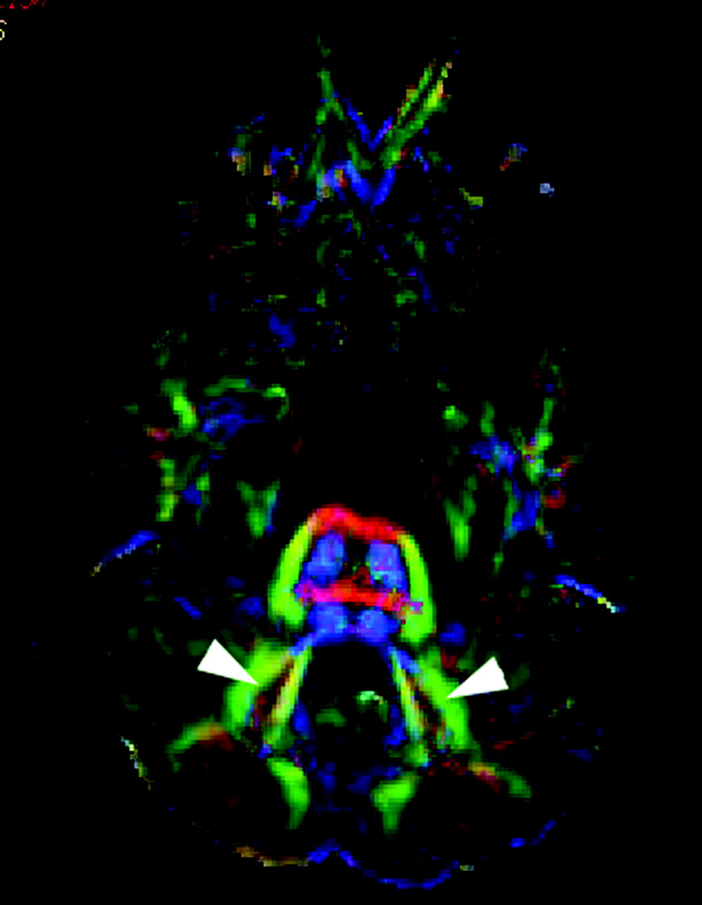
  • Fig 3.
    • Download figure
    • Open in new tab
    • Download powerpoint
    Fig 3.

    Color-coded FA-maps at the level of the dentate nuclei. In JS, the dentate nuclei are significantly lateralized (arrowheads).

  • Fig 4.
    • Download figure
    • Open in new tab
    • Download powerpoint
    Fig 4.

    Color-coded FA-maps of the decussation of the pyramidal tracts. A, In a healthy subject, the transverse orientation of the decussating fibers of the pyramidal tracts can be identified as a “red dot” within the caudal medulla (arrows). B, In JS, the “red dot” is missing, indicating that the pyramidal tracts do not cross within the caudal medulla. C, Fiber tractography displays the course of the pyramidal tracts (blue encoded) in a coronal projection. No crossing fibers could be identified, and the pyramidal tracts show a parallel course within the caudal medulla. A group of the noncrossing fibers within the superior cerebellar peduncles are also displayed on the left side (green encoded). An anatomic axial section is projected within the display for orientation purposes. D, In a healthy subject, fiber tractography displays the normal course of the pyramidal tracts (blue encoded) in a coronal projection. A partially red-encoded pyramidal decussation is seen at the level of the caudal medulla (large arrows). The red-encoded decussation of the superior cerebellar peduncles is seen at the level of the mesencephalon (arrowheads). In addition, multiple red-encoded crossing fibers are seen at the level of the pons (small arrows).

Tables

  • Figures
    • View popup
    Table 1:

    Clinical and genetic findings in 6 patients with Joubert syndrome (JS)

    Patient1234*5*6
    Age (y)272626181610
    OriginSwissSwissSwissTurkishTurkishSwiss
    Parental consanguinity+−−+++
        CNSAT, OMA, CIAT, OMA, CIAT, OMA, CIAT, OMA, CIAT, OMAb, CIAT, OMA, CI
    Features
        OcularPRnornorPRPRnor
        KidneynornornorNPHPNPHPnor
    Genetic formJBTS3not knownnot knownJBTS5JBTS5JBTS1
    • Note:— + indicates present; −, absent; AT, ataxia; OMA, ocular motor apraxia; CI, cognitive impairment; PR, pigmentary retinopathy; NPHP, nephronophthisis; nor, normal.

    • * Siblings.

    • View popup
    Table 2:

    Structural MR and diffusion tensor imaging findings in six patients with Joubert syndrome

    PatientVermisMTSSuperior Cerebellar PedunclesLocation of the Deep Cerebellar NucleiPyramidal Tract Decussation
    DecussationConfiguration
    1<1/3*+−HorizontalLateralized−
    2<1/3*+−HorizontalLateralized−
    3<1/3*+−HorizontalLateralized−
    4<1/3*+−HorizontalLateralized−
    5<2/3†+−HorizontalQuest, lateralized−
    6<1/3*+−HorizontalLateralized−
    • Note:—+ indicates present; −, absent; MTS, molar tooth sign; quest., questionably.

    • * Cerebellar vermis present only as far as the fissura prima.

    • † Cerebellar vermis present only as far as the fissura secunda.

PreviousNext
Back to top

In this issue

American Journal of Neuroradiology: 28 (10)
American Journal of Neuroradiology
Vol. 28, Issue 10
November 2007
  • Table of Contents
  • Index by author
Advertisement
Print
Download PDF
Email Article

Thank you for your interest in spreading the word on American Journal of Neuroradiology.

NOTE: We only request your email address so that the person you are recommending the page to knows that you wanted them to see it, and that it is not junk mail. We do not capture any email address.

Enter multiple addresses on separate lines or separate them with commas.
Diffusion Tensor Imaging in Joubert Syndrome
(Your Name) has sent you a message from American Journal of Neuroradiology
(Your Name) thought you would like to see the American Journal of Neuroradiology web site.
CAPTCHA
This question is for testing whether or not you are a human visitor and to prevent automated spam submissions.
Cite this article
A. Poretti, E. Boltshauser, T. Loenneker, E.M. Valente, F. Brancati, K. Il'Yasov, T.A.G.M. Huisman
Diffusion Tensor Imaging in Joubert Syndrome
American Journal of Neuroradiology Nov 2007, 28 (10) 1929-1933; DOI: 10.3174/ajnr.A0703

Citation Manager Formats

  • BibTeX
  • Bookends
  • EasyBib
  • EndNote (tagged)
  • EndNote 8 (xml)
  • Medlars
  • Mendeley
  • Papers
  • RefWorks Tagged
  • Ref Manager
  • RIS
  • Zotero
0 Responses
Respond to this article
Share
Bookmark this article
Diffusion Tensor Imaging in Joubert Syndrome
A. Poretti, E. Boltshauser, T. Loenneker, E.M. Valente, F. Brancati, K. Il'Yasov, T.A.G.M. Huisman
American Journal of Neuroradiology Nov 2007, 28 (10) 1929-1933; DOI: 10.3174/ajnr.A0703
del.icio.us logo Twitter logo Facebook logo Mendeley logo
  • Tweet Widget
  • Facebook Like
  • Google Plus One
Purchase

Jump to section

  • Article
    • Abstract
    • Patients and Methods
    • Results
    • Discussion
    • Conclusion
    • Acknowledgments
    • References
  • Figures & Data
  • Info & Metrics
  • Responses
  • References
  • PDF

Related Articles

  • No related articles found.
  • PubMed
  • Google Scholar

Cited By...

  • Pituitary Gland Duplication Syndrome: An International Imaging Analysis
  • Medullary Tegmental Cap Dysplasia: Fetal and Postnatal Presentations of a Unique Brainstem Malformation
  • Smoothened and ARL13B are critical in mouse for superior cerebellar peduncle targeting
  • Tractography of the Cerebellar Peduncles in Second- and Third-Trimester Fetuses
  • Anterior Mesencephalic Cap Dysplasia: Novel Brain Stem Malformative Features Associated with Joubert Syndrome
  • Arl13b regulates Shh signaling from both inside and outside the cilium
  • Diffusion Tractography Biomarkers of Pediatric Cerebellar Hypoplasia/Atrophy: Preliminary Results Using Constrained Spherical Deconvolution
  • Uncrossed epileptic seizures in Joubert syndrome
  • Joubert Syndrome and Related Disorders: Spectrum of Neuroimaging Findings in 75 Patients
  • Interpeduncular Heterotopia in Joubert Syndrome: A Previously Undescribed MR Finding
  • Human Genetic Disorders of Axon Guidance
  • Exceptions to the Valsalva doctrine
  • Eye Movement Abnormalities in Joubert Syndrome
  • Crossref (115)
  • Google Scholar

This article has been cited by the following articles in journals that are participating in Crossref Cited-by Linking.

  • Motile and non‐motile cilia in human pathology: from function to phenotypes
    Hannah M Mitchison, Enza Maria Valente
    The Journal of Pathology 2017 241 2
  • Joubert syndrome: congenital cerebellar ataxia with the molar tooth
    Marta Romani, Alessia Micalizzi, Enza Maria Valente
    The Lancet Neurology 2013 12 9
  • Primary Cilia in the Developing and Mature Brain
    Alicia Guemez-Gamboa, Nicole G. Coufal, Joseph G. Gleeson
    Neuron 2014 82 3
  • A developmental and genetic classification for midbrain-hindbrain malformations
    A. J. Barkovich, K. J. Millen, W. B. Dobyns
    Brain 2009 132 12
  • The corticospinal tract: Evolution, development, and human disorders
    Quentin Welniarz, Isabelle Dusart, Emmanuel Roze
    Developmental Neurobiology 2017 77 7
  • Cilia in the CNS: The Quiet Organelle Claims Center Stage
    Angeliki Louvi, Elizabeth A. Grove
    Neuron 2011 69 6
  • Arl13b in Primary Cilia Regulates the Migration and Placement of Interneurons in the Developing Cerebral Cortex
    Holden Higginbotham, Tae-Yeon Eom, Laura E. Mariani, Amelia Bachleda, Joshua Hirt, Vladimir Gukassyan, Corey L. Cusack, Cary Lai, Tamara Caspary, E.S. Anton
    Developmental Cell 2012 23 5
  • Clinical and molecular features of Joubert syndrome and related disorders
    Melissa A. Parisi
    American Journal of Medical Genetics Part C: Seminars in Medical Genetics 2009 151C 4
  • Congenital Abnormalities of the Posterior Fossa
    Thangamadhan Bosemani, Gunes Orman, Eugen Boltshauser, Aylin Tekes, Thierry A. G. M. Huisman, Andrea Poretti
    RadioGraphics 2015 35 1
  • Joubert Syndrome: Insights Into Brain Development, Cilium Biology, and Complex Disease
    Dan Doherty
    Seminars in Pediatric Neurology 2009 16 3

More in this TOC Section

  • Neuroimaging Delineation and Progression of SLSMD
  • fetal brain development of 10 weeks gestation
  • CHARGE fetal MRI clival cleft
Show more PEDIATRIC NEUROIMAGING

Similar Articles

Advertisement

Indexed Content

  • Current Issue
  • Accepted Manuscripts
  • Article Preview
  • Past Issues
  • Editorials
  • Editor's Choice
  • Fellows' Journal Club
  • Letters to the Editor
  • Video Articles

Cases

  • Case Collection
  • Archive - Case of the Week
  • Archive - Case of the Month
  • Archive - Classic Case

More from AJNR

  • Trainee Corner
  • Imaging Protocols
  • MRI Safety Corner
  • Book Reviews

Multimedia

  • AJNR Podcasts
  • AJNR Scantastics

Resources

  • Turnaround Time
  • Submit a Manuscript
  • Submit a Video Article
  • Submit an eLetter to the Editor/Response
  • Manuscript Submission Guidelines
  • Statistical Tips
  • Fast Publishing of Accepted Manuscripts
  • Graphical Abstract Preparation
  • Imaging Protocol Submission
  • Evidence-Based Medicine Level Guide
  • Publishing Checklists
  • Author Policies
  • Become a Reviewer/Academy of Reviewers
  • News and Updates

About Us

  • About AJNR
  • Editorial Board
  • Editorial Board Alumni
  • Alerts
  • Permissions
  • Not an AJNR Subscriber? Join Now
  • Advertise with Us
  • Librarian Resources
  • Feedback
  • Terms and Conditions
  • AJNR Editorial Board Alumni

American Society of Neuroradiology

  • Not an ASNR Member? Join Now

© 2025 by the American Society of Neuroradiology All rights, including for text and data mining, AI training, and similar technologies, are reserved.
Print ISSN: 0195-6108 Online ISSN: 1936-959X

Powered by HighWire