Advertisement

Main menu

  • Home
  • Content
    • Current Issue
    • Accepted Manuscripts
    • Article Preview
    • Past Issue Archive
    • Video Articles
    • AJNR Case Collection
    • Case of the Week Archive
    • Case of the Month Archive
    • Classic Case Archive
  • Special Collections
    • Advancing NeuroMRI with High-Relaxivity Contrast Agents
    • AJNR Awards
    • Low-Field MRI
    • Alzheimer Disease
    • ASNR Foundation Special Collection
    • Photon-Counting CT
    • View All
  • Multimedia
    • AJNR Podcasts
    • AJNR SCANtastic
    • Trainee Corner
    • MRI Safety Corner
    • Imaging Protocols
  • For Authors
    • Submit a Manuscript
    • Submit a Video Article
    • Submit an eLetter to the Editor/Response
    • Manuscript Submission Guidelines
    • Statistical Tips
    • Fast Publishing of Accepted Manuscripts
    • Graphical Abstract Preparation
    • Imaging Protocol Submission
    • Author Policies
  • About Us
    • About AJNR
    • Editorial Board
    • Editorial Board Alumni
  • More
    • Become a Reviewer/Academy of Reviewers
    • Subscribers
    • Permissions
    • Alerts
    • Feedback
    • Advertisers
    • ASNR Home

User menu

  • Alerts
  • Log in

Search

  • Advanced search
American Journal of Neuroradiology

American Journal of Neuroradiology

ASHNR American Society of Functional Neuroradiology ASHNR American Society of Pediatric Neuroradiology ASSR
  • Alerts
  • Log in

Advanced Search

  • Home
  • Content
    • Current Issue
    • Accepted Manuscripts
    • Article Preview
    • Past Issue Archive
    • Video Articles
    • AJNR Case Collection
    • Case of the Week Archive
    • Case of the Month Archive
    • Classic Case Archive
  • Special Collections
    • Advancing NeuroMRI with High-Relaxivity Contrast Agents
    • AJNR Awards
    • Low-Field MRI
    • Alzheimer Disease
    • ASNR Foundation Special Collection
    • Photon-Counting CT
    • View All
  • Multimedia
    • AJNR Podcasts
    • AJNR SCANtastic
    • Trainee Corner
    • MRI Safety Corner
    • Imaging Protocols
  • For Authors
    • Submit a Manuscript
    • Submit a Video Article
    • Submit an eLetter to the Editor/Response
    • Manuscript Submission Guidelines
    • Statistical Tips
    • Fast Publishing of Accepted Manuscripts
    • Graphical Abstract Preparation
    • Imaging Protocol Submission
    • Author Policies
  • About Us
    • About AJNR
    • Editorial Board
    • Editorial Board Alumni
  • More
    • Become a Reviewer/Academy of Reviewers
    • Subscribers
    • Permissions
    • Alerts
    • Feedback
    • Advertisers
    • ASNR Home
  • Follow AJNR on Twitter
  • Visit AJNR on Facebook
  • Follow AJNR on Instagram
  • Join AJNR on LinkedIn
  • RSS Feeds

AJNR Awards, New Junior Editors, and more. Read the latest AJNR updates


Improved Turnaround Times | Median time to first decision: 12 days

Research ArticleNeurointervention

Carotid Blowout Syndrome in Patients with Head-and-Neck Cancers: Reconstructive Management by Self-Expandable Stent-Grafts

F.-C. Chang, J.-F. Lirng, C.-B. Luo, W.-Y. Guo, M.M.H. Teng, S.-K. Tai and C.-Y. Chang
American Journal of Neuroradiology January 2007, 28 (1) 181-188;
F.-C. Chang
  • Find this author on Google Scholar
  • Find this author on PubMed
  • Search for this author on this site
J.-F. Lirng
  • Find this author on Google Scholar
  • Find this author on PubMed
  • Search for this author on this site
C.-B. Luo
  • Find this author on Google Scholar
  • Find this author on PubMed
  • Search for this author on this site
W.-Y. Guo
  • Find this author on Google Scholar
  • Find this author on PubMed
  • Search for this author on this site
M.M.H. Teng
  • Find this author on Google Scholar
  • Find this author on PubMed
  • Search for this author on this site
S.-K. Tai
  • Find this author on Google Scholar
  • Find this author on PubMed
  • Search for this author on this site
C.-Y. Chang
  • Find this author on Google Scholar
  • Find this author on PubMed
  • Search for this author on this site
  • Article
  • Figures & Data
  • Info & Metrics
  • Responses
  • References
  • PDF
Loading

Article Figures & Data

Figures

  • Tables
  • Fig 1.
    • Download figure
    • Open in new tab
    • Download powerpoint
    Fig 1.

    Patient 5.

    A, Left carotid angiogram shows a pseudoaneurysm in the bifurcation (arrow).

    B, After 3 fiber coils (arrowheads) were placed in the main trunk of the ECA, an 8 × 50-mm stent was deployed in the left carotid artery.

    C, Reconstructive CT angiography (curved multiplanar, reformatted images) of left carotid artery 2 months later shows complete obliteration of the pseudoaneurysm. Retained fiber coils in the thrombosed ECA with metallic artifact were also found (arrows).

    D and E, Contrast-enhanced axial CT scans of the head and neck 4 months later show septic thrombosis of the stent-graft with gas collection (arrow in panel D) and several brain abscesses (arrowheads in panel E).

  • Fig 2.
    • Download figure
    • Open in new tab
    • Download powerpoint
    Fig 2.

    Patient 1.

    A, Right carotid angiogram shows a ruptured carotid artery in the bifurcation (vertical arrows) and an active jet of extravasation (arrowheads) through a focal skin defect on the right side of the neck during the injection of contrast medium. Obvious long-segmental stenosis of the cervical ICA was also found (horizontal arrows).

    B, After the deployment of an 8 × 50-mm stent from the ICA to the CCA, acute thrombosis with total occlusion of the carotid artery occurred immediately.
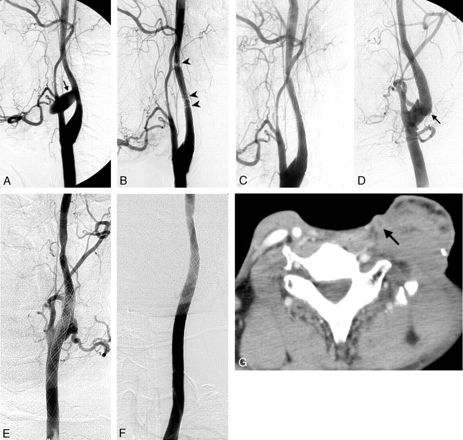
  • Fig 3.
    • Download figure
    • Open in new tab
    • Download powerpoint
    Fig 3.

    Patient 3. Left carotid angiograms. A, Pseudoaneurysm in the cervical ICA (arrow). B, Soon after a 7 × 30-mm stent was deployed in the cervical ICA, acute thrombosis was noted in the stent and its distal end (arrowheads). C, Acute thrombosis was lysed in 10 minutes by using an intravenous antiplatelet agent. D, Rebleeding due to a pseudoaneurysm in the carotid bulb was found 3 weeks later; the lesion (arrow) was just proximal to the first stent. E, A 9 × 50-mm stent was deployed in the carotid artery, which stopped the bleeding. F, Follow-up left carotid angiogram shows preserved carotid artery and complete obliteration of the pathologic lesion 1 week later. G, Six months later, axial contrast-enhanced CT scan of the neck shows thrombosis of the left carotid artery (arrow).

  • Fig 4.
    • Download figure
    • Open in new tab
    • Download powerpoint
    Fig 4.

    Patient 6.

    A, Right carotid angiogram shows a pseudoaneurysm in the right distal CCA (arrow).

    B, An 8 × 50-mm stent was deployed from the right carotid bulb to the right CCA.

    C, Another pseudoaneurysm in the right carotid bulb just distal to the distal end of the stent (arrow) was noted 2 months later.

    D, A second 8 × 50-mm stent was deployed in the right cervical ICA to overlap the first stent. Mottled gas collection (arrowheads) in the soft tissue indicates radiation necrosis.

    E, Right subclavian angiogram shows a pseudoaneurysm (arrow) in a branch of the right superior thyroid artery reconstituted from branches of the ipsilateral costocervical trunk via the ipsilateral ECA. The stent occludes the orifice of the right ECA.

    F, Direct percutaneous puncture of the main trunk of the right ECA with a spinal needle (arrowheads) was done by using a roadmap image. The needle contacted the right superior thyroid artery and showed the small pseudoaneurysm (arrow). The pseudoaneurysm was successfully embolized with a slow injection of a mixture of liquid adhesives (n-butyl cyanoacrylate and lipiodol).

Tables

  • Figures
  • Summary of patients with carotid blowout syndrome treated with self-expandable stent-grafts

    Patient No./Age (y)/SexCancer and Treatment HistoryPresentationBlowoutStent-Graft (mm)*Initial Complication†Follow-UpOutcome
    Onset (y)GroupLocation
    1/35/MMixed, submandibular gland; wide excision, R/T, C/TRecurrence, RN; lung, kidney metastasis5AcuteCBF8 × 50Asymptomatic acute ICA thrombosis and occlusionNot applicable19 d: rebleed; died
    2/43/MTonsillar; wide excision, R/T, C/TRecurrence, RN1AcuteCCA8 × 30NoneNot applicable2 d: rebleed; died
    3/52/MLaryngeal; total laryngectomy, R/T, C/TRecurrence, RN, PCF4AcuteICA7 × 30, 9 × 50Asymptomatic transient in-stent thrombosisRebleed wk 3, second stent-graft; patency on 1-mo CTA & 3-mo angiogram6-mo CT: asymptomatic carotid thrombosis; lived 20 mo
    4/49/MNasopharyngeal, R/T; second primary hypopharyngeal, R/T, C/TRecurrent hypopharyngeal cancer, RN12, 1ImpendingCBF8 × 50‡Embolic infarct, MCA territoryStent patency on 1-mo CT, sonography2-mo: died from progression
    5/52/MHypopharyngeal; R/T, C/TSlightly swollen soft tissue of neck1ImpendingCBF8 × 50‡NoneStent patency on 2-mo CTA4-mo: septic carotid thrombosis, brain, abscess; lived 14 mo
    6/65/MLaryngeal; R/TRN, PCF4AcuteCCA, CBF8 × 50, 8 × 50NoneRebleed at 2-mo, second stent-graft; rebleed at 3-mo, direct percutaneous ECA puncture for embolization; stent patency at 3-mo CT5-mo CT: asymptomatic carotid thrombosis; lived 13 mo
    7/44/MHypopharyngeal; total laryngopharyngectomy, C/T, R/TRecurrence, RN, PCF, lung metastasis1/2ThreatenedCCA9 × 70NoneStent patency on 2-wk sonography1-mo: died from lung metastasis
    8/54/MLower esophageal, primary hypopharyngeal; esophagectomy, laryngectomy, C/T, R/TRecurrence, RN, PCF1ThreatenedCCA9 × 70NoneStent patency on 2-wk CTA1.5-mo: died from progression, mediastinitis
    • Note.—CBF indicates carotid bifurcation; CCA, common carotid artery; C/T, chemotherapy; CTA, CT angiography; ECA, external carotid artery; MCA, middle cerebral artery; PCF, pharyngocutaneous fistula; RN, radiation necrosis; R/T, radiotherapy.

    • * Wallgraft; Boston Scientific Corporation.

    • † Immediate hemostasis was achieved in all patients.

    • ‡ Plus 3 fiber coils in the ECA

PreviousNext
Back to top

In this issue

American Journal of Neuroradiology: 28 (1)
American Journal of Neuroradiology
Vol. 28, Issue 1
January 2007
  • Table of Contents
  • Index by author
Advertisement
Print
Download PDF
Email Article

Thank you for your interest in spreading the word on American Journal of Neuroradiology.

NOTE: We only request your email address so that the person you are recommending the page to knows that you wanted them to see it, and that it is not junk mail. We do not capture any email address.

Enter multiple addresses on separate lines or separate them with commas.
Carotid Blowout Syndrome in Patients with Head-and-Neck Cancers: Reconstructive Management by Self-Expandable Stent-Grafts
(Your Name) has sent you a message from American Journal of Neuroradiology
(Your Name) thought you would like to see the American Journal of Neuroradiology web site.
CAPTCHA
This question is for testing whether or not you are a human visitor and to prevent automated spam submissions.
Cite this article
F.-C. Chang, J.-F. Lirng, C.-B. Luo, W.-Y. Guo, M.M.H. Teng, S.-K. Tai, C.-Y. Chang
Carotid Blowout Syndrome in Patients with Head-and-Neck Cancers: Reconstructive Management by Self-Expandable Stent-Grafts
American Journal of Neuroradiology Jan 2007, 28 (1) 181-188;

Citation Manager Formats

  • BibTeX
  • Bookends
  • EasyBib
  • EndNote (tagged)
  • EndNote 8 (xml)
  • Medlars
  • Mendeley
  • Papers
  • RefWorks Tagged
  • Ref Manager
  • RIS
  • Zotero
0 Responses
Respond to this article
Share
Bookmark this article
Carotid Blowout Syndrome in Patients with Head-and-Neck Cancers: Reconstructive Management by Self-Expandable Stent-Grafts
F.-C. Chang, J.-F. Lirng, C.-B. Luo, W.-Y. Guo, M.M.H. Teng, S.-K. Tai, C.-Y. Chang
American Journal of Neuroradiology Jan 2007, 28 (1) 181-188;
del.icio.us logo Twitter logo Facebook logo Mendeley logo
  • Tweet Widget
  • Facebook Like
  • Google Plus One

Jump to section

  • Article
    • Abstract
    • Methods
    • Results
    • Discussion
    • Conclusion
    • References
  • Figures & Data
  • Info & Metrics
  • Responses
  • References
  • PDF

Related Articles

  • No related articles found.
  • PubMed
  • Google Scholar

Cited By...

  • Endovascular management of intracranial carotid blowout syndrome in patients with head and neck cancer
  • Republished: Delayed extrusion of embolic coils into the airway after embolization of an external carotid artery pseudoaneurysm
  • Delayed extrusion of embolic coils into the airway after embolization of an external carotid artery pseudoaneurysm
  • Endovascular therapy of extracranial carotid artery pseudoaneurysms: case series and literature review
  • Acute Bleeding in the Head and Neck: Angiographic Findings and Endovascular Management
  • Covered stents safely utilized to prevent catastrophic hemorrhage in patients with advanced head and neck malignancy
  • Endovascular treatment of carotid blowout syndrome: who and how to treat
  • Crossref
  • Google Scholar

This article has not yet been cited by articles in journals that are participating in Crossref Cited-by Linking.

More in this TOC Section

  • Prophylactic Endovascular Treatments for CBS
  • Smoking & Aneurysm Outcomes: A Meta-Analysis
  • “Buddy-wire anchoring” technique for TVE
Show more NEUROINTERVENTION

Similar Articles

Advertisement

Indexed Content

  • Current Issue
  • Accepted Manuscripts
  • Article Preview
  • Past Issues
  • Editorials
  • Editor's Choice
  • Fellows' Journal Club
  • Letters to the Editor
  • Video Articles

Cases

  • Case Collection
  • Archive - Case of the Week
  • Archive - Case of the Month
  • Archive - Classic Case

More from AJNR

  • Trainee Corner
  • Imaging Protocols
  • MRI Safety Corner
  • Book Reviews

Multimedia

  • AJNR Podcasts
  • AJNR Scantastics

Resources

  • Turnaround Time
  • Submit a Manuscript
  • Submit a Video Article
  • Submit an eLetter to the Editor/Response
  • Manuscript Submission Guidelines
  • Statistical Tips
  • Fast Publishing of Accepted Manuscripts
  • Graphical Abstract Preparation
  • Imaging Protocol Submission
  • Evidence-Based Medicine Level Guide
  • Publishing Checklists
  • Author Policies
  • Become a Reviewer/Academy of Reviewers
  • News and Updates

About Us

  • About AJNR
  • Editorial Board
  • Editorial Board Alumni
  • Alerts
  • Permissions
  • Not an AJNR Subscriber? Join Now
  • Advertise with Us
  • Librarian Resources
  • Feedback
  • Terms and Conditions
  • AJNR Editorial Board Alumni

American Society of Neuroradiology

  • Not an ASNR Member? Join Now

© 2025 by the American Society of Neuroradiology All rights, including for text and data mining, AI training, and similar technologies, are reserved.
Print ISSN: 0195-6108 Online ISSN: 1936-959X

Powered by HighWire