Advertisement

Main menu

  • Home
  • Content
    • Current Issue
    • Accepted Manuscripts
    • Article Preview
    • Past Issue Archive
    • Video Articles
    • AJNR Case Collection
    • Case of the Week Archive
    • Case of the Month Archive
    • Classic Case Archive
  • Special Collections
    • AJNR Awards
    • Low-Field MRI
    • Alzheimer Disease
    • ASNR Foundation Special Collection
    • Photon-Counting CT
    • View All
  • Multimedia
    • AJNR Podcasts
    • AJNR SCANtastic
    • Trainee Corner
    • MRI Safety Corner
    • Imaging Protocols
  • For Authors
    • Submit a Manuscript
    • Submit a Video Article
    • Submit an eLetter to the Editor/Response
    • Manuscript Submission Guidelines
    • Statistical Tips
    • Fast Publishing of Accepted Manuscripts
    • Graphical Abstract Preparation
    • Imaging Protocol Submission
    • Author Policies
  • About Us
    • About AJNR
    • Editorial Board
    • Editorial Board Alumni
  • More
    • Become a Reviewer/Academy of Reviewers
    • Subscribers
    • Permissions
    • Alerts
    • Feedback
    • Advertisers
    • ASNR Home

User menu

  • Alerts
  • Log in

Search

  • Advanced search
American Journal of Neuroradiology

American Journal of Neuroradiology

ASHNR American Society of Functional Neuroradiology ASHNR American Society of Pediatric Neuroradiology ASSR
  • Alerts
  • Log in

Advanced Search

  • Home
  • Content
    • Current Issue
    • Accepted Manuscripts
    • Article Preview
    • Past Issue Archive
    • Video Articles
    • AJNR Case Collection
    • Case of the Week Archive
    • Case of the Month Archive
    • Classic Case Archive
  • Special Collections
    • AJNR Awards
    • Low-Field MRI
    • Alzheimer Disease
    • ASNR Foundation Special Collection
    • Photon-Counting CT
    • View All
  • Multimedia
    • AJNR Podcasts
    • AJNR SCANtastic
    • Trainee Corner
    • MRI Safety Corner
    • Imaging Protocols
  • For Authors
    • Submit a Manuscript
    • Submit a Video Article
    • Submit an eLetter to the Editor/Response
    • Manuscript Submission Guidelines
    • Statistical Tips
    • Fast Publishing of Accepted Manuscripts
    • Graphical Abstract Preparation
    • Imaging Protocol Submission
    • Author Policies
  • About Us
    • About AJNR
    • Editorial Board
    • Editorial Board Alumni
  • More
    • Become a Reviewer/Academy of Reviewers
    • Subscribers
    • Permissions
    • Alerts
    • Feedback
    • Advertisers
    • ASNR Home
  • Follow AJNR on Twitter
  • Visit AJNR on Facebook
  • Follow AJNR on Instagram
  • Join AJNR on LinkedIn
  • RSS Feeds

AJNR Awards, New Junior Editors, and more. Read the latest AJNR updates

Research ArticleSpine Imaging and Spine Image-Guided Interventions

Symptomatic Refractures after Vertebroplasty in Patients with Steroid-Induced Osteoporosis

M.I. Syed, N.A. Patel, S. Jan, A. Shaikh, B. Grunden and K. Morar
American Journal of Neuroradiology October 2006, 27 (9) 1938-1943;
M.I. Syed
  • Find this author on Google Scholar
  • Find this author on PubMed
  • Search for this author on this site
N.A. Patel
  • Find this author on Google Scholar
  • Find this author on PubMed
  • Search for this author on this site
S. Jan
  • Find this author on Google Scholar
  • Find this author on PubMed
  • Search for this author on this site
A. Shaikh
  • Find this author on Google Scholar
  • Find this author on PubMed
  • Search for this author on this site
B. Grunden
  • Find this author on Google Scholar
  • Find this author on PubMed
  • Search for this author on this site
K. Morar
  • Find this author on Google Scholar
  • Find this author on PubMed
  • Search for this author on this site
  • Article
  • Figures & Data
  • Info & Metrics
  • Responses
  • References
  • PDF
Loading

Article Figures & Data

Figures

  • Tables
  • Fig 1.
    • Download figure
    • Open in new tab
    • Download powerpoint
    Fig 1.

    Example of patient with steroid-induced osteoporosis. Lateral thoracic spine plain-film radiograph. Initial prevertebroplasty film demonstrates T6 compression fracture.

  • Fig 2.
    • Download figure
    • Open in new tab
    • Download powerpoint
    Fig 2.

    Follow-up of the same patient. Lateral thoracic spine plain-film radiograph. One-year follow-up film demonstrates that the patient has undergone vertebroplasties of T5 through T8 and presents with a new fracture of T10 at the time of the study.
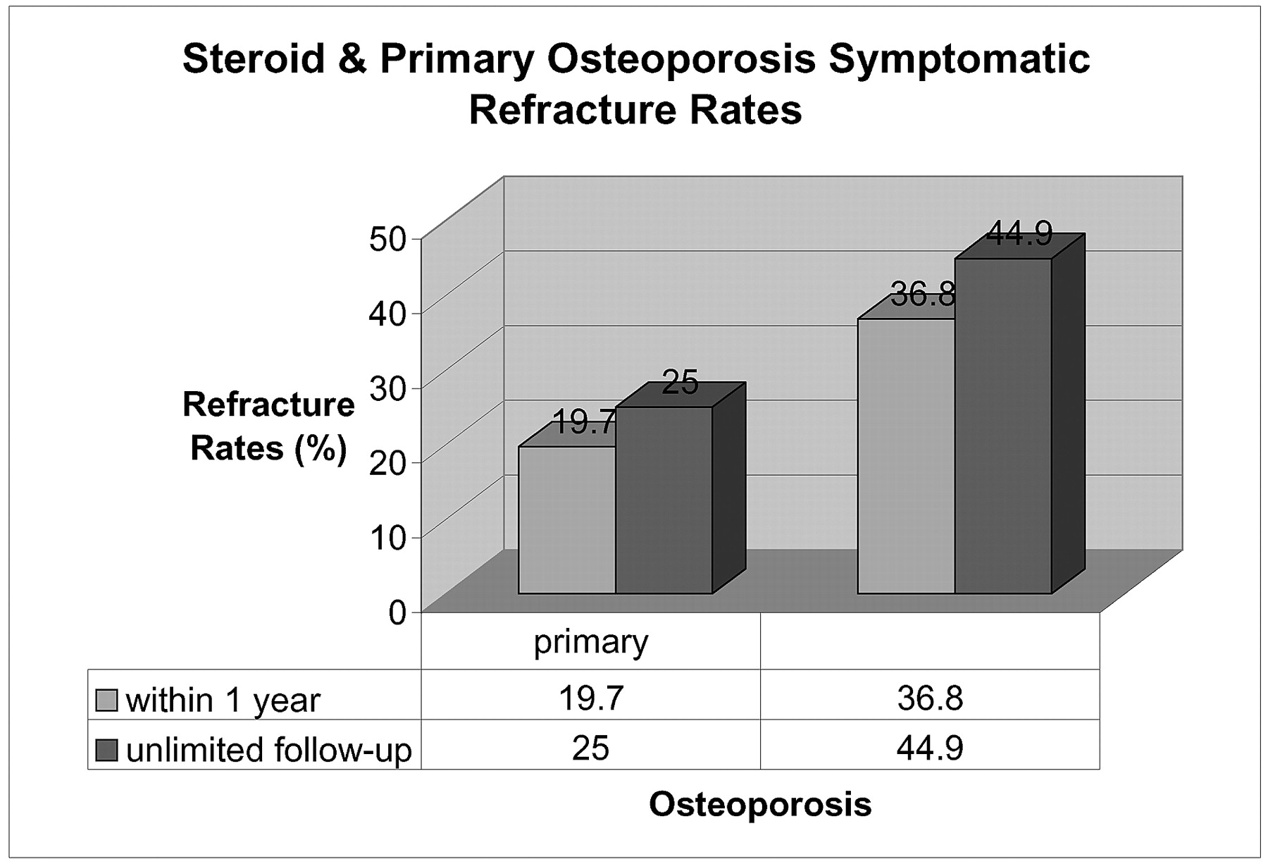
  • Fig 3.
    • Download figure
    • Open in new tab
    • Download powerpoint
    Fig 3.

    Primary and steroid-induced osteoporotic symptomatic refracture rates comparing 1-year follow-up with an extended follow-up
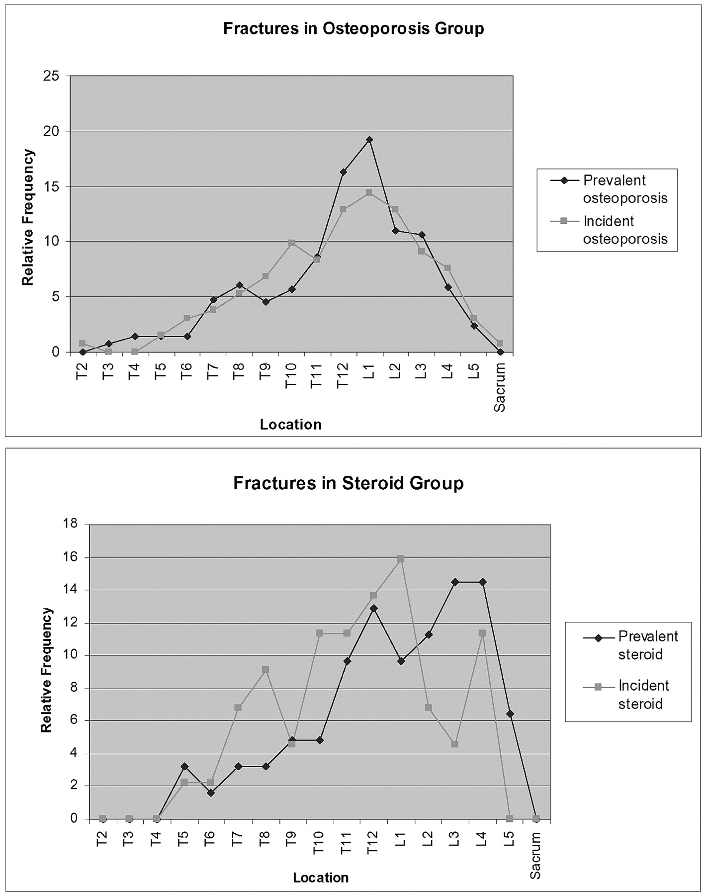
  • Fig 4.
    • Download figure
    • Open in new tab
    • Download powerpoint
    Fig 4.

    Graphs demonstrating relative frequencies of prevalent and incident fractures in both the steroid and osteoporosis groups.

Tables

  • Figures
    • View popup
    Table 1:

    Medical history of the 37 oral steroid-induced osteoporosis patients

    Medical HistoryNo. of Patients
    COPD18
    Rheumatoid arthritis5
    Asthma3
    Polymyalgia rheumatica3
    Giant cell arteritis2
    Mixed connective tissue disease1
    CNS vasculitis1
    Myasthenia gravis1
    Pulmonary fibrosis1
    Other2
    • Note:—COPD indicates chronic obstructive pulmonary disease; CNS, central nervous system.

    • View popup
    Table 2:

    Odds ratio (OR) by location of incident fracture: steroid-induced compared to primary

    Location of Incident Fracture
    T3–T11T12–L1L2–L5
    OR (95% CI)3.7 (1.56–8.62)3.0 (1.24–.7.06)1.5 (0.52–4.19)
    P value (group variable).0029.0146.4313
    • View popup
    Table 3:

    Mean number of incident fractures per patient by group and location

    LocationGroupMean No. of Incident Fractures per SubjectP-Value
    Any locationSteroid1.108.0025
    Primary0.397
    T3–T11Steroid0.568.0009
    Primary0.149
    T12–L1Steroid0.351.0025
    Primary0.114
    L2–L5Steroid0.189.2081
    Primary0.134
    • Note:—Because all median are equal to O, means have been reported instead.

    • View popup
    Table 4:

    Analysis of refractures within 1 year

    Percentage of Patients Having a Second Treatment Session Within 1 y of Initial ProcedurePercentage of Refractured Patients Having a Third Treatment Session Within 1 y of Initial ProcedureAverage Available Cement Volumes for Patients Refracuring Within 1 y of Initial Procedure (cc)Average Time Between Treatment Sessions for Refracturing Patients Within 1 y of Initial Procedures
    No. of Patients Having Initial ProcedureNo. Refractured (2nd Procedure)% RefracturingNo. Refractured (2nd Procedure)No. Refractured (3rd Procedure)% of Refractured Patients Having 3rd ProcedureΔt Between 1st and 2nd procedure wkΔt Between 2nd and 3rd procedure wk
    Primary osteoporosis3507220.6722331.9%5.18 ± 2.5811.312
    Steroid-induced osteoporosis371437.814642.9%5.97 ± 3.027.9310.8
    Total3878622.2862933.7%
    Relative risk1.841.34†
    P value.0163.3094
    • * Chi-squared analysis.

    • † Fisher exact test.

    • View popup
    Table 5:

    Prevalent vs incident fractures

    Vertebral LevelPrevalent Fractures in Steroid GroupIncident Fractures in Steroid Group (at 1 y)Prevalent Fractures in Primary Osteoporosis GroupIncident Fractures in Primary Osteoporosis Group (at 1 y)
    T20001
    T30040
    T40070
    T52172
    T61174
    T723245
    T824317
    T932239
    T10352913
    T11654411
    T12868317
    L1679819
    L2735617
    L3925412
    L4953010
    L540124
    Sacrum0001
PreviousNext
Back to top

In this issue

American Journal of Neuroradiology: 27 (9)
American Journal of Neuroradiology
Vol. 27, Issue 9
October 2006
  • Table of Contents
  • Index by author
Advertisement
Print
Download PDF
Email Article

Thank you for your interest in spreading the word on American Journal of Neuroradiology.

NOTE: We only request your email address so that the person you are recommending the page to knows that you wanted them to see it, and that it is not junk mail. We do not capture any email address.

Enter multiple addresses on separate lines or separate them with commas.
Symptomatic Refractures after Vertebroplasty in Patients with Steroid-Induced Osteoporosis
(Your Name) has sent you a message from American Journal of Neuroradiology
(Your Name) thought you would like to see the American Journal of Neuroradiology web site.
CAPTCHA
This question is for testing whether or not you are a human visitor and to prevent automated spam submissions.
Cite this article
M.I. Syed, N.A. Patel, S. Jan, A. Shaikh, B. Grunden, K. Morar
Symptomatic Refractures after Vertebroplasty in Patients with Steroid-Induced Osteoporosis
American Journal of Neuroradiology Oct 2006, 27 (9) 1938-1943;

Citation Manager Formats

  • BibTeX
  • Bookends
  • EasyBib
  • EndNote (tagged)
  • EndNote 8 (xml)
  • Medlars
  • Mendeley
  • Papers
  • RefWorks Tagged
  • Ref Manager
  • RIS
  • Zotero
0 Responses
Respond to this article
Share
Bookmark this article
Symptomatic Refractures after Vertebroplasty in Patients with Steroid-Induced Osteoporosis
M.I. Syed, N.A. Patel, S. Jan, A. Shaikh, B. Grunden, K. Morar
American Journal of Neuroradiology Oct 2006, 27 (9) 1938-1943;
del.icio.us logo Twitter logo Facebook logo Mendeley logo
  • Tweet Widget
  • Facebook Like
  • Google Plus One

Jump to section

  • Article
    • Abstract
    • Methods
    • Statistical Methods
    • Results
    • Discussion
    • Conclusions
    • References
  • Figures & Data
  • Info & Metrics
  • Responses
  • References
  • PDF

Related Articles

  • No related articles found.
  • PubMed
  • Google Scholar

Cited By...

  • Subsequent Fracture after Percutaneous Vertebroplasty Can Be Predicted on Preoperative Multidetector Row CT
  • Crossref
  • Google Scholar

This article has not yet been cited by articles in journals that are participating in Crossref Cited-by Linking.

More in this TOC Section

  • Cone Beam CT Myelography
  • Cone Beam CTM for detecting CSF-venous Fistulas
  • Post-Procedural Brachial Neuritis Features
Show more Spine Imaging and Spine Image-Guided Interventions

Similar Articles

Advertisement

Indexed Content

  • Current Issue
  • Accepted Manuscripts
  • Article Preview
  • Past Issues
  • Editorials
  • Editor's Choice
  • Fellows' Journal Club
  • Letters to the Editor
  • Video Articles

Cases

  • Case Collection
  • Archive - Case of the Week
  • Archive - Case of the Month
  • Archive - Classic Case

More from AJNR

  • Trainee Corner
  • Imaging Protocols
  • MRI Safety Corner

Multimedia

  • AJNR Podcasts
  • AJNR Scantastics

Resources

  • Turnaround Time
  • Submit a Manuscript
  • Submit a Video Article
  • Submit an eLetter to the Editor/Response
  • Manuscript Submission Guidelines
  • Statistical Tips
  • Fast Publishing of Accepted Manuscripts
  • Graphical Abstract Preparation
  • Imaging Protocol Submission
  • Evidence-Based Medicine Level Guide
  • Publishing Checklists
  • Author Policies
  • Become a Reviewer/Academy of Reviewers
  • News and Updates

About Us

  • About AJNR
  • Editorial Board
  • Editorial Board Alumni
  • Alerts
  • Permissions
  • Not an AJNR Subscriber? Join Now
  • Advertise with Us
  • Librarian Resources
  • Feedback
  • Terms and Conditions
  • AJNR Editorial Board Alumni

American Society of Neuroradiology

  • Not an ASNR Member? Join Now

© 2025 by the American Society of Neuroradiology All rights, including for text and data mining, AI training, and similar technologies, are reserved.
Print ISSN: 0195-6108 Online ISSN: 1936-959X

Powered by HighWire