
Article Figures & Data
Figures
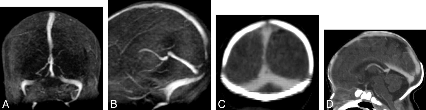
- Fig 1.
Coronal (A) and sagittal (B) views from maximum intensity projection (MIP) of 2D TOF MRV demonstrate signal intensity void in the posterior aspect of superior sagittal sinus and medial aspect of transverse sinuses. On the coronal (C) and sagittal (D) reformatted images of the CTV, there is normal flow seen in the superior sagittal sinus and transverse sinuses, which confirms that the signal intensity void seen in the superior sagittal and transverse sinuses is artifactual.
- Fig 2.
A, Axial T2 shows loss of the normal flow void in the right transverse sinus (arrows). B, Coronal view of MIP from 2D TOF MRV shows loss of signal intensity in the right and left transverse sinuses. C, Sagittal view of MIP from 2D TOF MRV shows loss of signal intensity in the posterior aspect of superior sagittal sinus (arrow), which is clearly demonstrated despite image degradation by motion. The appearance is suspicious of artifactual flow gap in the transverse sinuses and superior sagittal sinus caused by in-plane saturation, possibly exacerbated by skull molding. Coronal (D) and sagittal (E) reformatted images of CTV show that the transverse and superior sagittal sinuses are patent. This confirms that the flow gaps seen in the venous sinuses on TOF MRV are artifactual. There is, however, persistent narrowing of the superior sagittal sinus in the region where the lambdoid sutures converge (arrowhead), which suggests that skull molding may play a role in narrowing the caliber of the superior sagittal sinus, thereby increasing the likelihood of demonstrating flow gaps in the superior sagittal sinus on 2D TOF MRV in neonates.
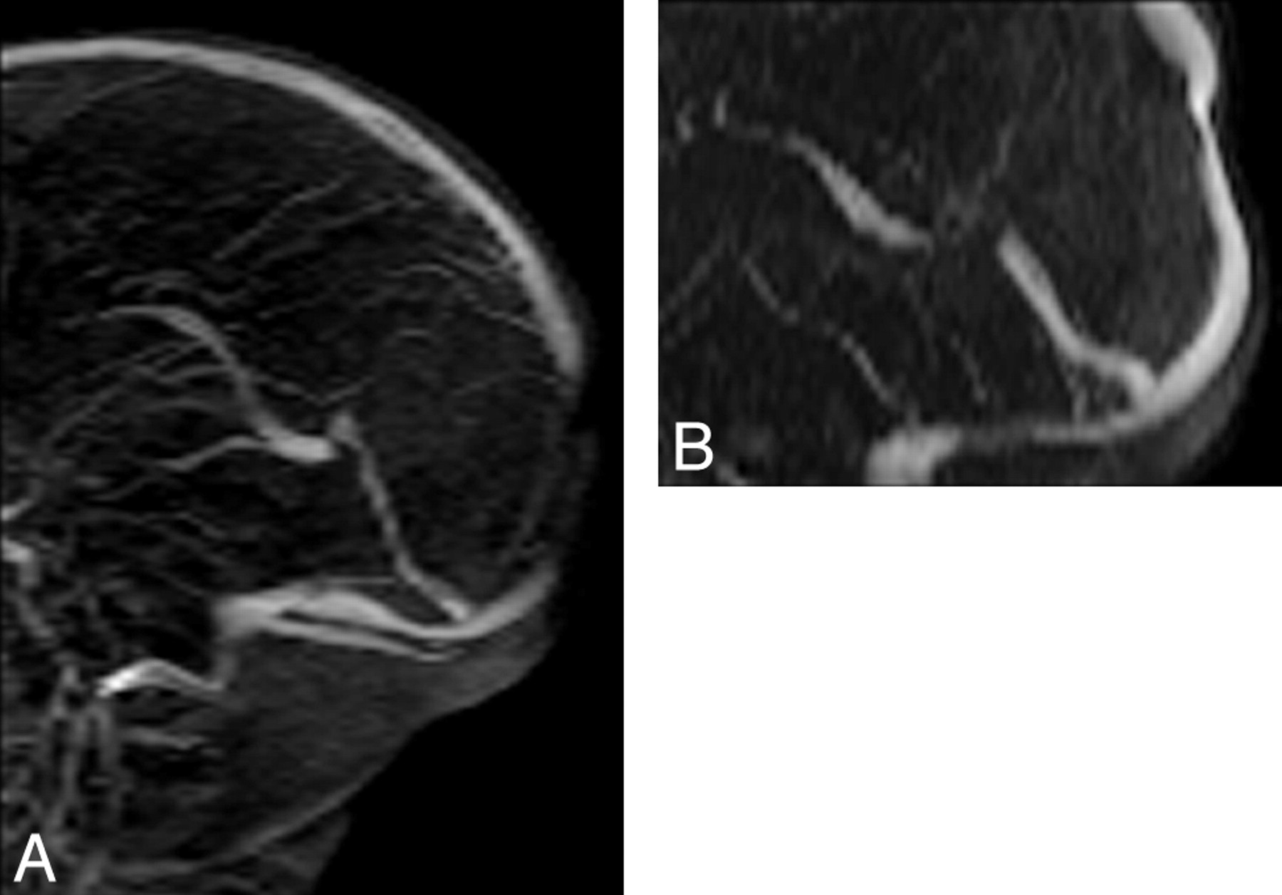
- Fig 3.
A, Flow gap is seen in the posterior aspect of superior sagittal sinus on the coronal 2D TOF MRV. This flow gap is artifactual as a result of in-plane saturation of the imaging plane. This effect is negated by changing the plane of imaging. B, On the axial 2D TOF MRV, there is flow in the superior sagittal sinus. There is short segment stenosis of the superior sagittal sinus on axial 2D TOF MRV, in the region of the convergence of lambdoid suture, possibly as a result of skull molding and pressure effect in the supine position.
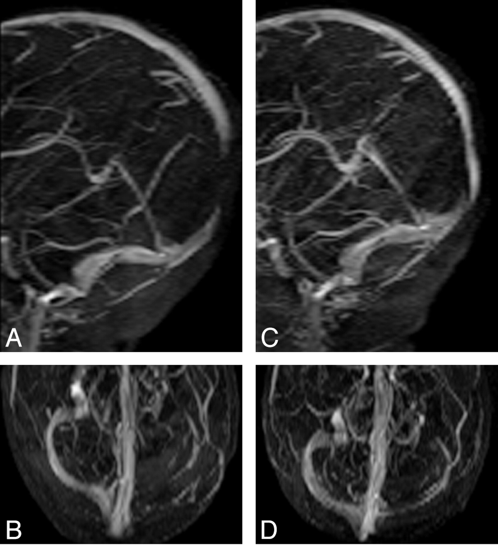
- Fig 4.
A and B, MIP images of coronal 2D TOF MRV acquired with TE of 5.5 ms demonstrate flow gap in the superior sagittal sinus and left transverse sinus. The flow gap is artifactual as a result of in-plane saturation. C and D, MIP images of coronal 2D TOF MRV acquired with TE of 4.9 ms. Flow gap due to in-plane saturation is minimized by using shorter TE (4.9 ms) and thereby improves visualization of superior sagittal and transverse sinus. The left transverse sinus is smaller compared with the right transverse sinus but patent, possibly due to congenitally smaller caliber left transverse sinus.


Tables
Flow void, stenosis, or absence of venous sinuses detected in 9 neonates who have had coronal 2D time-of-flight MR venography (TOF MRV) and CT venography (CTV)
Coronal 2D TOF MRV ON MIP Images Coronal 2D TOF MRV on Source Images CTV Superior sagittal sinus Narrowing 2 2 2 Flow gap 6 3 0 Right transverse sinus Flow gap 3 2 0 Absent 1 1 1 Left transverse sinus Flow gap 6 4 0 Right sigmoid sinus Flow gap 3 2 0 Left sigmoid sinus Flow gap 2 1 0 Right internal jugular vein Flow gap 0 0 0 Left internal jugular vein Flow gap 0 0 0 Note:—MIP indicates maximum intensity projections.


