
Article Figures & Data
Figures
- Fig 1.
Midline sagittal SS-FSE T2-weighted image demonstrates the normal appearance of the corpus callosum in a 26-gestational-week-old fetus (arrow). Obtaining a nonoblique midline sagittal image is critical for evaluating the corpus callosum. Note the uniform thickness of the corpus callosum.
- Fig 2.
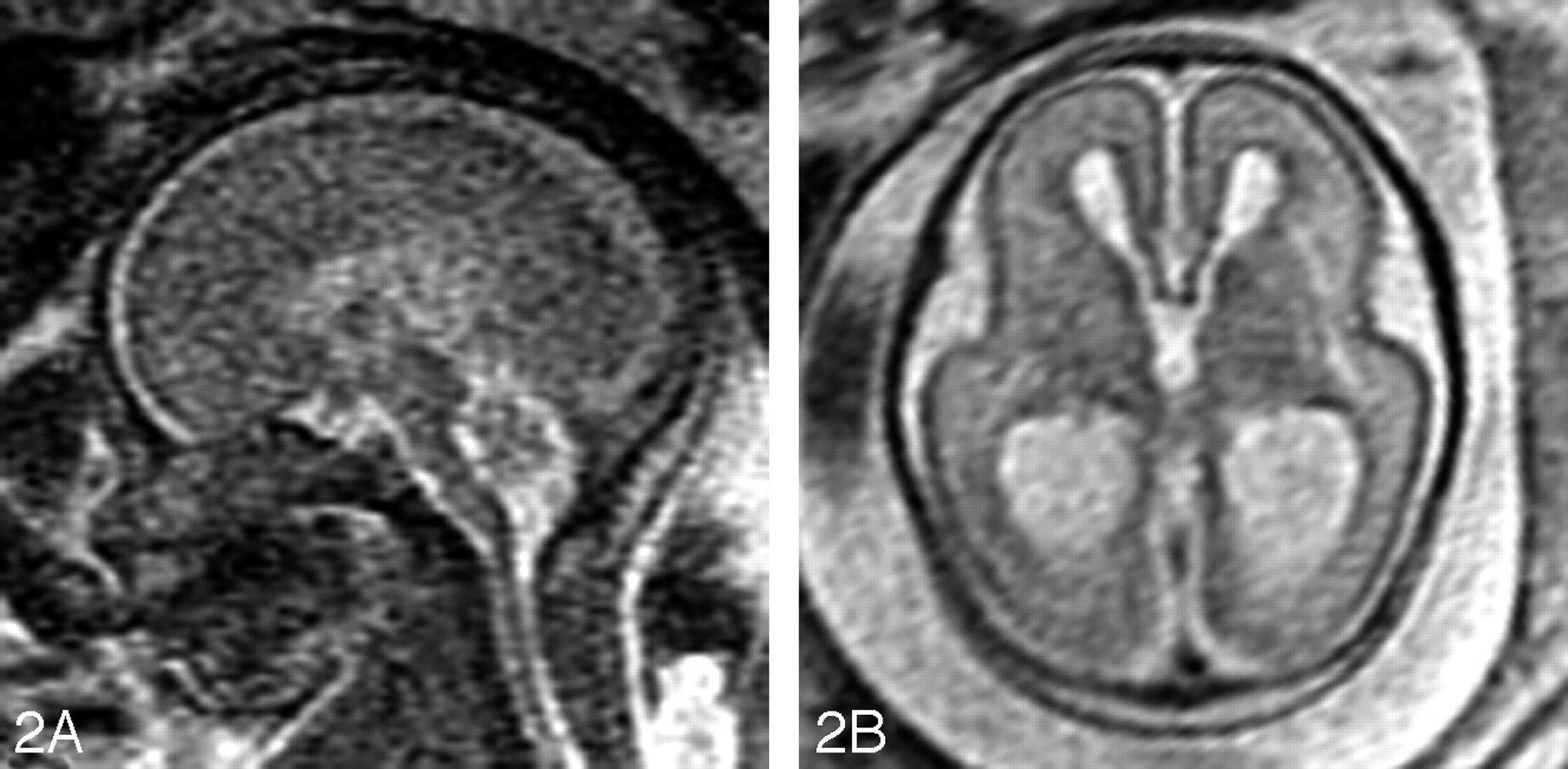
A, Sagittal SS-FSE T2-weighted image in a 25-gestational-week-old fetus demonstrates agenesis of the corpus callosum as well as a small pons.
B, Axial SS-FSE T2-weighted image demonstrates abnormally shallow Sylvian fissures for gestational age.
- Fig 3.
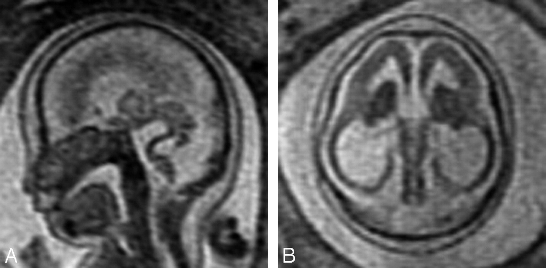
A, Axial SS-FSE T2-weighted image in a 19-gestational-week-old fetus referred for callosal agenesis demonstrates thinning of the right frontal parenchyma with several abnormal infoldings of the developing cortex (arrow) consistent with polymicrogyria.
B, Coronal SS-FSE T2-weighted image confirms multiple abnormal cortical infoldings (black arrow) and also demonstrates several areas of nodularity (white arrow) along the dilated right frontal horn consistent with periventricular nodular heterotopia. Microphthalmia of the right globe (arrowhead) is also present.
C, Axial SS-FSE T2-weighted image demonstrates abnormal morphology of the fourth ventricle with small, dysgenetic cerebellar hemispheres bilaterally. This constellation of findings led to the prenatal diagnosis of Aicardi syndrome.
- Fig 4.
Sagittal SS-FSE T2-weighted image in a 33-gestational-week-old fetus demonstrates a large cisterna magna. The vermis is identified and is normal in size, excluding the diagnosis of a Dandy-Walker malformation.
- Fig 5.
A, Sagittal SS-FSE T2-weighted image in a 29-gestational-week-old fetus demonstrates hypoplasia of the vermis.
B, Coronal SS-FSE T2-weighted image demonstrates small, slightly dysmorphic left cerebellar hemisphere (arrow).
- Fig 6.
A, Sagittal SS-FSE T2-weighted image in a 23-gestational-week-old fetus demonstrates abnormally thin brain stem with kinking at pontomesencephalic junction. Callosal and vermian agenesis was detected by prenatal sonography and is also present on the MR imaging.
B, Axial SS-FSE T2-weighted image demonstrates abnormally thin and homogeneous parenchyma with absent Sylvian fissures. The deep gray nuclei appear small and dysplastic as well.
This constellation of findings led to the prenatal diagnosis of Walker-Warburg syndrome.
- Fig 7.
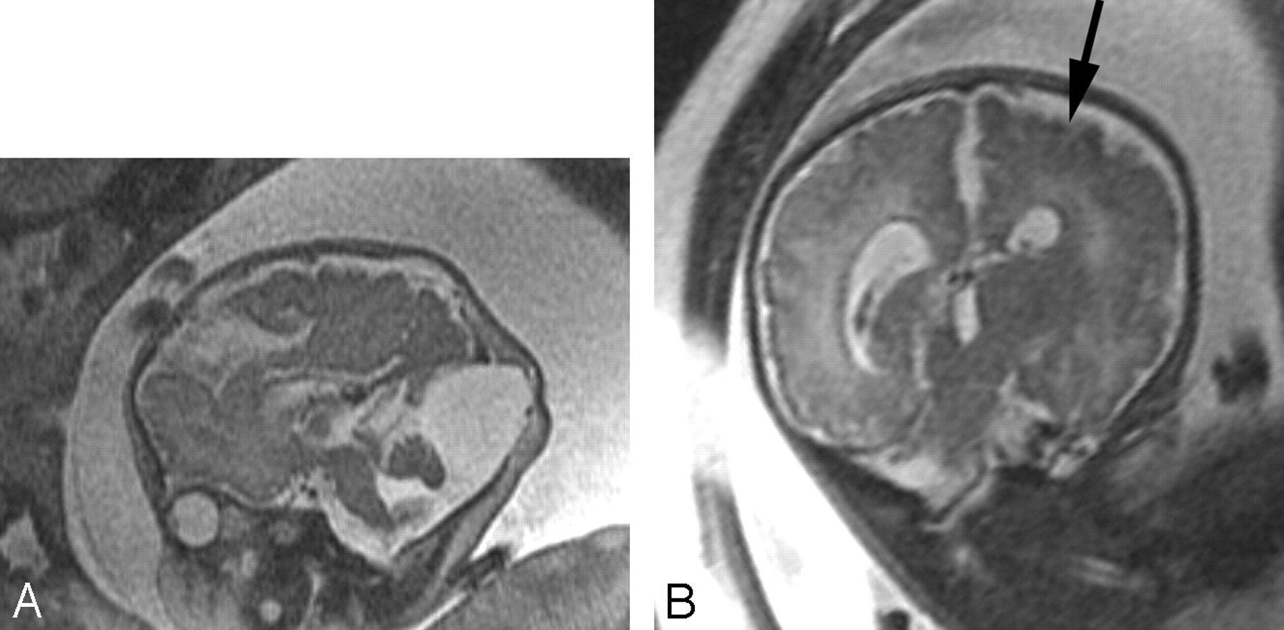
A, Sagittal SS-FSE T2-weighted image in 39-gestational-week-old fetus demonstrates vermian hypoplasia and enlarged posterior fossa with cyst, consistent with a Dandy-Walker malformation. An occipital meningocele is also identified.
B, Coronal SS-FSE T2-weighted image demonstrates diffusely abnormal sulcal pattern for gestational age, with areas of polymicrogyria (arrow). There is associated diminished white matter volume and ventriculomegaly.
- Fig 8.
A, Axial SS-FSE T2-weighted image demonstrates an area of increased signal intensity with peripheral decreased signal intensity in the right cerebellar hemisphere (arrow).
B, Axial FMP-SPGR T1-weighted image at corresponding level shows areas of increased signal intensity consistent with cerebellar hemorrhage (arrow). Bilobed appearance of the hematoma on both T1- and T2-weighted images suggests possible involvement of the cerebellar vermis.
- Fig 9.
SSFSE T2-weighted image of 24-gestational-week-old fetus with a large hypointense hematoma in the posterior fossa on both axial (A) and coronal (B) views. The hematoma displaces the cerebellum anteriorly and appears subdural in location. An ovoid more hypointense area is concerning for an abnormal vessel. A dural arteriovenous fistula was found at autopsy.
- Fig 10.
Coronal SSFSE T2-weighted image in a 33-gestational-week-old fetus demonstrates bilateral open lip schizencephalic defects (arrows). The adjacent sulcal pattern is abnormal. The septum pellucidum is absent.
- Fig 11.
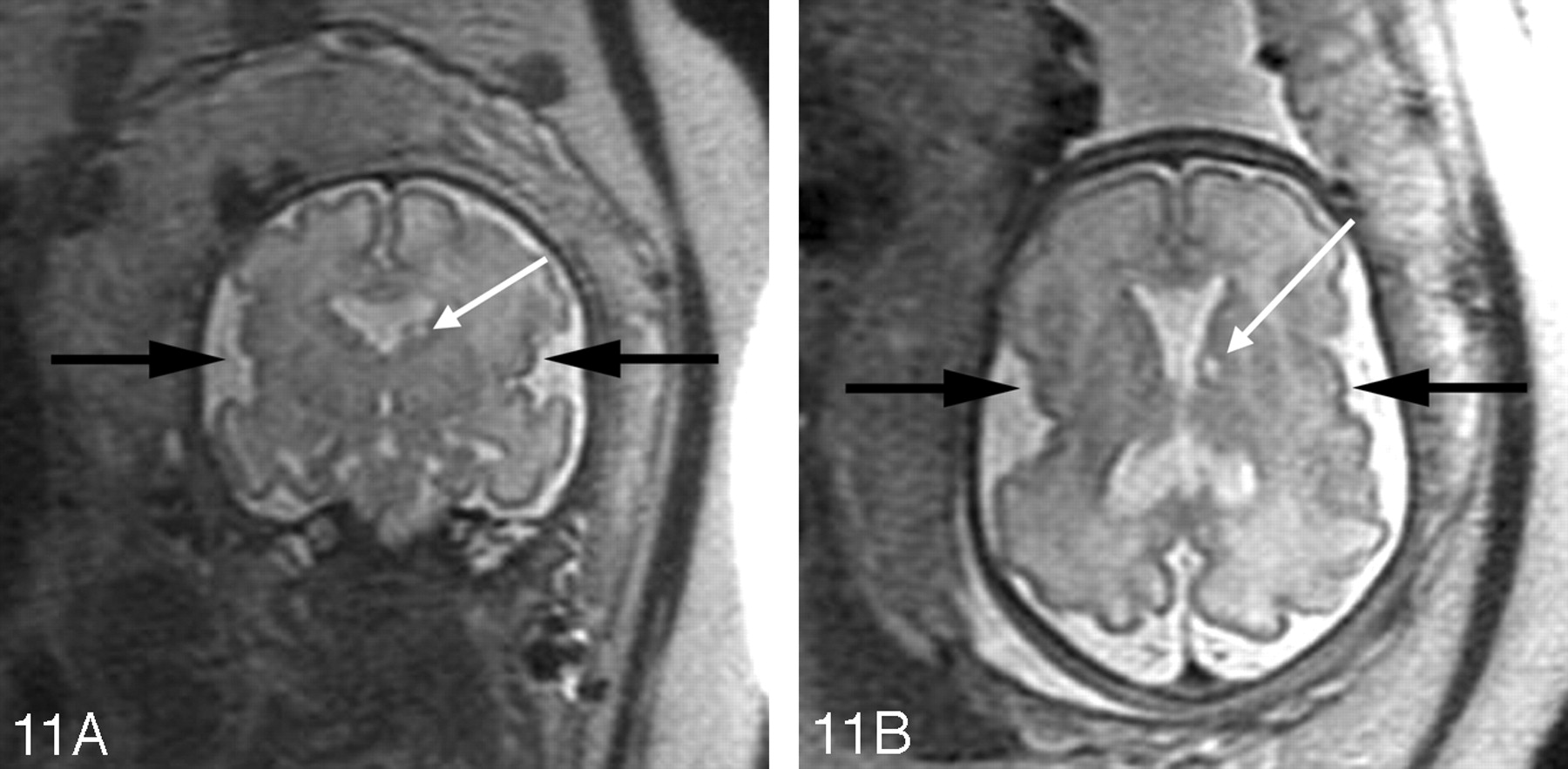
A, Coronal SS-FSE T2-weighted image in a 30-gestational-week-old fetus demonstrates dysplastic-appearing Sylvian fissures with multiple abnormal small infoldings of the cortex (black arrows) consistent with perisylvian polymicrogyria. Absence of the septum pellucidum is present, and was noted on the prenatal sonogram. A hyperintense focus (white arrow) is seen adjacent to the left lateral ventricle in the region of the caudate head, consistent with an area of injury.
B, Axial images confirms perisylvian polymicrogyria (black arrows) and periventricular cyst (white arrow).
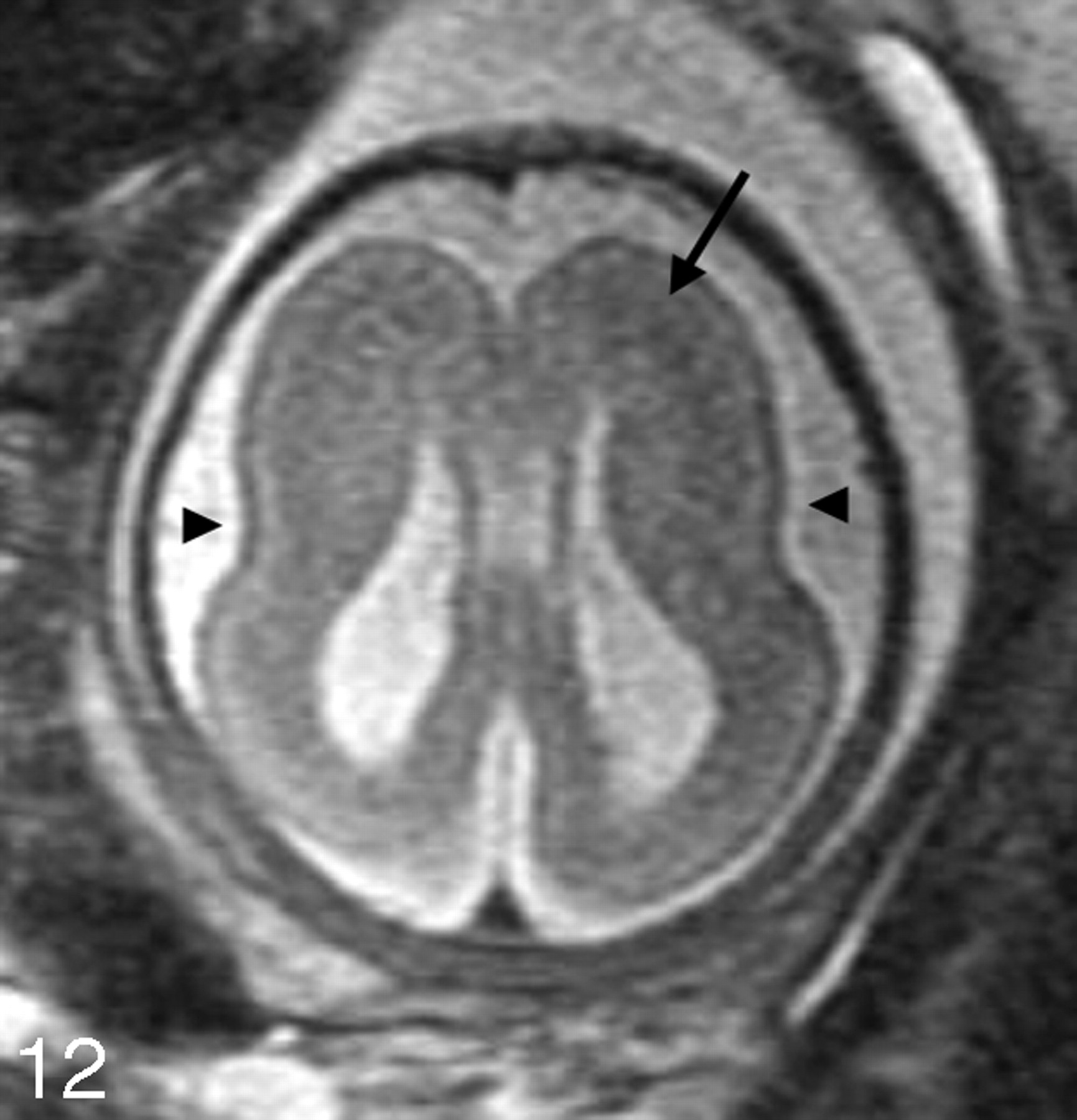
- Fig 12.
Axial SS-FSE T2-weighted image in a 34-gestational-week-old fetus demonstrates minimal formation of the Sylvian fissures (arrowheads) consistent with classical lissencephaly. A thick band of low signal intensity (arrow) is seen in the developing fetal white matter consistent with arrested migration of neurons.
- Fig 13.
Coronal SS-FSE T2-weighted image demonstrates a 24-week twin gestation complicated by twin-twin transfusion syndrome requiring in utero laser ablation of placental vessels. The smaller, donor, twin has focal area of hyperintensity (arrow) adjacent to the left frontal horn, with ex vacuo enlargement of the frontal horn. Periventricular white matter injury with coagulative necrosis was found at autopsy.
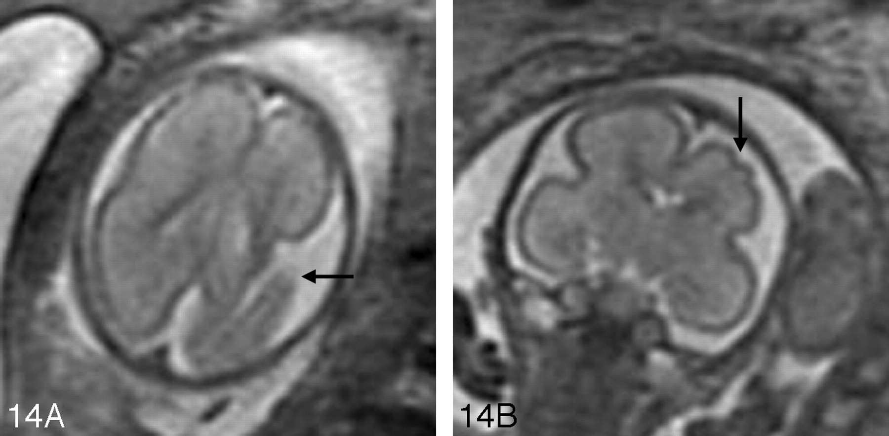
- Fig 14.
A, Axial SS-FSE T2-weighted image of a 23-gestational-week-old fetus in a monochorionic twin pregnancy complicated by co-twin demise. There is a large area of encephalomalacia (arrow) involving the left frontal and parietal lobes.
B, Coronal SS-FSE T2-weighted image demonstrates several infoldings of the developing cortex (arrow) consistent with polymicrogyria. Findings were confirmed by postnatal MR imaging.
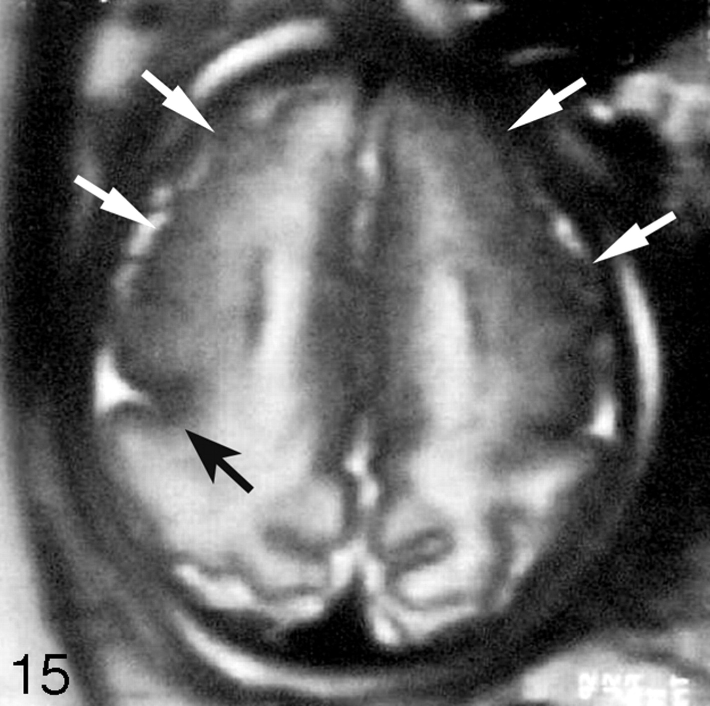
- Fig 15.
Axial SS-FSE T2-weighted image of a 30-gestational-week-old fetus with congenital cytomegalovirus infection. The image shows polymicrogyria, manifested as a deep infolding of abnormal thick cortex (black arrow) in the right posterior frontal lobe and as too many, abnormally shallow sulci over both frontal convexities (white arrows). In addition, there is abnormal hyperintensity of the right frontal white matter.
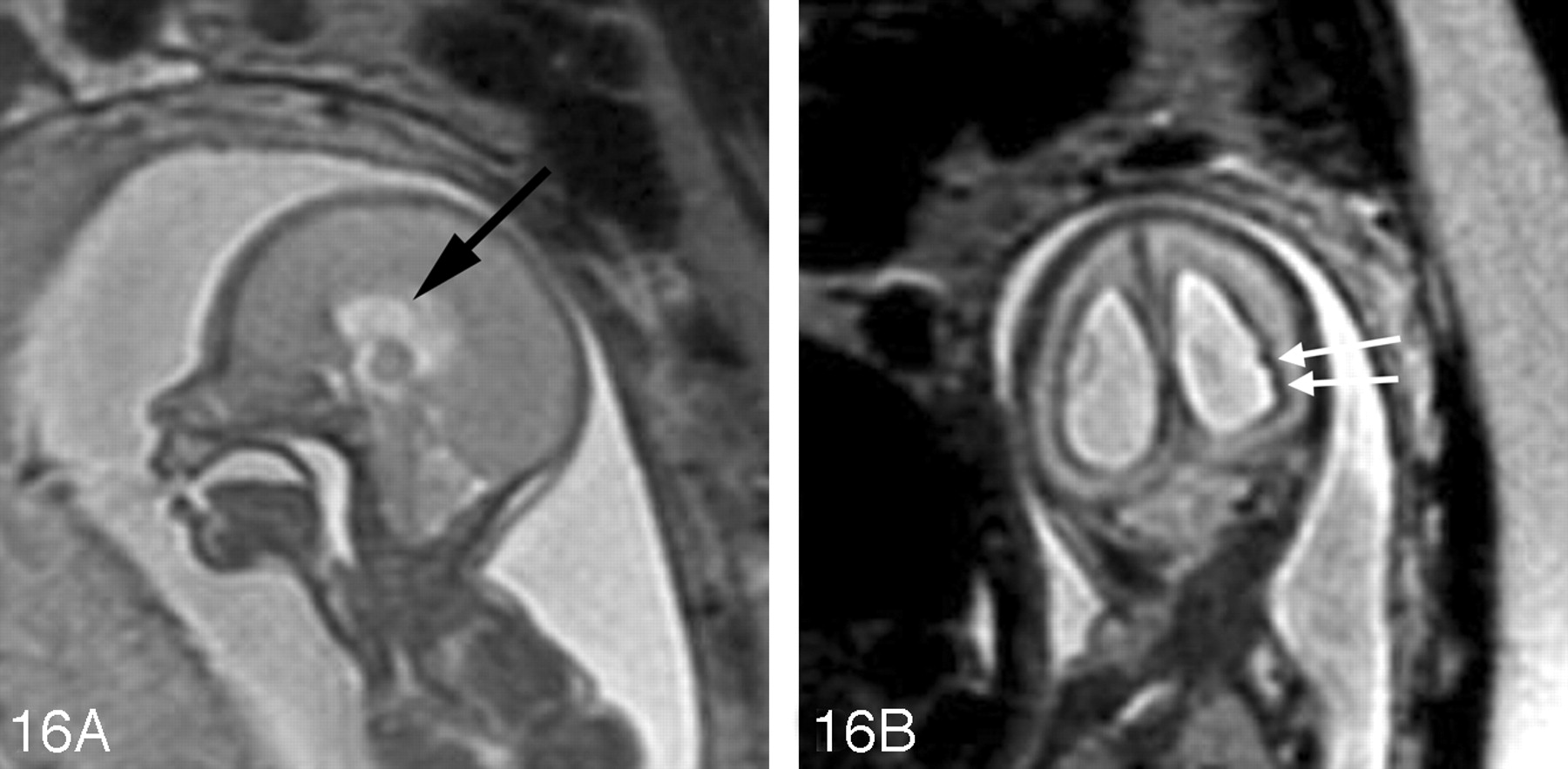
- Fig 16.
A, Sagittal SS-FSE T2-weighted image demonstrates a small posterior fossa with displacement of the cerebellar vermis inferiorly, consistent with a Chiari II malformation in a 22-gestational-week-old fetus. The corpus callosum is hypogenetic (arrow). There is diffuse effacement of the subarachnoid spaces, which is typically observed in these cases.
B, Coronal SS-FSE T2-weighted image demonstrates 2 small hypointense nodules (arrows) along the lateral margin of the atrium of the left lateral ventricle, consistent with periventricular nodular heterotopia. The ventricles are enlarged. Findings were confirmed by postnatal MR imaging.
- Fig 17.
Sagittal SS-FSE T2-weighted image in a 23-gestational-week-old fetus demonstrates multiple bony anomalies in the cervicothoracic region and lumbar spine. There is an abnormally high termination of the spinal cord (arrow) with marked narrowing of the bony spinal canal at the upper and midlumbar spine (A).
B, The lower lumbar bony spinal canal is more normal in caliber and contains abnormal neural tissue (arrow) in the sacral region. Findings are consistent with segmental spinal dysgenesis, and were confirmed by postnatal imaging.


In this issue


Jump to section
Related Articles
Cited By...
- PW-GAN: Pseudo-Warping Field Guided GAN for Unsupervised Denoising of Fetal Brain MRI Images
- The Role of Magnetic Resonance Imaging in Diagnosing Fetal Brain Pathologies
- Development of Gestational Age-Based Fetal Brain and Intracranial Volume Reference Norms Using Deep Learning
- Prenatal Evaluation of Intracranial Hemorrhage on Fetal MRI: A Retrospective Review
- Spinal Imaging Findings of Open Spinal Dysraphisms on Fetal and Postnatal MRI
- Hindbrain Herniation in Chiari II Malformation on Fetal and Postnatal MRI
- MR Imaging of the Pituitary Gland and Postsphenoid Ossification in Fetal Specimens
- Evaluation of Subependymal Gray Matter Heterotopias on Fetal MRI
- Motion-Compensation Techniques in Neonatal and Fetal MR Imaging
- High-Resolution In Utero 3D MR Imaging of Inner Ear Microstructures in Fetal Sheep
- Corpus Callosum Length by Gestational Age as Evaluated by Fetal MR Imaging
- Local Tissue Growth Patterns Underlying Normal Fetal Human Brain Gyrification Quantified In Utero
- Assessment of Sulcation of the Fetal Brain in Cases of Isolated Agenesis of the Corpus Callosum Using In Utero MR Imaging
- Novel Presentation of Aicardi Syndrome With Agenesis of the Corpus Callosum and an Orbital Cyst
- Chiari Malformations
- What Does Magnetic Resonance Imaging Add to the Prenatal Sonographic Diagnosis of Ventriculomegaly?