Advertisement

Main menu

  • Home
  • Content
    • Current Issue
    • Accepted Manuscripts
    • Article Preview
    • Past Issue Archive
    • Video Articles
    • AJNR Case Collection
    • Case of the Week Archive
    • Case of the Month Archive
    • Classic Case Archive
  • Special Collections
    • AJNR Awards
    • Low-Field MRI
    • Alzheimer Disease
    • ASNR Foundation Special Collection
    • Photon-Counting CT
    • View All
  • Multimedia
    • AJNR Podcasts
    • AJNR SCANtastic
    • Trainee Corner
    • MRI Safety Corner
    • Imaging Protocols
  • For Authors
    • Submit a Manuscript
    • Submit a Video Article
    • Submit an eLetter to the Editor/Response
    • Manuscript Submission Guidelines
    • Statistical Tips
    • Fast Publishing of Accepted Manuscripts
    • Graphical Abstract Preparation
    • Imaging Protocol Submission
    • Author Policies
  • About Us
    • About AJNR
    • Editorial Board
    • Editorial Board Alumni
  • More
    • Become a Reviewer/Academy of Reviewers
    • Subscribers
    • Permissions
    • Alerts
    • Feedback
    • Advertisers
    • ASNR Home

User menu

  • Alerts
  • Log in

Search

  • Advanced search
American Journal of Neuroradiology

American Journal of Neuroradiology

ASHNR American Society of Functional Neuroradiology ASHNR American Society of Pediatric Neuroradiology ASSR
  • Alerts
  • Log in

Advanced Search

  • Home
  • Content
    • Current Issue
    • Accepted Manuscripts
    • Article Preview
    • Past Issue Archive
    • Video Articles
    • AJNR Case Collection
    • Case of the Week Archive
    • Case of the Month Archive
    • Classic Case Archive
  • Special Collections
    • AJNR Awards
    • Low-Field MRI
    • Alzheimer Disease
    • ASNR Foundation Special Collection
    • Photon-Counting CT
    • View All
  • Multimedia
    • AJNR Podcasts
    • AJNR SCANtastic
    • Trainee Corner
    • MRI Safety Corner
    • Imaging Protocols
  • For Authors
    • Submit a Manuscript
    • Submit a Video Article
    • Submit an eLetter to the Editor/Response
    • Manuscript Submission Guidelines
    • Statistical Tips
    • Fast Publishing of Accepted Manuscripts
    • Graphical Abstract Preparation
    • Imaging Protocol Submission
    • Author Policies
  • About Us
    • About AJNR
    • Editorial Board
    • Editorial Board Alumni
  • More
    • Become a Reviewer/Academy of Reviewers
    • Subscribers
    • Permissions
    • Alerts
    • Feedback
    • Advertisers
    • ASNR Home
  • Follow AJNR on Twitter
  • Visit AJNR on Facebook
  • Follow AJNR on Instagram
  • Join AJNR on LinkedIn
  • RSS Feeds

AJNR Awards, New Junior Editors, and more. Read the latest AJNR updates

Research ArticleBrain

First Results in an MR Imaging-Compatible Canine Model of Acute Stroke

A. Shaibani, S. Khawar, W. Shin, T.A. Cashen, B. Schirf, M. Rohany, S. Kakodkar and T.J. Carroll
American Journal of Neuroradiology September 2006, 27 (8) 1788-1793;
A. Shaibani
  • Find this author on Google Scholar
  • Find this author on PubMed
  • Search for this author on this site
S. Khawar
  • Find this author on Google Scholar
  • Find this author on PubMed
  • Search for this author on this site
W. Shin
  • Find this author on Google Scholar
  • Find this author on PubMed
  • Search for this author on this site
T.A. Cashen
  • Find this author on Google Scholar
  • Find this author on PubMed
  • Search for this author on this site
B. Schirf
  • Find this author on Google Scholar
  • Find this author on PubMed
  • Search for this author on this site
M. Rohany
  • Find this author on Google Scholar
  • Find this author on PubMed
  • Search for this author on this site
S. Kakodkar
  • Find this author on Google Scholar
  • Find this author on PubMed
  • Search for this author on this site
T.J. Carroll
  • Find this author on Google Scholar
  • Find this author on PubMed
  • Search for this author on this site
  • Article
  • Figures & Data
  • Info & Metrics
  • Responses
  • References
  • PDF
Loading

Article Figures & Data

Figures

  • Tables
  • Fig 1.
    • Download figure
    • Open in new tab
    • Download powerpoint
    Fig 1.

    Right internal carotid artery injection delineating the canine circle of Willis (A) before and (B) after injection of an autologous blood clot. Note the conspicuous absence of the middle cerebral artery on the right side after the injection confirming the stroke.

  • Fig 2.
    • Download figure
    • Open in new tab
    • Download powerpoint
    Fig 2.

    The evolution of the stroke as demonstrated by water diffusion changes in 2 coronal sections acquired in the first hour post ictus. b = 1000 images (A) and ADC images (B) demonstrate the increased diffusion contrast and reduced ADC in the region affected by the stroke (arrows).
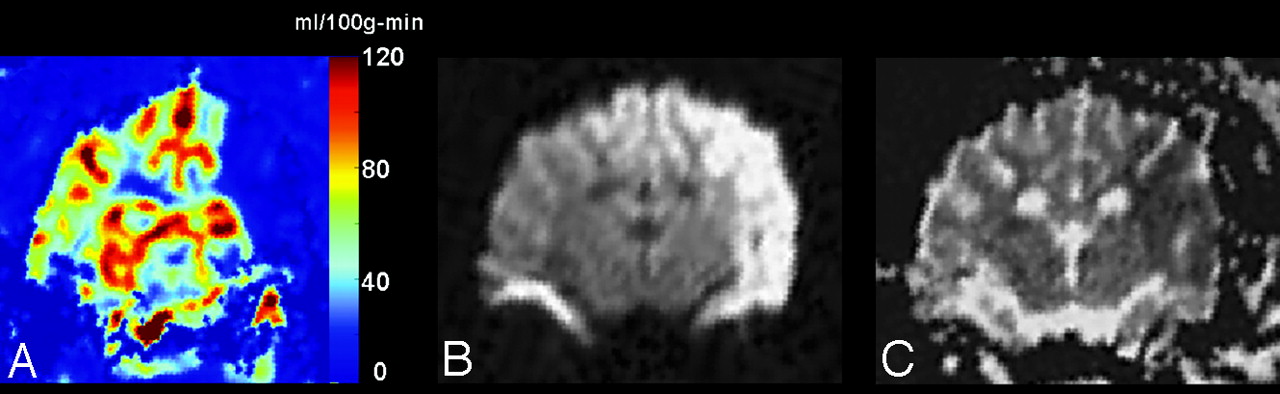
  • Fig 3.
    • Download figure
    • Open in new tab
    • Download powerpoint
    Fig 3.

    A, An image of quantitative CBF acquired 30 minutes post ictus demonstrates a large region of hypoperfusion (qCBF <10 mL/100 g-min) in the left MCA territory.

    B, Diffusion-weighted images (b = 1000) acquired after 2 hours show that, in this case, the territory of hypoperfusion observed immediately after the stroke goes on to become a pronounced diffusion abnormality.

    C, ADC values confirm the region of diffusion seen in the b = 1000 image.

  • Fig 4.
    • Download figure
    • Open in new tab
    • Download powerpoint
    Fig 4.

    Thrombolysis achieved partial restoration of cerebral perfusion in 1 animal.

    A, Pretreatment time to peak of a single coronal section demonstrates a large region of prolonged TTP in the left MCA territory (arrows).

    B, The ADC image acquired at 2 hours confirms a region of reduced ADC.

    C, The corresponding TTP acquired upon completion of the infusion of rtPA shows marked improvement of flow in the affected territory. Quantitative flow images calculated before (D) and after (E) treatment show that qCBF in the region affected by the stroke has been restored to a value that is above the known threshold for ischemic stroke.

Tables

  • Figures
    • View popup
    Table 1:

    MR imaging protocol

    Perfusion Weighted*Diffusion Weighted*
    TR (ms)1150† (1200)3000 (1600)
    TE (ms)52 (52)97 (91)
    Field of view (mm)148 × 148148 × 148
    Matrix128 × 128128 × 128
    No. of sections9–1015
    Thickness (mm)5.05.0
    Echo-planar imaging factor128128
    b value (s/mm2)n/a0, 500, 1000
    Bandwidth (Hz/pixel)12601220
    • * Values of the 1.5T experimental are shown in parentheses.

    • † Fifty phases acquired.

    • View popup
    Table 2:

    Experimental conditions

    DescriptionStroke Region (mL/100 g/min)Normal Contralateral (mL/100 g/min)
    3T, stroke2.72 ± 1.2142.93 ± 16.70
    3T stroke*,†15.09 ± 4.7149.56 ± 12.75
    3T, stroke*3.81 ± 3.7746.51 ± 11.68
    3T stroke*6.22 ± 3.9627.42 ± 10.67
    1.5T, stroke8.28 ± 2.3144.83 ± 8.08
    Mean value7.22 ± 4.9042.25 ± 8.64
    P value<.0007
    3T, control51.84 ± 13.2659.50 ± 14.48
    • * Treated with recombinant tissue plasminogen activator.

    • † Recanalization was observed.

PreviousNext
Back to top

In this issue

American Journal of Neuroradiology: 27 (8)
American Journal of Neuroradiology
Vol. 27, Issue 8
September 2006
  • Table of Contents
  • Index by author
Advertisement
Print
Download PDF
Email Article

Thank you for your interest in spreading the word on American Journal of Neuroradiology.

NOTE: We only request your email address so that the person you are recommending the page to knows that you wanted them to see it, and that it is not junk mail. We do not capture any email address.

Enter multiple addresses on separate lines or separate them with commas.
First Results in an MR Imaging-Compatible Canine Model of Acute Stroke
(Your Name) has sent you a message from American Journal of Neuroradiology
(Your Name) thought you would like to see the American Journal of Neuroradiology web site.
CAPTCHA
This question is for testing whether or not you are a human visitor and to prevent automated spam submissions.
Cite this article
A. Shaibani, S. Khawar, W. Shin, T.A. Cashen, B. Schirf, M. Rohany, S. Kakodkar, T.J. Carroll
First Results in an MR Imaging-Compatible Canine Model of Acute Stroke
American Journal of Neuroradiology Sep 2006, 27 (8) 1788-1793;

Citation Manager Formats

  • BibTeX
  • Bookends
  • EasyBib
  • EndNote (tagged)
  • EndNote 8 (xml)
  • Medlars
  • Mendeley
  • Papers
  • RefWorks Tagged
  • Ref Manager
  • RIS
  • Zotero
0 Responses
Respond to this article
Share
Bookmark this article
First Results in an MR Imaging-Compatible Canine Model of Acute Stroke
A. Shaibani, S. Khawar, W. Shin, T.A. Cashen, B. Schirf, M. Rohany, S. Kakodkar, T.J. Carroll
American Journal of Neuroradiology Sep 2006, 27 (8) 1788-1793;
del.icio.us logo Twitter logo Facebook logo Mendeley logo
  • Tweet Widget
  • Facebook Like
  • Google Plus One

Jump to section

  • Article
    • Abstract
    • Materials and Methods
    • Results
    • Discussion
    • Conclusion
    • References
  • Figures & Data
  • Info & Metrics
  • Responses
  • References
  • PDF

Related Articles

  • No related articles found.
  • PubMed
  • Google Scholar

Cited By...

  • Image-Guided Method in the Rat for Inducing Cortical or Striatal Infarction and for Controlling Cerebral Blood Flow Under MRI
  • Preclinical acute ischemic stroke modeling
  • Quantitative Evaluation of C-Arm CT Cerebral Blood Volume in a Canine Model of Ischemic Stroke
  • C-Arm CT Measurement of Cerebral Blood Volume in Ischemic Stroke: An Experimental Study in Canines
  • Novel Microcatheters for Selective Intra-Arterial Injection of Fluid in the Rat Brain
  • Crossref
  • Google Scholar

This article has not yet been cited by articles in journals that are participating in Crossref Cited-by Linking.

More in this TOC Section

  • Quiet PROPELLER MRI Techniques Match the Quality of Conventional PROPELLER Brain Imaging Techniques
  • Predictors of Reperfusion in Patients with Acute Ischemic Stroke
  • Enhanced Axonal Metabolism during Early Natalizumab Treatment in Relapsing-Remitting Multiple Sclerosis
Show more Brain

Similar Articles

Advertisement

Indexed Content

  • Current Issue
  • Accepted Manuscripts
  • Article Preview
  • Past Issues
  • Editorials
  • Editor's Choice
  • Fellows' Journal Club
  • Letters to the Editor
  • Video Articles

Cases

  • Case Collection
  • Archive - Case of the Week
  • Archive - Case of the Month
  • Archive - Classic Case

More from AJNR

  • Trainee Corner
  • Imaging Protocols
  • MRI Safety Corner
  • Book Reviews

Multimedia

  • AJNR Podcasts
  • AJNR Scantastics

Resources

  • Turnaround Time
  • Submit a Manuscript
  • Submit a Video Article
  • Submit an eLetter to the Editor/Response
  • Manuscript Submission Guidelines
  • Statistical Tips
  • Fast Publishing of Accepted Manuscripts
  • Graphical Abstract Preparation
  • Imaging Protocol Submission
  • Evidence-Based Medicine Level Guide
  • Publishing Checklists
  • Author Policies
  • Become a Reviewer/Academy of Reviewers
  • News and Updates

About Us

  • About AJNR
  • Editorial Board
  • Editorial Board Alumni
  • Alerts
  • Permissions
  • Not an AJNR Subscriber? Join Now
  • Advertise with Us
  • Librarian Resources
  • Feedback
  • Terms and Conditions
  • AJNR Editorial Board Alumni

American Society of Neuroradiology

  • Not an ASNR Member? Join Now

© 2025 by the American Society of Neuroradiology All rights, including for text and data mining, AI training, and similar technologies, are reserved.
Print ISSN: 0195-6108 Online ISSN: 1936-959X

Powered by HighWire