
Article Figures & Data
Figures
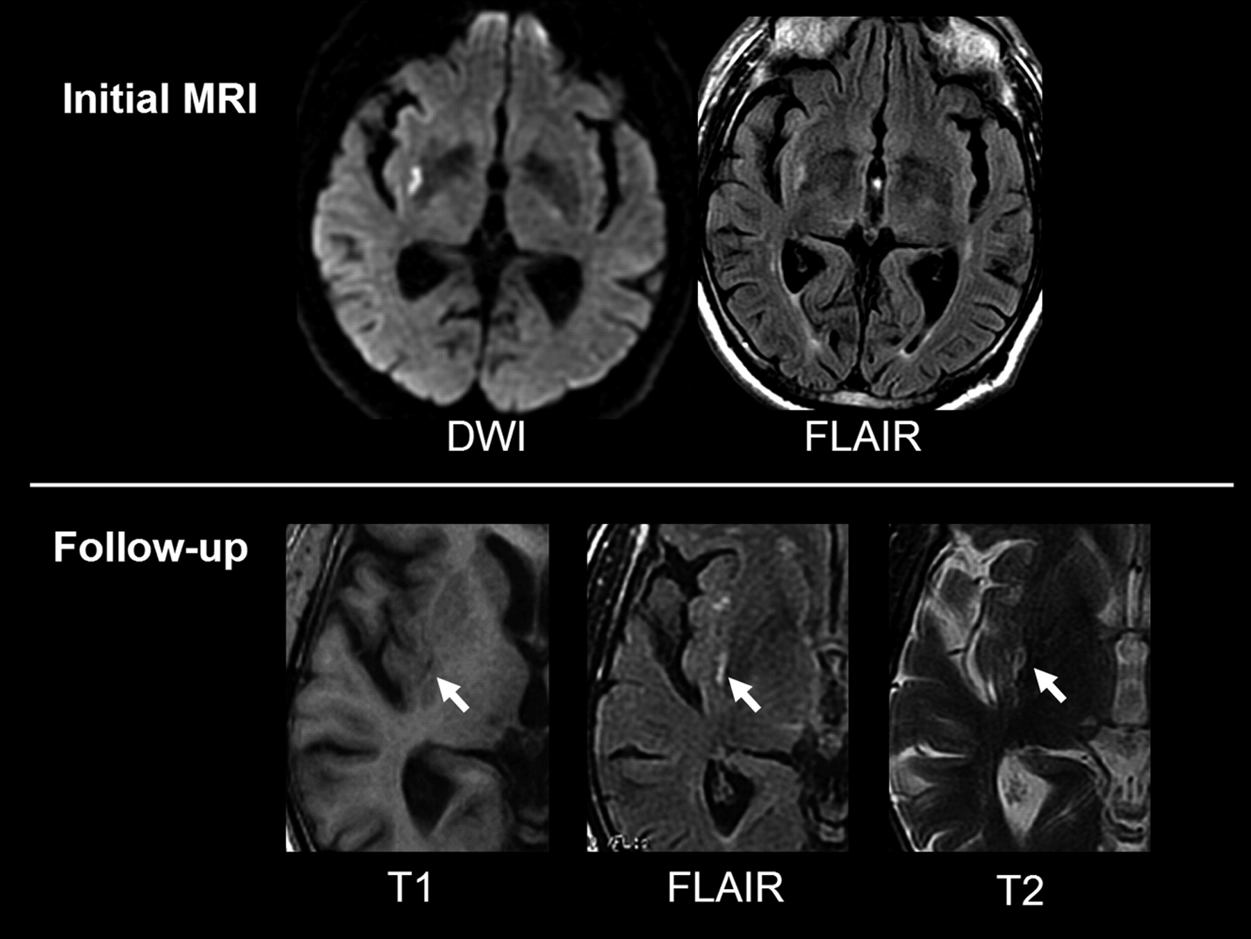
- Fig 1.
A 67-year-old man with a left sensory-motor deficit for 30 minutes. Initial MR imaging (3 days after onset) demonstrates a small DWI and FLAIR hyperintensity in the deep right middle cerebral artery (MCA) territory. On follow-up MR imaging (14 months after onset), focal signal intensity changes on all sequences indicate permanent injury in the corresponding area.
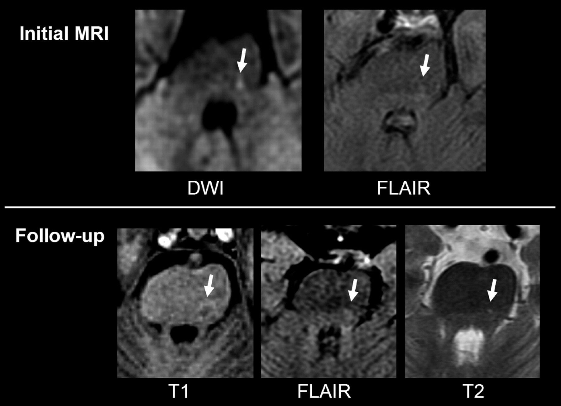
- Fig 2.
A 63-year-old man with a right sensory deficit for 10 hours. Initial MR imaging (63 hours after onset) demonstrates a focal DWI and FLAIR hyperintensity in the left brain stem (arrow). On follow-up MR imaging (6 months after onset), a small permanent injury can be seen as a dark signal intensity on T1-weighted sequence and a bright signal intensity on FLAIR/T2-weighted sequence in the corresponding area.
- Fig 3.
A 55-year-old man with sensory-motor deficit of the upper right limb for 90 minutes. Initial MR imaging (10 hours after onset) demonstrates a focal DWI hyperintensity with mild FLAIR signal intensity changes in the left primary motor cortex, matching clinical symptoms. On follow-up MR imaging (8 months after onset), no permanent injury can be identified.
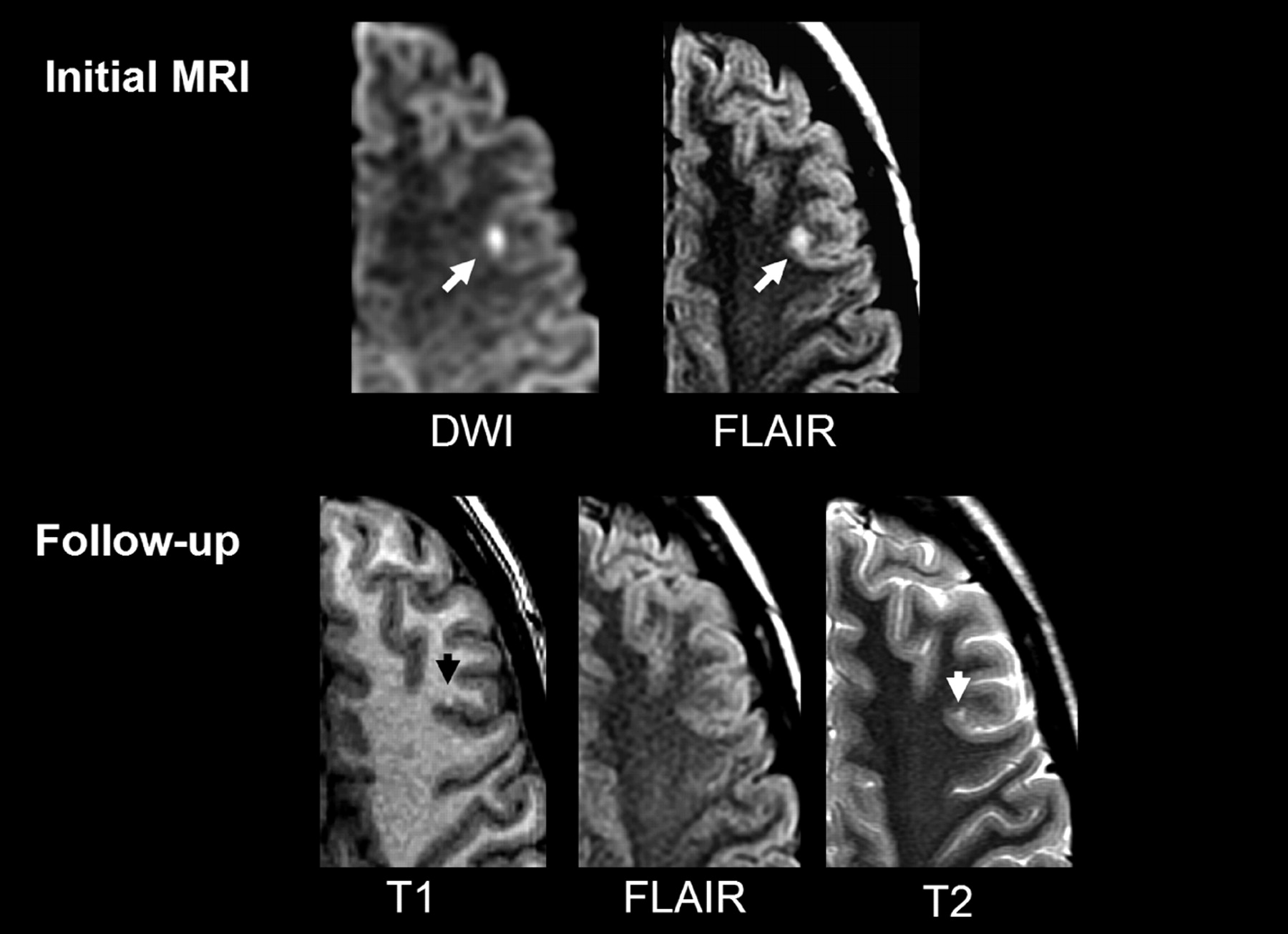
- Fig 4.
A 21-year-old woman with a right sensory-motor deficit and aphasia for 60 minutes. Initial MR imaging (4 days after onset) shows a punctate cortical DWI/FLAIR hyperintensity in the left superficial middle cerebral artery (MCA) territory. On follow-up MR imaging (15 months after onset), a small permanent injury can be seen as a bright cortical dot on T1-weighted sequence with mild atrophy on T2-weighted sequence. Note that no signal intensity change is seen on FLAIR.


Tables
- Table 1:
Characteristics of transient ischemic attack (TIA) patients with and without infarction on follow-up MR imaging (MRI)
Reversible (n = 7) Infarction (n = 26) All Patients (n = 33) P Value Male, % 5 (71%) 18 (70%) 23 (69%) 1 Age, y (mean ± SD) 58 ± 15 61 ± 17 60 ± 16 .5 Symptoms Duration, min (mean ± SD) 76 ± 85 188 ± 253 201 ± 262 .2 Duration <60 min 3 (43%) 10 (38%) 13 (39%) 1 Multiple TIA events 3 (43%) 10 (38%) 13 (39%) 1 Identified cause, % 3 (43%) 12 (46%) 15 (45%) 1 Initial MRI Delay from onset MRI, h 21 ± 10 33 ± 36 30 ± 33 .9 Solitary lesions 6 (86%) 11 (42%) 17 (51.5%) .08 Follow-up MRI Delay from onset MRI, mo 11.6 ± 3.1 10.4 ± 5.5 10.6 ± 5 .75 Note:— Patients with multiple lesions of different outcome are classed in the infarction group.
- Table 2:
Quantitative diffusion-weighted MR imaging (DWI)–derived variables of the 59 transient ischemic attack (TIA) lesions according to imaging outcome
Reversible (n = 14) Infarction (n = 45) P Value DWI volume, cm3 (mean ± SD) 0.21 ± 0.21 0.91 ± 1.7 .003 Absolute ADC, 10−6 mm2/s (mean ± SD) 722 ± 118 631 ± 135 .022 rADC (mean ± SD) 91 ± 9% 79 ± 15% .001 Note:— rADC corresponds to apparent diffusion coefficient (ADC) within the TIA lesion divided by the mirror ADC value.
In this issue


Jump to section
Related Articles
Cited By...
- Small cortical grey matter lesions show no persistent infarction in transient ischaemic attack? A prospective cohort study
- Have Stroke Neurologists Entered the Arena of Stroke-Related Cognitive Dysfunctions?: Not Yet, but They Should!
- DWI Reversal Is Associated with Small Infarct Volume in Patients with TIA and Minor Stroke
- Displacement of Sensory Maps and Disorganization of Motor Cortex After Targeted Stroke in Mice
- The Massachusetts General Hospital acute stroke imaging algorithm: an experience and evidence based approach
- Acute silent cerebral ischemia and infarction during acute anemia in children with and without sickle cell disease
- Prevalence of MRI-defined recent silent ischemia and associated bleeding risk with thrombolysis
- Acute Perfusion and Diffusion Abnormalities Predict Early New MRI Lesions 1 Week After Minor Stroke and Transient Ischemic Attack
- Reporting standards for endovascular repair of saccular intracranial cerebral aneurysms
- How Much Would Performing Diffusion-Weighted Imaging for All Transient Ischemic Attacks Increase MRI Utilization?
- Rapid resolution of diffusion weighted MRI abnormality in a patient with a stuttering stroke
- Reporting Standards for Endovascular Repair of Saccular Intracranial Cerebral Aneurysms
- Definition and Evaluation of Transient Ischemic Attack: A Scientific Statement for Healthcare Professionals From the American Heart Association/American Stroke Association Stroke Council; Council on Cardiovascular Surgery and Anesthesia; Council on Cardiovascular Radiology and Intervention; Council on Cardiovascular Nursing; and the Interdisciplinary Council on Peripheral Vascular Disease: The American Academy of Neurology affirms the value of this statement as an educational tool for neurologists.
- Reporting Standards for Endovascular Repair of Saccular Intracranial Cerebral Aneurysms
- DWI Lesions and TIA Etiology Improve the Prediction of Stroke After TIA
- Proposal for a Universal Definition of Cerebral Infarction