Advertisement

Main menu

  • Home
  • Content
    • Current Issue
    • Accepted Manuscripts
    • Article Preview
    • Past Issue Archive
    • Video Articles
    • AJNR Case Collection
    • Case of the Week Archive
    • Case of the Month Archive
    • Classic Case Archive
  • Special Collections
    • AJNR Awards
    • Low-Field MRI
    • Alzheimer Disease
    • ASNR Foundation Special Collection
    • Photon-Counting CT
    • View All
  • Multimedia
    • AJNR Podcasts
    • AJNR SCANtastic
    • Trainee Corner
    • MRI Safety Corner
    • Imaging Protocols
  • For Authors
    • Submit a Manuscript
    • Submit a Video Article
    • Submit an eLetter to the Editor/Response
    • Manuscript Submission Guidelines
    • Statistical Tips
    • Fast Publishing of Accepted Manuscripts
    • Graphical Abstract Preparation
    • Imaging Protocol Submission
    • Author Policies
  • About Us
    • About AJNR
    • Editorial Board
    • Editorial Board Alumni
  • More
    • Become a Reviewer/Academy of Reviewers
    • Subscribers
    • Permissions
    • Alerts
    • Feedback
    • Advertisers
    • ASNR Home

User menu

  • Alerts
  • Log in

Search

  • Advanced search
American Journal of Neuroradiology

American Journal of Neuroradiology

ASHNR American Society of Functional Neuroradiology ASHNR American Society of Pediatric Neuroradiology ASSR
  • Alerts
  • Log in

Advanced Search

  • Home
  • Content
    • Current Issue
    • Accepted Manuscripts
    • Article Preview
    • Past Issue Archive
    • Video Articles
    • AJNR Case Collection
    • Case of the Week Archive
    • Case of the Month Archive
    • Classic Case Archive
  • Special Collections
    • AJNR Awards
    • Low-Field MRI
    • Alzheimer Disease
    • ASNR Foundation Special Collection
    • Photon-Counting CT
    • View All
  • Multimedia
    • AJNR Podcasts
    • AJNR SCANtastic
    • Trainee Corner
    • MRI Safety Corner
    • Imaging Protocols
  • For Authors
    • Submit a Manuscript
    • Submit a Video Article
    • Submit an eLetter to the Editor/Response
    • Manuscript Submission Guidelines
    • Statistical Tips
    • Fast Publishing of Accepted Manuscripts
    • Graphical Abstract Preparation
    • Imaging Protocol Submission
    • Author Policies
  • About Us
    • About AJNR
    • Editorial Board
    • Editorial Board Alumni
  • More
    • Become a Reviewer/Academy of Reviewers
    • Subscribers
    • Permissions
    • Alerts
    • Feedback
    • Advertisers
    • ASNR Home
  • Follow AJNR on Twitter
  • Visit AJNR on Facebook
  • Follow AJNR on Instagram
  • Join AJNR on LinkedIn
  • RSS Feeds

AJNR Awards, New Junior Editors, and more. Read the latest AJNR updates

Research ArticleBRAIN

Relationship between Variations in the Circle of Willis and Flow Rates in Internal Carotid and Basilar Arteries Determined by Means of Magnetic Resonance Imaging with Semiautomated Lumen Segmentation: Reference Data from 125 Healthy Volunteers

H. Tanaka, N. Fujita, T. Enoki, K. Matsumoto, Y. Watanabe, K. Murase and H. Nakamura
American Journal of Neuroradiology September 2006, 27 (8) 1770-1775;
H. Tanaka
  • Find this author on Google Scholar
  • Find this author on PubMed
  • Search for this author on this site
N. Fujita
  • Find this author on Google Scholar
  • Find this author on PubMed
  • Search for this author on this site
T. Enoki
  • Find this author on Google Scholar
  • Find this author on PubMed
  • Search for this author on this site
K. Matsumoto
  • Find this author on Google Scholar
  • Find this author on PubMed
  • Search for this author on this site
Y. Watanabe
  • Find this author on Google Scholar
  • Find this author on PubMed
  • Search for this author on this site
K. Murase
  • Find this author on Google Scholar
  • Find this author on PubMed
  • Search for this author on this site
H. Nakamura
  • Find this author on Google Scholar
  • Find this author on PubMed
  • Search for this author on this site
  • Article
  • Figures & Data
  • Info & Metrics
  • Responses
  • References
  • PDF
Loading

Article Figures & Data

Figures

  • Tables
  • Fig 1.
    • Download figure
    • Open in new tab
    • Download powerpoint
    Fig 1.

    MR examination and flow measurement procedures for a representative case (23-year-old woman).

    A, Lateral view of 3D time-of-flight MR angiography. The white line indicates the section used for the phase-contrast MR imaging.

    B, Axial view of 3D time-of-flight MR angiography. The variation in the circle of Willis in this case was assessed as hypoplasia of the precommunicating segment of the right anterior cerebral artery (right A1 hypoplasia) based on diameter measurements obtained from the original MR angiography images.

    C, As the first step of flow measurement, an operator selected 3 pixels from each artery. These pixels are shown as red dots in this phase-contrast image. The inset in the bottom right corner shows a magnified image of the basilar artery for instance.

    D, From these selected pixels, vessel lumens (clusters of red dots) were automatically identified by pulsatility-based segmentation.17 To compensate for eddy current-induced error, 3 static regions of interest (clusters of blue dots) were also determined automatically. This compensation was based on the assumption that the error induced by the eddy current was a linear function of space.14 Eventually volume flow rate of each arterywas automatically obtained.

  • Fig 2.
    • Download figure
    • Open in new tab
    • Download powerpoint
    Fig 2.

    Classification of the anatomic variations in the circle of Willis. In the “textbook” type, both the precommunicating segment of the anterior cerebral artery (A1) and that of the posterior cerebral artery (P1) were normal in size. The next group included both right and left A1 hypoplasia. Because no significant difference between cerebral arteries on the right and left sides has been established,5,18 we combined right and left A1 hypoplasia into A1 hypoplasia. The next group included right and left P1 hypoplasia, which again were treated as a single category, P1 hypoplasia. “Other” type included a combination of A1 hypoplasia and P1 hypoplasia, bilateral P1 hypoplasia, as well as other unclassified variations. ACA indicates anterior cerebral artery; ACo, anterior communicating artery; MCA, middle cerebral artery; ICA, internal cerebral artery; PCo, posterior communicating artery; PCA, posterior cerebral artery; BA, basilar artery
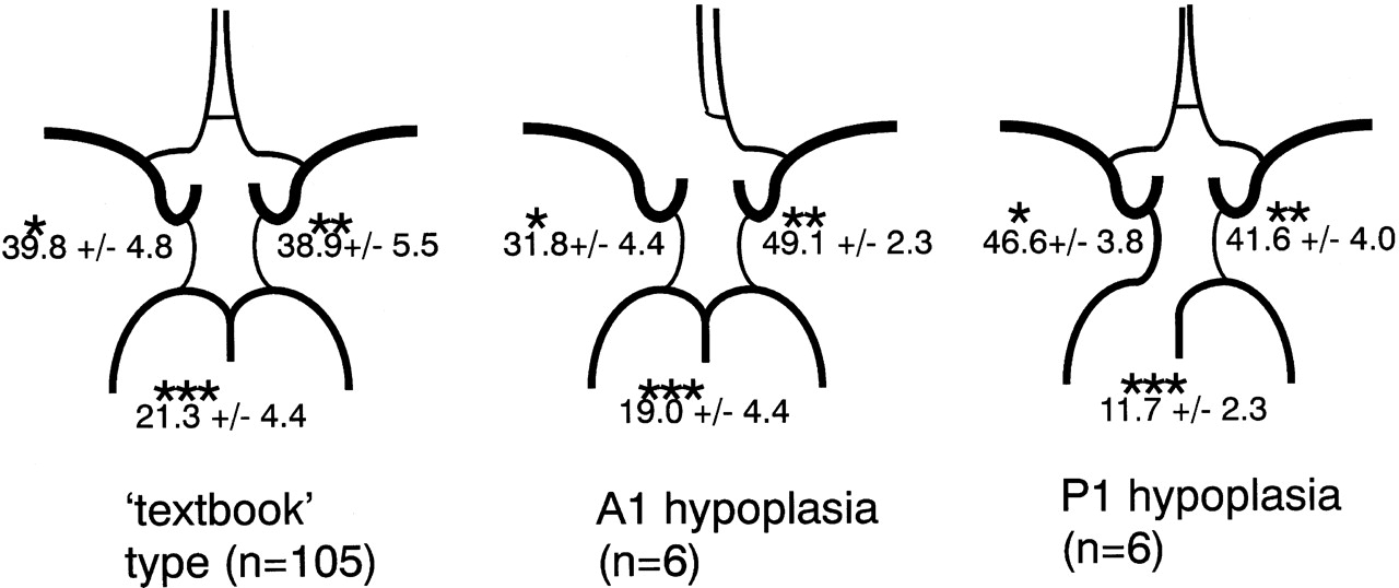
  • Fig 3.
    • Download figure
    • Open in new tab
    • Download powerpoint
    Fig 3.

    Relative contribution of proximal arteries to total volume flow in variations in the circle of Willis. Values signify mean percentage ± SD. The upper left value corresponds to the relative contribution of the right internal carotid artery in the “textbook” type or of the internal carotid artery ipsilateral to hypoplastic A1 or P1 in the other variations. The upper right value corresponds to the relative contribution of the left internal carotid artery in the “textbook” type, or of the internal carotid artery contralateral to hypoplastic A1 or P1 in the other variations. The value at the bottom corresponds to the relative contribution of the basilar artery.

    * The value for A1 hypoplasia variation was significantly smaller than those for “textbook” type and P1 hypoplasia variation. The value for P1 hypoplasia variation was significantly larger than that for “textbook” type.

    ** The value for A1 hypoplasia variation was significantly larger than that for “textbook” type.

    *** The value for P1 hypoplasia variation was significantly smaller than that for “textbook” type.
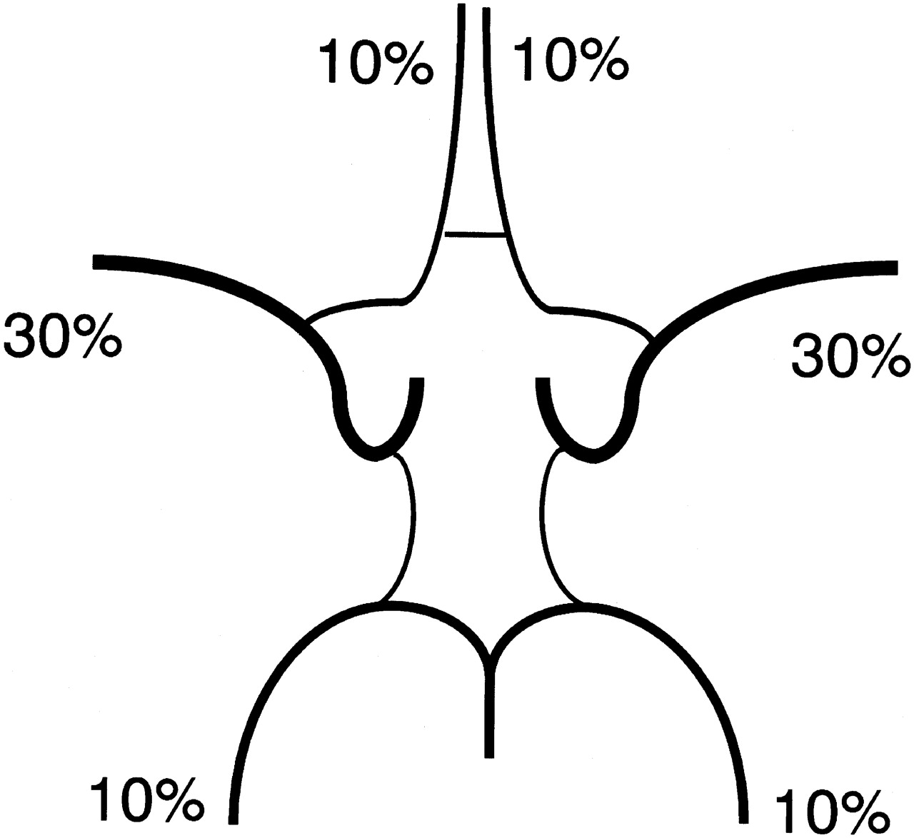
  • Fig 4.
    • Download figure
    • Open in new tab
    • Download powerpoint
    Fig 4.

    Estimated volume flow ratio of the anterior, middle, and posterior cerebral artery. These values were estimated from Fig 3 and approximated to multiples of 10%.

Tables

  • Figures
    • View popup
    Table 1:

    Diameters of A1 and P1 and volume flow rates of 6 subjects with A1 hypolasia (nos. 1–6) and 6 with P1 hypoplasia (nos. 7–12)

    Subject No.Diameter (mm)Volume Flow Rates (mL/min) (Relative Percentage Contribution (%))
    Ipsilateral to Hypolastic A1 or P1Contralateral to Hypoplastic A1 or P1ICA Ipsilateral to Hypoplastic A1 or P1ICA Contralateral to Hypoplastic A1 or P1Basilar Artery
    A1P1A1P1
    10.0*2.31.4 + 1.3†2.4267 (29)468 (51)188 (20)
    20.71.02.01.7305 (38)405 (51)88 (11)
    30.92.02.72.4257 (32)380 (47)166 (21)
    40.81.41.52.1212 (35)274 (45)119 (20)
    50.0*1.82.32.0179 (26)348 (50)169 (24)
    60.0*1.92.72.0199 (31)325 (51)116 (18)
    71.50.82.22.1438 (48)349 (38)120 (13)
    81.80.92.21.4298 (44)303 (45)76 (11)
    91.30.91.72.0379 (52)254 (35)91 (13)
    102.10.82.01.1296 (48)266 (43)52 (8)
    111.40.0*2.01.8300 (42)312 (43)108 (15)
    121.50.92.51.9422 (45)418 (45)95 (10)
    • Note:— A1 indicates precommunicating segment of the anterior cerebral artery; P1, precommunicating segment of the posterior cerebral artery; ICA, internal carotid artery. An artery with a diameter ≤0.9 mm was considered to be hypoplastic.

    • * The relevant artery was not depicted.

    • † Duplicate A1s were observed.

    • View popup
    Table 2:

    Variations in the circle of Willis and volume flow rates

    Variations in the Circle of Willis“Textbook” Type (n = 105)A1 Hypoplasia (n = 6)P1 Hypoplasia (n = 6)
    Total volume flow (mL/min) (mean ± SD)781 ± 151744 ± 119763 ± 129
    Right internal carotid artery or internal carotid artery ipsilateral to hypoplastic A1 or P1311 ± 73236 ± 48355 ± 66
    Left internal carotid artery or internal carotid artery contralateral to hypoplastic A1 to P1304 ± 74367 ± 67317 ± 60
    Basilar artery165 ± 43141 ± 3990 ± 24
    • Note:— A1 indicates precommunicating segment of the anterior cerebral artery; P1, precommunicating segment of the posterior cerebral artery.

PreviousNext
Back to top

In this issue

American Journal of Neuroradiology: 27 (8)
American Journal of Neuroradiology
Vol. 27, Issue 8
September 2006
  • Table of Contents
  • Index by author
Advertisement
Print
Download PDF
Email Article

Thank you for your interest in spreading the word on American Journal of Neuroradiology.

NOTE: We only request your email address so that the person you are recommending the page to knows that you wanted them to see it, and that it is not junk mail. We do not capture any email address.

Enter multiple addresses on separate lines or separate them with commas.
Relationship between Variations in the Circle of Willis and Flow Rates in Internal Carotid and Basilar Arteries Determined by Means of Magnetic Resonance Imaging with Semiautomated Lumen Segmentation: Reference Data from 125 Healthy Volunteers
(Your Name) has sent you a message from American Journal of Neuroradiology
(Your Name) thought you would like to see the American Journal of Neuroradiology web site.
CAPTCHA
This question is for testing whether or not you are a human visitor and to prevent automated spam submissions.
Cite this article
H. Tanaka, N. Fujita, T. Enoki, K. Matsumoto, Y. Watanabe, K. Murase, H. Nakamura
Relationship between Variations in the Circle of Willis and Flow Rates in Internal Carotid and Basilar Arteries Determined by Means of Magnetic Resonance Imaging with Semiautomated Lumen Segmentation: Reference Data from 125 Healthy Volunteers
American Journal of Neuroradiology Sep 2006, 27 (8) 1770-1775;

Citation Manager Formats

  • BibTeX
  • Bookends
  • EasyBib
  • EndNote (tagged)
  • EndNote 8 (xml)
  • Medlars
  • Mendeley
  • Papers
  • RefWorks Tagged
  • Ref Manager
  • RIS
  • Zotero
0 Responses
Respond to this article
Share
Bookmark this article
Relationship between Variations in the Circle of Willis and Flow Rates in Internal Carotid and Basilar Arteries Determined by Means of Magnetic Resonance Imaging with Semiautomated Lumen Segmentation: Reference Data from 125 Healthy Volunteers
H. Tanaka, N. Fujita, T. Enoki, K. Matsumoto, Y. Watanabe, K. Murase, H. Nakamura
American Journal of Neuroradiology Sep 2006, 27 (8) 1770-1775;
del.icio.us logo Twitter logo Facebook logo Mendeley logo
  • Tweet Widget
  • Facebook Like
  • Google Plus One

Jump to section

  • Article
    • Abstract
    • Materials and Methods
    • Results
    • Discussion
    • References
  • Figures & Data
  • Info & Metrics
  • Responses
  • References
  • PDF

Related Articles

  • No related articles found.
  • PubMed
  • Google Scholar

Cited By...

  • Variations in the Circle of Willis in a large population sample using 3D TOF angiography: The Tromso Study
  • The Role Of Circle Of Willis Anatomy Variations In Cardio-embolic Stroke - A Patient-specific Simulation Based Study
  • Aneurysmal Parent Artery-Specific Inflow Conditions for Complete and Incomplete Circle of Willis Configurations
  • Low prevalence of fetal-type posterior cerebral artery in patients with basilar tip aneurysms
  • Association of aneurysms and variation of the A1 segment
  • The role of circle of Willis anomalies in cerebral aneurysm rupture
  • ACCF/ACR/AHA/NASCI/SCMR 2010 Expert Consensus Document on Cardiovascular Magnetic Resonance: A Report of the American College of Cardiology Foundation Task Force on Expert Consensus Documents
  • ACCF/ACR/AHA/NASCI/SCMR 2010 Expert Consensus Document on Cardiovascular Magnetic Resonance: A Report of the American College of Cardiology Foundation Task Force on Expert Consensus Documents
  • Embolus Trajectory Through a Physical Replica of the Major Cerebral Arteries
  • Impact of posterior communicating artery on basilar artery steno-occlusive disease
  • Crossref
  • Google Scholar

This article has not yet been cited by articles in journals that are participating in Crossref Cited-by Linking.

More in this TOC Section

  • Multimodal CT Provides Improved Performance for Lacunar Infarct Detection
  • Optimal MRI Sequence for Identifying Occlusion Location in Acute Stroke: Which Value of Time-Resolved Contrast-Enhanced MRA?
  • Evaluating the Effects of White Matter Multiple Sclerosis Lesions on the Volume Estimation of 6 Brain Tissue Segmentation Methods
Show more BRAIN

Similar Articles

Advertisement

Indexed Content

  • Current Issue
  • Accepted Manuscripts
  • Article Preview
  • Past Issues
  • Editorials
  • Editor's Choice
  • Fellows' Journal Club
  • Letters to the Editor
  • Video Articles

Cases

  • Case Collection
  • Archive - Case of the Week
  • Archive - Case of the Month
  • Archive - Classic Case

More from AJNR

  • Trainee Corner
  • Imaging Protocols
  • MRI Safety Corner
  • Book Reviews

Multimedia

  • AJNR Podcasts
  • AJNR Scantastics

Resources

  • Turnaround Time
  • Submit a Manuscript
  • Submit a Video Article
  • Submit an eLetter to the Editor/Response
  • Manuscript Submission Guidelines
  • Statistical Tips
  • Fast Publishing of Accepted Manuscripts
  • Graphical Abstract Preparation
  • Imaging Protocol Submission
  • Evidence-Based Medicine Level Guide
  • Publishing Checklists
  • Author Policies
  • Become a Reviewer/Academy of Reviewers
  • News and Updates

About Us

  • About AJNR
  • Editorial Board
  • Editorial Board Alumni
  • Alerts
  • Permissions
  • Not an AJNR Subscriber? Join Now
  • Advertise with Us
  • Librarian Resources
  • Feedback
  • Terms and Conditions
  • AJNR Editorial Board Alumni

American Society of Neuroradiology

  • Not an ASNR Member? Join Now

© 2025 by the American Society of Neuroradiology All rights, including for text and data mining, AI training, and similar technologies, are reserved.
Print ISSN: 0195-6108 Online ISSN: 1936-959X

Powered by HighWire