
Article Figures & Data
Figures
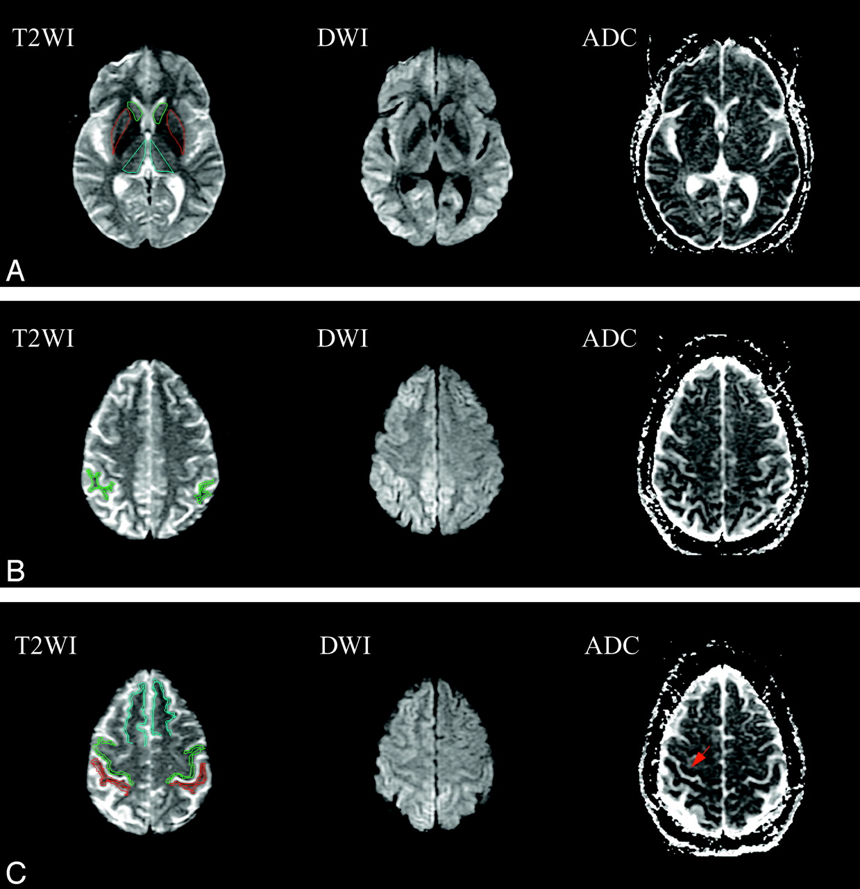
- Fig 1.
ROI in head of caudate nucleus (green), putamen (red), thalamus (cyan) (A), supramarginal gyri (SMG) (B), precentral gyri (PMC) (green), postcentral gyri (PSC) (red), and superior frontal gyri (SFG) (cyan) (C) on EPI B0 T2-weighted images (T2WI), and the corresponding diffusion-weighted images (DWI) and apparent diffusion coefficient maps (ADC). Bilateral abnormal hyperintensity can be observed on DWI with corresponding decreased intensity on ADC maps in thalamus (A) frontal lobes (B), and parietal lobes (C). Characteristic sparing of the PMC and PSC are seen in best in C, where these areas of Rolandic cortex appear normal. Hypointensity can be seen in the right Rolandic cortex on the ADC maps (arrow).
- Fig 2.
ADCs measured in gray matter ROI in patients with CJD (white bars) are significantly lower (14% on average) than ADCs in the corresponding ROI of control patients (shaded bars) in all areas including both commonly affected areas of the frontal lobes (SFG) and parietal lobes (SMG) and typically spared frontal (PMC) and parietal (PSC) Rolandic cortex (individual ROI P value, 0.001–0.05).
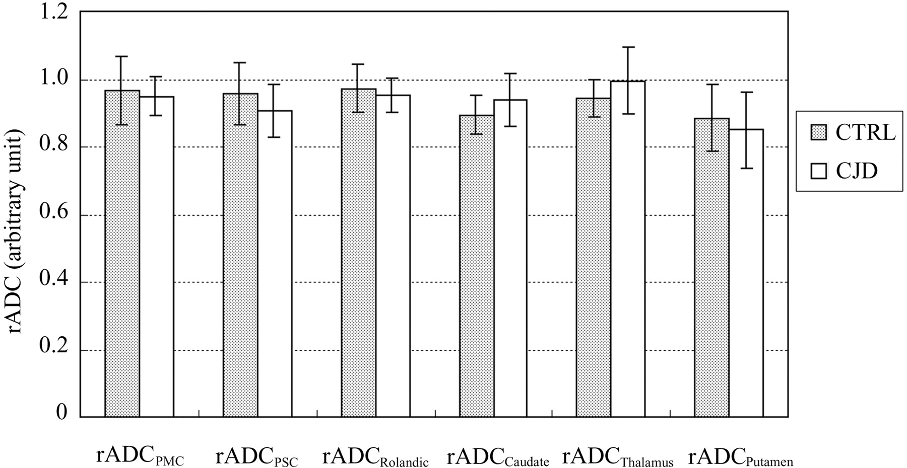
- Fig 3.
rADC ratios between frontal (PMC) and parietal (PSC) Rolandic cortex ROI and commonly affected adjacent frontal (SFG) and parietal (SMG) lobe neocortical ROI in patients with CJD (white bars) do not differ significantly from the equivalent rADC ratios in control patients (shaded bars), confirming that gray matter diffusivity is abnormal to a similar degree in Rolandic cortex and other cortical areas in early CJD, despite apparent “sparing” of Rolandic cortex on DWI (Fig 1C). Likewise, rADC ratios in the deep nuclei do not differ between patients with CJD and control patients, suggesting that the degree of involvement of the caudate, putamen, and thalamus is similar to that of neocortex (individual P value range: 0.25–0.70).


In this issue


Jump to section
Related Articles
Cited By...
- Multicentre multiobserver study of diffusion-weighted and fluid-attenuated inversion recovery MRI for the diagnosis of sporadic Creutzfeldt-Jakob disease: a reliability and agreement study
- Diffusion-weighted MRI hyperintensity patterns differentiate CJD from other rapid dementias
- High-b-Value Diffusion MR Imaging and Basal Nuclei Apparent Diffusion Coefficient Measurements in Variant and Sporadic Creutzfeldt-Jakob Disease
- Brain-water diffusion coefficients reflect the severity of inherited prion disease
- Enhanced Detection of Diffusion Reductions in Creutzfeldt-Jakob Disease at a Higher B Factor