
Article Figures & Data
Figures
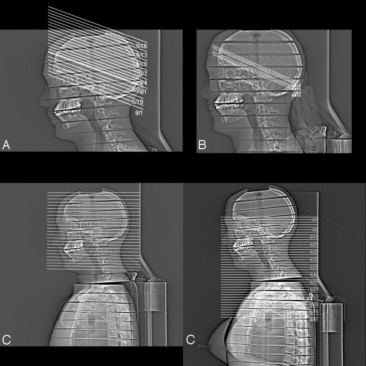
- Fig 1.
Topogram of Alderson-Rando-phantom demonstrating scan areas for standard CT of the head (A), CTP (B), as well as intracranial CTA (C), and cervical CTA (D), respectively.

Tables
Standards CT CT Angiography CT Perfusion Base Cerebrum Intracranial Cervical Protocol I Protocol II Protocol III Collimation (mm) 20 × 1.2 20 × 1.2 64 × 0.6 64 × 0.6 20 × 1.2 20 × 1.2 20 × 1.2 Table feed (mm) 24 24 45,9 69,7 0 0 0 Pitch 1 1 1.2 1.8 0 0 0 Tube current (mAseff) 350 300 160 115 270 200 200 Electrical mA 350 300 383 417 270 200 200 Potential (kV) 120 120 100 120 80 80 120 Rot. time (s) 1 1 0.5 0.33 1 1 1 Scan length (mm) 48 95 250 280 24 24 24 Scan time (s) 6.25 4.58 40 40 40 CTDIvol total (mGy) 39.86 34.16 12.79 7.9 432 320 970 DLP total (mGy x cm) 203 348 386 277 1037 768 2330 Note:—Table feed indicates table movement per second. Pitch corresponds to the dose-relevant pitch (table feed per total collimation considering the number of detector rows acquired simultaneously). CTDI indicates computed tomography dose index; DLP, dose–length product.
- Table 2:
Organ doses and effective doses as measured by lithium fluoride thermoluminescent dosimeters in several positions for different scan protocols
Standard CT CT Angiography CT Perfusion Intracranial Cervical Protocol I Protocol II Protocol III Organ doses (mGy) Cerebrum (overall) 22.2 11.7 9.8 117.9 82.5 325.2 Cerebrum (highest dose) 35.2 9.2 10.3 164.9 114.1 444.2 Skin (total) 11.7 3.1 0.7 26.9 20.2 126.9 Skin (highest dose) 36.2 16.1 19.2 112.3 87.2 404.8 Bone marrow (total) 2.2 2.7 4.3 4.2 4.2 20.7 Bone marrow (skull) 15.8 9.9 10 21.4 21.9 118.1 Eye lens 5.4 15.2 13 13.7 7.9 35.7 Thyroid 1.1 19.7 19.2 2.4 2.4 9.2 Esophagus 0.7 4.8 7.5 2.0 1.2 4.7 Lung 0.4 0.6 4.0 0.7 0.4 1.0 Gonads 0.2 0.3 0.2 0.2 0.2 0.2 Effective dose (mSv)* 1.7 1.9 2.8 1.2/1.3 1.1/1.2 5.0 * Data for effective doses are given for male and female subjects if different (male/female).
In this issue


Jump to section
Related Articles
Cited By...
- Clinical Applications of Conebeam CTP Imaging in Cerebral Disease: A Systematic Review
- Overview of Imaging Modalities in Stroke
- Remote control of neural function by X-ray-induced scintillation
- Evolution from total variation to nonlinear sparsifying transform for sparse-view CT image reconstruction
- The feasibility of low-concentration contrast and low tube voltage in computed tomography perfusion imaging: an animal study
- Safety of Computed Tomographic Angiography in the Evaluation of Patients With Acute Stroke: A Single-Center Experience
- Dynamic Angiography and Perfusion Imaging Using Flat Detector CT in the Angiography Suite: A Pilot Study in Patients with Acute Middle Cerebral Artery Occlusions
- Investigating intracerebral haemorrhage
- Radiation Doses of Cerebral Blood Volume Measurements Using C-Arm CT: A Phantom Study
- Appropriate Use of CT Perfusion following Aneurysmal Subarachnoid Hemorrhage: A Bayesian Analysis Approach
- Emergency Noninvasive Angiography for Acute Intracerebral Hemorrhage
- Defining Intravenous Recombinant Tissue Plasminogen Activator Failure
- A Comparison of Radiation Exposure between Diagnostic CTA and DSA Examinations of Cerebral and Cervicocerebral Vessels
- Dual-Energy CT in the Evaluation of Intracerebral Hemorrhage of Unknown Origin: Differentiation between Tumor Bleeding and Pure Hemorrhage
- Feasibility of Cerebral Blood Volume Mapping by Flat Panel Detector CT in the Angiography Suite: First Experience in Patients with Acute Middle Cerebral Artery Occlusions
- CT Perfusion in Acute Ischemic Stroke: A Comparison of 2-Second and 1-Second Temporal Resolution
- Radiation dose evaluation in multidetector-row CT imaging for acute stroke with an anthropomorphic phantom
- Cumulative Radiation Dose in Patients Admitted with Subarachnoid Hemorrhage: A Prospective Study Using a Self-Developing Film Badge
- Dose Exposure of Patients Undergoing Comprehensive Stroke Imaging by Multidetector-Row CT: Comparison of 320-Detector Row and 64-Detector Row CT Scanners
- Comparison of Image Quality and Radiation Dose between Fixed Tube Current and Combined Automatic Tube Current Modulation in Craniocervical CT Angiography
- Steroid-Responsive Large Vessel Vasculitis: Application of Whole-Brain 320-Detector Row Dynamic Volume CT Angiography and Perfusion
- 320-slice CT neuroimaging: initial clinical experience and image quality evaluation
- Advances in Imaging 2007
- Global Hemispheric CT Hypoperfusion May Differentiate Headache With Associated Neurological Deficits and Lymphocytosis From Acute Stroke