Advertisement

Main menu

  • Home
  • Content
    • Current Issue
    • Accepted Manuscripts
    • Article Preview
    • Past Issue Archive
    • Video Articles
    • AJNR Case Collection
    • Case of the Week Archive
    • Case of the Month Archive
    • Classic Case Archive
  • Special Collections
    • AJNR Awards
    • Low-Field MRI
    • Alzheimer Disease
    • ASNR Foundation Special Collection
    • Photon-Counting CT
    • View All
  • Multimedia
    • AJNR Podcasts
    • AJNR SCANtastic
    • Trainee Corner
    • MRI Safety Corner
    • Imaging Protocols
  • For Authors
    • Submit a Manuscript
    • Submit a Video Article
    • Submit an eLetter to the Editor/Response
    • Manuscript Submission Guidelines
    • Statistical Tips
    • Fast Publishing of Accepted Manuscripts
    • Graphical Abstract Preparation
    • Imaging Protocol Submission
    • Author Policies
  • About Us
    • About AJNR
    • Editorial Board
    • Editorial Board Alumni
  • More
    • Become a Reviewer/Academy of Reviewers
    • Subscribers
    • Permissions
    • Alerts
    • Feedback
    • Advertisers
    • ASNR Home

User menu

  • Alerts
  • Log in

Search

  • Advanced search
American Journal of Neuroradiology

American Journal of Neuroradiology

ASHNR American Society of Functional Neuroradiology ASHNR American Society of Pediatric Neuroradiology ASSR
  • Alerts
  • Log in

Advanced Search

  • Home
  • Content
    • Current Issue
    • Accepted Manuscripts
    • Article Preview
    • Past Issue Archive
    • Video Articles
    • AJNR Case Collection
    • Case of the Week Archive
    • Case of the Month Archive
    • Classic Case Archive
  • Special Collections
    • AJNR Awards
    • Low-Field MRI
    • Alzheimer Disease
    • ASNR Foundation Special Collection
    • Photon-Counting CT
    • View All
  • Multimedia
    • AJNR Podcasts
    • AJNR SCANtastic
    • Trainee Corner
    • MRI Safety Corner
    • Imaging Protocols
  • For Authors
    • Submit a Manuscript
    • Submit a Video Article
    • Submit an eLetter to the Editor/Response
    • Manuscript Submission Guidelines
    • Statistical Tips
    • Fast Publishing of Accepted Manuscripts
    • Graphical Abstract Preparation
    • Imaging Protocol Submission
    • Author Policies
  • About Us
    • About AJNR
    • Editorial Board
    • Editorial Board Alumni
  • More
    • Become a Reviewer/Academy of Reviewers
    • Subscribers
    • Permissions
    • Alerts
    • Feedback
    • Advertisers
    • ASNR Home
  • Follow AJNR on Twitter
  • Visit AJNR on Facebook
  • Follow AJNR on Instagram
  • Join AJNR on LinkedIn
  • RSS Feeds

AJNR Awards, New Junior Editors, and more. Read the latest AJNR updates

OtherPediatric Neuroimaging

Parry-Romberg Syndrome with a Clinically Silent White Matter Lesion

A. Okumura, T. Ikuta, T. Tsuji, T. Kato, H. Fukatsu, S. Naganawa, K. Kato and K. Watanabe
American Journal of Neuroradiology September 2006, 27 (8) 1729-1731;
A. Okumura
  • Find this author on Google Scholar
  • Find this author on PubMed
  • Search for this author on this site
T. Ikuta
  • Find this author on Google Scholar
  • Find this author on PubMed
  • Search for this author on this site
T. Tsuji
  • Find this author on Google Scholar
  • Find this author on PubMed
  • Search for this author on this site
T. Kato
  • Find this author on Google Scholar
  • Find this author on PubMed
  • Search for this author on this site
H. Fukatsu
  • Find this author on Google Scholar
  • Find this author on PubMed
  • Search for this author on this site
S. Naganawa
  • Find this author on Google Scholar
  • Find this author on PubMed
  • Search for this author on this site
K. Kato
  • Find this author on Google Scholar
  • Find this author on PubMed
  • Search for this author on this site
K. Watanabe
  • Find this author on Google Scholar
  • Find this author on PubMed
  • Search for this author on this site
  • Article
  • Figures & Data
  • Info & Metrics
  • Responses
  • References
  • PDF
Loading

Abstract

SUMMARY: We performed a detailed neuroimaging study in a patient with Parry-Romberg syndrome. Proton MR spectroscopy demonstrated normal spectral patterns, though conventional MR imaging revealed high-intensity areas in the entire white matter in the left hemisphere. Single-photon emission tomography showed increased perfusion in the cortex of the affected hemisphere. Pyramidal tracts and optic radiations were preserved on diffusion tensor tractography. We will correlate these neuroimaging findings with normal psychomotor development in our patient.

Parry-Romberg syndrome is characterized by a progressive and self-limited hemifacial atrophy of the skin and subcutaneous tissue and sometimes underlying bony structures of the face and cranium. Common neuroimaging findings of this syndrome are calcification on CT and high-intensity areas on MR imaging.1–9 We report a patient with Parry-Romberg syndrome who had achieved normal psychomotor development despite extensive white matter lesions. We performed precise neuroradiologic studies including single-photon emission CT (SPECT), proton MR spectroscopy (1H-MR spectroscopy), and diffusion tensor imaging (DTI).

Case Report

A 22-month-old boy was referred to our hospital because of facial hemiatrophy and MR imaging abnormalities. He was the first child of healthy nonconsanguineous parents. At 4 months of age, the parents noticed asymmetry of the patient’s face. The skin around the left eyelid and maxillary region was atrophic. Facial hemiatrophy progressed during the following 12 months. He was diagnosed with Parry-Romberg syndrome at 18 months of age. However, his developmental milestones remained normal.

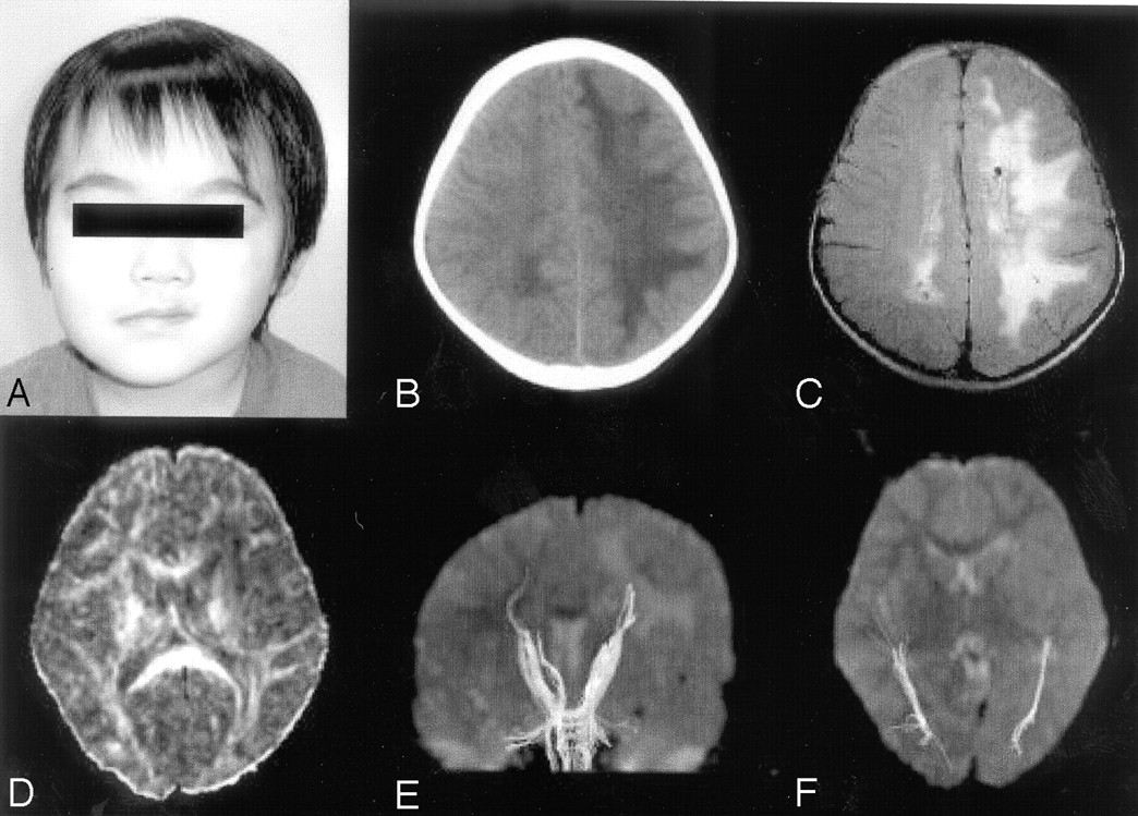
The patient presented at 22 months of age because of widespread white matter lesions in the left hemisphere that showed decreased signal intensities on T1-weighted images and increased signal intensities on T2-weighted and fluid-attenuated inversion recovery (FLAIR) images. A similar white matter lesion was also seen in the occipital areas in the right hemisphere. Findings of a neurologic examination and his psychomotor development were normal at the presentation, though left facial hemiatrophy was observed (Fig 1A). Findings of electroencephalograms and brain stem auditory-evoked potentials were also normal. CT at 34 months of age showed marked low attenuation in the entire white matter in the left hemisphere (Fig 1B). Calcification was not detected.

Fig 1.
  • Download figure
  • Open in new tab
  • Download powerpoint
Fig 1.

Photograph, CT, and MR imaging of the patient. A, Photograph at 34 months of age. Skin and soft-tissue atrophy of the left periorbital and premaxillary areas are recognized. B, CT scan obtained at 34 months of age. Low attenuation is shown in the entire white matter in the left hemisphere. C, FLAIR image obtained at 44 months of age. Abnormal high-intensity areas are observed in the entire white matter in the left hemisphere and in the occipital region in the right hemisphere. Small cysts are seen in the bilateral deep white matter. D, FA map. Decrease of anisotropy is observed in the white matter in the left hemisphere. E, Tractography of pyramidal tracts. Pyramidal tracts are clearly depicted in both hemispheres. F, Tractography of optic radiations, which are also depicted symmetrically.

Serial MR imaging was performed at 33, 44, and 60 months of age. All images demonstrated marked T1 and T2 prolongation in the entire white matter in the left hemisphere and in the occipital region in the right hemisphere (Fig 1C). The extent of the white matter lesions had not changed until 60 months of age. Ventriculomegaly or cortical abnormalities had not been observed until the last study, though small cysts in the bilateral deep white matter were newly recognized on the second and the third scans. MR angiography performed at 33 and 44 months of age did not show any vascular abnormalities.

SPECT was performed with technetium Tc99m hexamethylpropyleneamine oxime by using GCA-9300A (Toshiba, Tokyo, Japan) at 31 months of age. On visual inspection, blood perfusion was decreased in the entire white matter in the left hemisphere. In contrast, it was relatively increased in the cerebral cortex in the affected hemisphere. Quantitative regions of interest analysis also showed that blood flow within the white matter in the left hemisphere was 55% of the corresponding region of the right hemisphere, whereas that within the cortex in the left frontal lobe was 113% of the contralateral hemisphere (Fig 2).

Fig 2.
  • Download figure
  • Open in new tab
  • Download powerpoint
Fig 2.

SPECT findings. Blood perfusion is decreased in the white matter in the left hemisphere, whereas it is increased in the cerebral cortex in the ipsilateral hemisphere.

1H-MR spectroscopy was performed on a 3T MR imaging scanner (Trio, Siemens, Erlangen, Germany) at 60 months of age. 2D chemical shift imaging with a point-resolved 1H spectroscopy sequence was used, covering bilateral deep white matter with scan parameters as follows: TR/TE, 1700/30 ms; 3 number of averaging; voxel size, 10 × 10 × 15 mm; matrix, 8 × 8. 1H-MR spectroscopy showed normal spectral patterns in the white matter in the frontal and parietal regions in the left hemisphere as well as in the corresponding regions in the right hemisphere (Fig 3).

Fig 3.
  • Download figure
  • Open in new tab
  • Download powerpoint
Fig 3.

Proton MR spectroscopy. Normal spectral patterns are observed in both hemispheres.

DTI was performed on a 3T MR imaging scanner by using a Stejskal-Tanner sequence with single-shot spin-echo type echo-planar imaging (TR/TE, 7000/75 ms; matrix size, 128 × 128; field of view, 256 mm; 60 axial sections; 2-mm section thickness). We collected diffusion-weighted images along 6 different directions with a b value of 700 seconds/mm2, as well as an image without diffusion weighting (b = 0 seconds/mm2). The brain fiber tracking was performed by using the free software dTV (version 1.5, http://www.utradiology.umin.jp/people/masutani/dTV.htm) for DTI analysis developed by the Image Computing and Analysis Laboratory, Department of Radiology, the University of Tokyo Hospital, Japan.

A fractional anisotropy (FA) map showed remarkable decrease of anisotropy in the white matter in the left hemisphere (Fig 1D). However, tractography demonstrated excellent resolution of white matter tracts in both hemispheres. Pyramidal tracts and optic radiations were clearly depicted without apparent asymmetry (Fig 1E, -F).

At the last follow-up at 65 months of age, our patient’s psychomotor development was still normal. Findings of neurologic examinations were also normal. Neurologic symptoms such as migraine, seizure, or facial pain had never been observed.

Discussion

The outstanding feature of our patient is normal psychomotor development, though conventional MR imaging showed extensive white matter lesions. The conventional MR imaging findings in our patient are consistent with several previous reports.2–7 However, SPECT, 1H-MR spectroscopy, and DTI provided some new findings in our patient, which correlated with normal psychomotor development.

SPECT showed relatively increased regional blood flow in the cortex of the affected hemisphere, whereas it was remarkably reduced in the white matter of the same hemisphere. On the other hand, DeFelipe et al6 reported that SPECT showed an extensive hypoperfusion in the parietooccipital area in the affected hemisphere in a patient with this syndrome and temporal lobe epilepsy. We believe that the difference between these patients is explained by the presence or absence of significant cortical dysfunction. Relatively increased blood flow in our patient is related to the preserved cortical function, despite widespread white matter abnormalities. As a result, he can achieve normal psychomotor development and remain free of neurologic symptoms.

1H-MR spectroscopy findings were noteworthy in our patient, revealing no abnormalities in the affected white matter. Several changes in levels of metabolites, such as N-acetylaspartate, choline, and lactate, are usually observed in various degenerative, ischemic, or inflammatory brain disorders, but normal 1H-MR spectroscopy findings suggest that neuronal or axonal damage is rather mild, if present, despite extensive changes on conventional MR imaging. This is also related to the preserved brain function in our patient.

DTI findings were also remarkable in our patient. DTI is an imaging method that reflects the molecular movement of water. Diffusion of water molecules is restricted by cell membranes in cerebral tissue. Water molecules can diffuse relatively freely parallel but, only to a limited extent, perpendicular to tracts. The multiple myelin laminae are closely related to restriction of water molecules in the white matter. In our patient, widespread decrease of FA was recognized in the white matter corresponding to the abnormal high-intensity areas on conventional MR imaging. This finding suggests that MR imaging abnormalities may be attributable to impaired myelination. On the other hand, pyramidal tracts and optic radiations were symmetrically depicted on tractography. This suggests that white matter pathways are likely to be preserved in our patient. Axonal injuries may be mild, if present, though widespread white matter abnormalities were observed on both conventional MR imaging and DTI.

In summary, SPECT, 1H-MR spectroscopy, and DTI suggested the preserved cortical function and white matter pathways in our patient, though conventional MR imaging revealed extensive white matter abnormalities. These advanced neuroimaging techniques are useful in understanding the functional aspects of the brain in patients with conventional MR imaging abnormalities.

References

  1. ↵
    Fry JA, Alvarellos A, Fink CW, et al. Intracranial findings in progressive facial hemiatrophy. J Rheumatol 1992;19:956–58
    PubMed
  2. ↵
    Moko SB, Mistry Y, Blandin de Chalain TM. Parry-Romberg syndrome: intracranial MRI appearances. J Craniomaxillofac Surg 2003;31:321–24
    PubMed
  3. Taylor HM, Robinson R, Cox T. Progressive facial hemiatrophy: MRI appearances. Dev Med Child Neurol 1997;39:484–86
    PubMed
  4. Cory RC, Clayman DA, Faillace WJ, et al. Clinical and radiologic findings in progressive facial hemiatrophy (Parry-Romberg syndrome). AJNR Am J Neuroradiol 1997;18:751–57
    Abstract/FREE Full Text
  5. Shah JR, Juhasz C, Kupsky WJ, et al. Rasmussen encephalitis associated with Parry-Romberg syndrome. Neurology 2003;61:395–97
    Abstract/FREE Full Text
  6. ↵
    DeFelipe J, Segura T, Arellano JI, et al. Neuropathological findings in a patient with epilepsy and the Parry-Romberg syndrome. Epilepsia 2001;42:1198–203
    PubMed
  7. ↵
    Yano T, Sawaishi Y, Toyono M, et al. Progressive facial hemiatrophy after epileptic seizures. Pediatr Neurol 2000;23:164–66
    CrossRefPubMed
  8. Terstegge K, Kunath B, Felber S, et al. MR of brain involvement in progressive facial hemiatrophy (Romberg disease): reconsideration of a syndrome. AJNR Am J Neuroradiol 1994;15:145–50
    Abstract/FREE Full Text
  9. ↵
    Sathornsumetee S, Schanberg L, Rabinovich E, et al. Parry-Romberg syndrome with fatal brain stem involvement. J Pediatr 2005;146:429–31
    CrossRefPubMed
  • Received September 8, 2005.
  • Accepted after revision September 23, 2005.
  • Copyright © American Society of Neuroradiology
View Abstract
PreviousNext
Back to top

In this issue

American Journal of Neuroradiology: 27 (8)
American Journal of Neuroradiology
Vol. 27, Issue 8
September 2006
  • Table of Contents
  • Index by author
Advertisement
Print
Download PDF
Email Article

Thank you for your interest in spreading the word on American Journal of Neuroradiology.

NOTE: We only request your email address so that the person you are recommending the page to knows that you wanted them to see it, and that it is not junk mail. We do not capture any email address.

Enter multiple addresses on separate lines or separate them with commas.
Parry-Romberg Syndrome with a Clinically Silent White Matter Lesion
(Your Name) has sent you a message from American Journal of Neuroradiology
(Your Name) thought you would like to see the American Journal of Neuroradiology web site.
CAPTCHA
This question is for testing whether or not you are a human visitor and to prevent automated spam submissions.
Cite this article
A. Okumura, T. Ikuta, T. Tsuji, T. Kato, H. Fukatsu, S. Naganawa, K. Kato, K. Watanabe
Parry-Romberg Syndrome with a Clinically Silent White Matter Lesion
American Journal of Neuroradiology Sep 2006, 27 (8) 1729-1731;

Citation Manager Formats

  • BibTeX
  • Bookends
  • EasyBib
  • EndNote (tagged)
  • EndNote 8 (xml)
  • Medlars
  • Mendeley
  • Papers
  • RefWorks Tagged
  • Ref Manager
  • RIS
  • Zotero
0 Responses
Respond to this article
Share
Bookmark this article
Parry-Romberg Syndrome with a Clinically Silent White Matter Lesion
A. Okumura, T. Ikuta, T. Tsuji, T. Kato, H. Fukatsu, S. Naganawa, K. Kato, K. Watanabe
American Journal of Neuroradiology Sep 2006, 27 (8) 1729-1731;
del.icio.us logo Twitter logo Facebook logo Mendeley logo
  • Tweet Widget
  • Facebook Like
  • Google Plus One

Jump to section

  • Article
    • Abstract
    • Case Report
    • Discussion
    • References
  • Figures & Data
  • Info & Metrics
  • Responses
  • References
  • PDF

Related Articles

  • No related articles found.
  • PubMed
  • Google Scholar

Cited By...

  • Brain Abnormalities and Epilepsy in Patients with Parry-Romberg Syndrome
  • Case 1: Alopecia in a Healthy 9-year-old Girl
  • Parry Romberg Syndrome: 7 Cases and Literature Review
  • Crossref
  • Google Scholar

This article has not yet been cited by articles in journals that are participating in Crossref Cited-by Linking.

More in this TOC Section

  • fetal brain development of 10 weeks gestation
  • CHARGE fetal MRI clival cleft
  • Neuroimaging Delineation and Progression of SLSMD
Show more Pediatric Neuroimaging

Similar Articles

Advertisement

Indexed Content

  • Current Issue
  • Accepted Manuscripts
  • Article Preview
  • Past Issues
  • Editorials
  • Editor's Choice
  • Fellows' Journal Club
  • Letters to the Editor
  • Video Articles

Cases

  • Case Collection
  • Archive - Case of the Week
  • Archive - Case of the Month
  • Archive - Classic Case

More from AJNR

  • Trainee Corner
  • Imaging Protocols
  • MRI Safety Corner

Multimedia

  • AJNR Podcasts
  • AJNR Scantastics

Resources

  • Turnaround Time
  • Submit a Manuscript
  • Submit a Video Article
  • Submit an eLetter to the Editor/Response
  • Manuscript Submission Guidelines
  • Statistical Tips
  • Fast Publishing of Accepted Manuscripts
  • Graphical Abstract Preparation
  • Imaging Protocol Submission
  • Evidence-Based Medicine Level Guide
  • Publishing Checklists
  • Author Policies
  • Become a Reviewer/Academy of Reviewers
  • News and Updates

About Us

  • About AJNR
  • Editorial Board
  • Editorial Board Alumni
  • Alerts
  • Permissions
  • Not an AJNR Subscriber? Join Now
  • Advertise with Us
  • Librarian Resources
  • Feedback
  • Terms and Conditions
  • AJNR Editorial Board Alumni

American Society of Neuroradiology

  • Not an ASNR Member? Join Now

© 2025 by the American Society of Neuroradiology All rights, including for text and data mining, AI training, and similar technologies, are reserved.
Print ISSN: 0195-6108 Online ISSN: 1936-959X

Powered by HighWire