Advertisement

Main menu

  • Home
  • Content
    • Current Issue
    • Accepted Manuscripts
    • Article Preview
    • Past Issue Archive
    • Video Articles
    • AJNR Case Collection
    • Case of the Week Archive
    • Case of the Month Archive
    • Classic Case Archive
  • Special Collections
    • AJNR Awards
    • Low-Field MRI
    • Alzheimer Disease
    • ASNR Foundation Special Collection
    • Photon-Counting CT
    • View All
  • Multimedia
    • AJNR Podcasts
    • AJNR SCANtastic
    • Trainee Corner
    • MRI Safety Corner
    • Imaging Protocols
  • For Authors
    • Submit a Manuscript
    • Submit a Video Article
    • Submit an eLetter to the Editor/Response
    • Manuscript Submission Guidelines
    • Statistical Tips
    • Fast Publishing of Accepted Manuscripts
    • Graphical Abstract Preparation
    • Imaging Protocol Submission
    • Author Policies
  • About Us
    • About AJNR
    • Editorial Board
    • Editorial Board Alumni
  • More
    • Become a Reviewer/Academy of Reviewers
    • Subscribers
    • Permissions
    • Alerts
    • Feedback
    • Advertisers
    • ASNR Home

User menu

  • Alerts
  • Log in

Search

  • Advanced search
American Journal of Neuroradiology

American Journal of Neuroradiology

ASHNR American Society of Functional Neuroradiology ASHNR American Society of Pediatric Neuroradiology ASSR
  • Alerts
  • Log in

Advanced Search

  • Home
  • Content
    • Current Issue
    • Accepted Manuscripts
    • Article Preview
    • Past Issue Archive
    • Video Articles
    • AJNR Case Collection
    • Case of the Week Archive
    • Case of the Month Archive
    • Classic Case Archive
  • Special Collections
    • AJNR Awards
    • Low-Field MRI
    • Alzheimer Disease
    • ASNR Foundation Special Collection
    • Photon-Counting CT
    • View All
  • Multimedia
    • AJNR Podcasts
    • AJNR SCANtastic
    • Trainee Corner
    • MRI Safety Corner
    • Imaging Protocols
  • For Authors
    • Submit a Manuscript
    • Submit a Video Article
    • Submit an eLetter to the Editor/Response
    • Manuscript Submission Guidelines
    • Statistical Tips
    • Fast Publishing of Accepted Manuscripts
    • Graphical Abstract Preparation
    • Imaging Protocol Submission
    • Author Policies
  • About Us
    • About AJNR
    • Editorial Board
    • Editorial Board Alumni
  • More
    • Become a Reviewer/Academy of Reviewers
    • Subscribers
    • Permissions
    • Alerts
    • Feedback
    • Advertisers
    • ASNR Home
  • Follow AJNR on Twitter
  • Visit AJNR on Facebook
  • Follow AJNR on Instagram
  • Join AJNR on LinkedIn
  • RSS Feeds

AJNR Awards, New Junior Editors, and more. Read the latest AJNR updates

OtherNeurointervention

Intra-Arterial Digital Subtraction Angiography Through the Ear Artery for Experimental Aneurysm Imaging

Y.H. Ding, D. Dai, M.A. Danielson, R. Kadirvel, D.A. Lewis, H.J. Cloft and D.F. Kallmes
American Journal of Neuroradiology September 2006, 27 (8) 1700-1702;
Y.H. Ding
  • Find this author on Google Scholar
  • Find this author on PubMed
  • Search for this author on this site
D. Dai
  • Find this author on Google Scholar
  • Find this author on PubMed
  • Search for this author on this site
M.A. Danielson
  • Find this author on Google Scholar
  • Find this author on PubMed
  • Search for this author on this site
R. Kadirvel
  • Find this author on Google Scholar
  • Find this author on PubMed
  • Search for this author on this site
D.A. Lewis
  • Find this author on Google Scholar
  • Find this author on PubMed
  • Search for this author on this site
H.J. Cloft
  • Find this author on Google Scholar
  • Find this author on PubMed
  • Search for this author on this site
D.F. Kallmes
  • Find this author on Google Scholar
  • Find this author on PubMed
  • Search for this author on this site
  • Article
  • Figures & Data
  • Info & Metrics
  • Responses
  • References
  • PDF
Loading

Abstract

SUMMARY: Digital subtraction angiography through the central artery of the ear was performed to show elastase-induced aneurysms in 34 rabbits. Twenty-eight (82%) aneurysms in which common origins of the bilateral common carotid artery (CCA) were found were well shown. All 6 other aneurysms not well shown had separate origins of the CCAs from the aortic arch (P < .001). This method can be used for angiographic follow-up when there is a common origin of the CCAs.

Currently, conventional intra-arterial digital subtraction angiography (IADSA) through the femoral artery is the standard technique to examine the dimensions of an aneurysm in rabbits,1–4 though it is invasive and expensive. As a less-invasive, simple, and economic technique, IADSA through the central artery of the ear was reported recently.5 We set out to validate the use of IADSA through the ear artery for evaluation of elastase-induced aneurysm models in rabbits.

Description of the Technique

Elastase-induced saccular aneurysms created at the origin of the right common carotid artery (RCCA) in 34 New Zealand white rabbits (body weight, 3–4 kg) were analyzed in this study, including 8 uncoiled aneurysms and 26 aneurysms previously embolized with platinum coils. All procedures were approved by our Institutional Animal Care and Use Committee. Detailed procedures for aneurysm creation have been described.6 Briefly, the stump of the RCCA was converted to an “aneurysm” cavity by distal ligation and intraluminal incubation of porcine elastase. Aneurysm embolization, when performed, was done from a transfemoral approach, as previously described in detail.1–2

IADSA

IADSA through the central ear artery of the left ear was performed in all 34 aneurysms. The left ear was chosen because the contrast can flow retrogradely into the aortic arch through left common carotid artery (LCCA) (Fig 1). On the right side, the RCCA was occluded during aneurysm creation, so retrograde flow to the aortic arch would be difficult.

Fig 1.
  • Download figure
  • Open in new tab
  • Download powerpoint
Fig 1.

Ear IADSA showing contrast from the left ear artery (left block arrow) to the parent artery of the aneurysm.

A 24-gauge angiocatheter was placed in the central artery of the left ear. Digital subtraction angiography (DSA) (Advantx, GE Healthcare, Milwaukee, Wis) at 2 frames per second was performed after injection of 7 mL of iodinated contrast material (iohexol, Omnipaque 300, GE Healthcare, Piscataway, NJ) through the catheter for 2 seconds; the injection rate was approximately 3 mL/s. Radiopaque sizing markers, consisting of metallic spheres ranging from 2–7 mm in diameter, were placed over the chest, as close as possible to the brachiocephalic artery but not obscuring the region of the aneurysm.

Conventional IADSA from a transfemoral approach was also performed in 22 of the 34 aneurysms. Briefly, a cut-down was performed over the right common femoral artery, and a 5F sheath was placed in the artery. The side-port of the sheath was flushed with heparinized saline; then, a bolus of heparin (100 U/kg) was delivered through the sheath. A 5F Envoy catheter (Cordis Endovascular, Miami Lakes, Fla) was advanced into the brachiocephalic artery through the sheath under fluoroscopy. DSA at 2 frames per second was performed with a hand injection of 5 mL of iodinated contrast material (Omnipaque 300) at a rate of approximately 3 mL/s.

Intravenous DSA (IVDSA) was performed in the other 12 aneurysms. Detailed procedures for IVDSA have been described elsewhere.7

Image Quality Evaluation

Image quality of the central ear artery DSA was evaluated. Two categories were classified for consideration: grade A, in which both the aneurysm and parent artery were clearly shown; and grade B, in which any portion of the aneurysm and/or parent artery was not well shown. All DSA images for any given subject were viewed separately by the 2 experienced observers, an experienced neuroradiologist (D.F.K.) and a trained research fellow (Y.H.D). Agreement was reached between the 2 observers for all aneurysms.

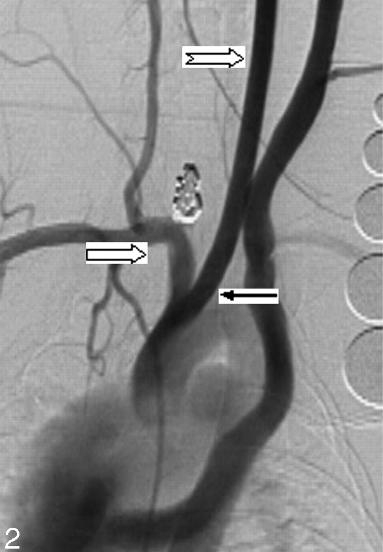
Twenty-eight (82%) of the 34 cases were considered grade A, or good, image quality (Fig 2). Six (18%) of 34 aneurysms were judged grade B, or poor, image quality. Of these 6 cases, 2 had poor opacification of the aneurysm neck region, 3 had poor opacification of the parent artery, and 1 had poor opacification of both the parent artery and neck region (Fig 3).

Fig 2.
  • Download figure
  • Open in new tab
  • Download powerpoint
Fig 2.

Ear IADSA. Both embolized aneurysm and parent artery (right block arrow) are well shown. Note that more contrast fills the LCCA (notched right arrow) because contrast material is injected through the left ear artery. In addition, LCCA and RCCA originate at the same location from the aorta (black arrow).

Fig 3.
  • Download figure
  • Open in new tab
  • Download powerpoint
Fig 3.

IADSA through the left ear artery. Both embolized aneurysm and parent artery (right block arrow) are not well shown. Note that the LCCA (notched right arrow) is seriously opacified. On the contrary, the parent artery is very faint. LCCA and RCCA, originating separately from the aorta (black arrow).

Anatomic Evaluation

There were 2 types of vessel origination: type A, with a common origin for the RCCA and LCCA; and type B, with separate origins of the RCCA and LCCA from the aortic arch. All 28 aneurysms with grade A image quality had type A anatomic configuration (Fig 2). In all 6 cases with poor opacification (grade B), anatomic configuration type B was shown (Fig 3).

Statistical Analysis

The relationship between image quality and anatomic configuration was compared by using the Fisher exact test. Image quality was coordinated with vessel origination (P < .001).

Discussion

Elastase-induced aneurysms in rabbits have been proved to be an excellent model for preclinical device development and basic neurovascular research.7–11 Unfortunately, because of the small size of the femoral artery, placement of arterial catheters usually requires ligation of the access vessel. Thus, serial studies by using conventional angiographic follow-up are either extremely difficult or impossible. The current study showed that most aneurysms can be well shown by IADSA through the central artery of the left ear.

The observation that separate origin of the carotid arteries leads to poor image quality could be predicted on the basis of the pattern of contrast opacification with ear artery DSA. The contrast fills retrograde down the LCCA into the aortic arch. If the origin of the LCCA is separate and downstream from the RCCA, then opacification of the aneurysm is poor. In the cases of common origins of the carotid arteries, the contrast traverses the brachiocephalic artery before entering the aortic arch. Then the contrast is carried antegrade to opacify the aneurysm.

IADSA through the central artery of the ear artery offers numerous advantages compared with IADSA from a transfemoral approach. IADSA through the ear artery is less invasive and economic, and most important, it can be repeated many times in each rabbit subject. Thus, this method is especially useful for studies using multiple time point follow-ups. In addition, it offers high concentrations of intravascular contrast, which we consider superior to that achieved by using IVDSA.

There are several technical limitations that decrease the accuracy of IADSA through the ear artery. In most cases, the concentration of intravascular contrast is less than that achieved with transfemoral angiography, with the catheter placed in the brachiocephalic artery. Another factor is motion artifact, which may be substantial in some cases because contrast may irritate the subject during injection. Third, anatomy variation, such as separate origin of the carotid arteries from the aortic arch, impacts the image quality. Previous series of ear artery DSA noted favorable anatomy in approximately 60% of 23 rabbits.5 Our series, which was slightly larger, had favorable arch anatomy for ear DSA in more than 80% of cases. Variation may depend on the source of the rabbits, though the New Zealand white rabbit represents a highly out-bred species.

Other investigators have used noninvasive methods such as CT angiography and MR angiography for follow-up of experimental aneurysms in rabbits.12 Also, in intraoperative angiograms obtained clinically, the superficial temporal artery may be accessed to allow retrograde opacification of the carotid arterial system.13 We offer IADSA through the ear artery as a complementary technique, especially in laboratories without MR and CT scanners available for animal research.

Conclusion

Angiographic follow-up of right-sided elastase-induced aneurysms in rabbits can be performed by using the central artery of the left ear when there is a common origin of the CCAs from the aortic arch.

Footnotes

  • This study was supported by NIH grant NS42646.

References

  1. ↵
    Kallmes DF, Fujiwara NH. New expandable hydrogel-platinum coil hybrid device for aneurysm embolization. AJNR Am J Neuroradiol 2002;23:1580–88
    Abstract/FREE Full Text
  2. ↵
    Kallmes DF, Fujiwara NH, Yuen D, et al. A collagen-based coil for embolization of saccular aneurysms in a New Zealand white rabbit model. AJNR Am J Neuroradiol 2003;24:591–96
    Abstract/FREE Full Text
  3. Karamessini MT, Kagadis GC, Petsas T, et al. CT angiography with three-dimensional techniques for the early diagnosis of intracranial aneurysms: comparison with intra-arterial DSA and the surgical findings. Eur J Radiol 2004;49:212–23
    CrossRefPubMed
  4. ↵
    Dammert S, Krings T, Moller-Hartmann W, et al. Detection of intracranial aneurysms with multislice CT: comparison with conventional angiography. Neuroradiology 2004;46:427–34. Epub 2004 Apr 23
    PubMed
  5. ↵
    Miskolczi L, Nemes B, Cesar L, et al. Contrast injection via the central artery of the left ear in rabbits: a new technique to simplify follow-up studies. AJNR Am J Neuroradiol 2005;26:1964–66
    Abstract/FREE Full Text
  6. ↵
    Altes TA, Cloft HJ, Short JG, et al. 1999 ARRS Executive Council Award: creation of saccular aneurysms in the rabbit—a model suitable for testing endovascular devices. AJR Am J Roentgenol 2000;174:349–54
    CrossRefPubMed
  7. ↵
    Fujiwara NH, Cloft HJ, Marx WF, et al. Serial angiography in an elastase-induced aneurysm model in rabbits: evidence for progressive aneurysm enlargement after creation. AJNR Am J Neuroradiol 2001;22:698–703
    Abstract/FREE Full Text
  8. Short JG, Fujiwara, NH, Marx WF, et al. Elastase-induced saccular aneurysms in rabbits: comparison of geometric features with those of human aneurysms. AJNR Am J Neuroradiol 2001;22:1833–37
    Abstract/FREE Full Text
  9. Kallmes DF, Fujiwara NH, Berr SS, et al. Elastase-induced saccular aneurysms in rabbits: a dose-escalation study. AJNR Am J Neuroradiol 2002;23:295–98
    Abstract/FREE Full Text
  10. Kallmes DF, Williams AD, Cloft HJ, et al. Platinum coil-mediated implantation of growth factor-secreting endovascular tissue grafts: an in vivo study. Radiology 1998;207:519–23
    PubMed
  11. ↵
    De Gast AN, Altes TA, Marx WF, et al. Transforming growth factor beta-coated platinum coils for endovascular treatment of aneurysms: an animal study. Neurosurgery 2001;49:690–94
    CrossRefPubMed
  12. ↵
    Doerfler A, Becker W, Wanke I, et al. Multimodal imaging in the elastase-induced aneurysm model in rabbits: a comparative study using serial DSA, MRA and CTA. Rofo Fortschr Gebb Rontgenstr Neuen Bildgeb Verfahr 2004;176:590–96
  13. ↵
    Lee MC, MacDonald RL. Intraoperative cerebral angiography: superficial temporal artery method and results. Neurosurgery 2003;53:1067–74
    PubMed
  • Received July 29, 2005.
  • Accepted after revision April 2, 2006.
  • Copyright © American Society of Neuroradiology
View Abstract
PreviousNext
Back to top

In this issue

American Journal of Neuroradiology: 27 (8)
American Journal of Neuroradiology
Vol. 27, Issue 8
September 2006
  • Table of Contents
  • Index by author
Advertisement
Print
Download PDF
Email Article

Thank you for your interest in spreading the word on American Journal of Neuroradiology.

NOTE: We only request your email address so that the person you are recommending the page to knows that you wanted them to see it, and that it is not junk mail. We do not capture any email address.

Enter multiple addresses on separate lines or separate them with commas.
Intra-Arterial Digital Subtraction Angiography Through the Ear Artery for Experimental Aneurysm Imaging
(Your Name) has sent you a message from American Journal of Neuroradiology
(Your Name) thought you would like to see the American Journal of Neuroradiology web site.
CAPTCHA
This question is for testing whether or not you are a human visitor and to prevent automated spam submissions.
Cite this article
Y.H. Ding, D. Dai, M.A. Danielson, R. Kadirvel, D.A. Lewis, H.J. Cloft, D.F. Kallmes
Intra-Arterial Digital Subtraction Angiography Through the Ear Artery for Experimental Aneurysm Imaging
American Journal of Neuroradiology Sep 2006, 27 (8) 1700-1702;

Citation Manager Formats

  • BibTeX
  • Bookends
  • EasyBib
  • EndNote (tagged)
  • EndNote 8 (xml)
  • Medlars
  • Mendeley
  • Papers
  • RefWorks Tagged
  • Ref Manager
  • RIS
  • Zotero
0 Responses
Respond to this article
Share
Bookmark this article
Intra-Arterial Digital Subtraction Angiography Through the Ear Artery for Experimental Aneurysm Imaging
Y.H. Ding, D. Dai, M.A. Danielson, R. Kadirvel, D.A. Lewis, H.J. Cloft, D.F. Kallmes
American Journal of Neuroradiology Sep 2006, 27 (8) 1700-1702;
del.icio.us logo Twitter logo Facebook logo Mendeley logo
  • Tweet Widget
  • Facebook Like
  • Google Plus One

Jump to section

  • Article
    • Abstract
    • Description of the Technique
    • Discussion
    • Conclusion
    • Footnotes
    • References
  • Figures & Data
  • Info & Metrics
  • Responses
  • References
  • PDF

Related Articles

  • No related articles found.
  • PubMed
  • Google Scholar

Cited By...

  • No citing articles found.
  • Crossref
  • Google Scholar

This article has not yet been cited by articles in journals that are participating in Crossref Cited-by Linking.

More in this TOC Section

  • A Retrospective Study in Tentorial DAVFs
  • Proximal Protection Devices for Carotid Stenting
  • Rescue Reentry in Carotid Near-Occlusion
Show more NEUROINTERVENTION

Similar Articles

Advertisement

Indexed Content

  • Current Issue
  • Accepted Manuscripts
  • Article Preview
  • Past Issues
  • Editorials
  • Editor's Choice
  • Fellows' Journal Club
  • Letters to the Editor
  • Video Articles

Cases

  • Case Collection
  • Archive - Case of the Week
  • Archive - Case of the Month
  • Archive - Classic Case

More from AJNR

  • Trainee Corner
  • Imaging Protocols
  • MRI Safety Corner
  • Book Reviews

Multimedia

  • AJNR Podcasts
  • AJNR Scantastics

Resources

  • Turnaround Time
  • Submit a Manuscript
  • Submit a Video Article
  • Submit an eLetter to the Editor/Response
  • Manuscript Submission Guidelines
  • Statistical Tips
  • Fast Publishing of Accepted Manuscripts
  • Graphical Abstract Preparation
  • Imaging Protocol Submission
  • Evidence-Based Medicine Level Guide
  • Publishing Checklists
  • Author Policies
  • Become a Reviewer/Academy of Reviewers
  • News and Updates

About Us

  • About AJNR
  • Editorial Board
  • Editorial Board Alumni
  • Alerts
  • Permissions
  • Not an AJNR Subscriber? Join Now
  • Advertise with Us
  • Librarian Resources
  • Feedback
  • Terms and Conditions
  • AJNR Editorial Board Alumni

American Society of Neuroradiology

  • Not an ASNR Member? Join Now

© 2025 by the American Society of Neuroradiology All rights, including for text and data mining, AI training, and similar technologies, are reserved.
Print ISSN: 0195-6108 Online ISSN: 1936-959X

Powered by HighWire