
Article Figures & Data
Figures
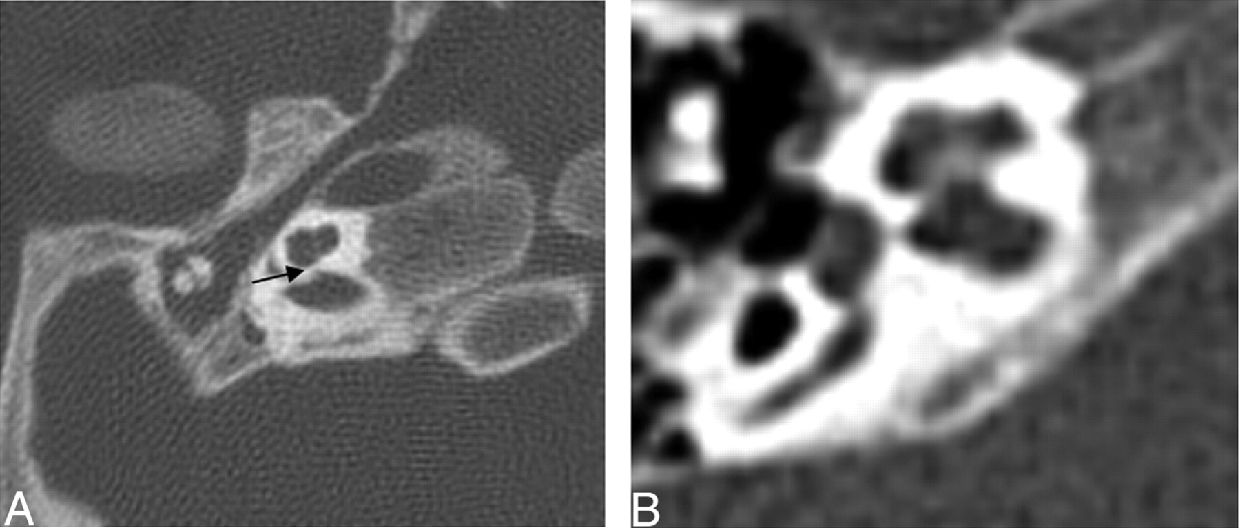
- Fig. 1.
Axial CT at the level of the cochlea demonstrates cochlear aperture atresia or trapped cochlea. The modiolus is dysplastic as well. Left, there is ossification over the cochlear aperture (black arrow), which is normally widely patent and occupied by the cochlear nerve (right).
- Fig. 2.
Axial CT of the cochlea. Left: Dysplastic posterior strut of the modiolus (black arrows) with lack of the normally ossified crown-like structure seen on the right.
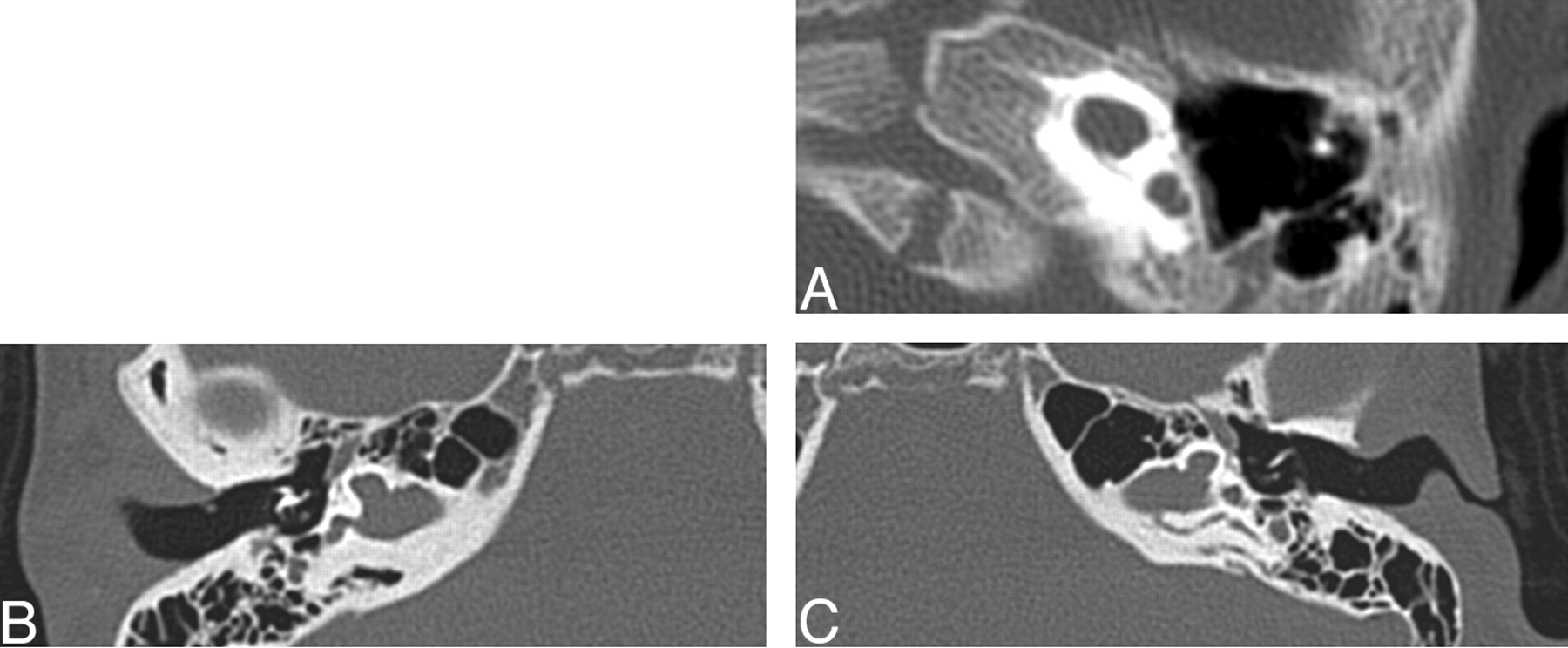
- Fig. 3.
Three axial CT images at the level of the cochlea demonstrating examples of cochlea that are amorphous and lack definable turns, internal septation, or a modiolus. The lower 2 cases demonstrate gross hypoplasia.
- Fig. 4.
Axial CT of the cochlea. Left, Apical turn hypoplasia. Right: Normal cochlea.
- Fig. 5.
Axial images at the level of the vestibule. Top, Bilateral absent semicircular canals with isolated vestibules. Bottom, Normally formed semicircular canals.
- Fig. 6.
Axial CT at the level of the vestibule. The black arrow points to a dysplastic vestibule. The middle ear cavity is small and nonpneumatized, and the head of the malleus is ankylosed to the anterior epitympanic wall.
- Fig. 7.
Axial CT images of the middle ear. Left, Small middle ear cavity in patient with CHARGE syndrome with ankylosis of the dysplastic ossicles to each other and to the epitympanic wall. Right, Normal middle ear cavity.
- Fig. 8.
Axial CT at the level of the round window. Left, The black arrow points to the small round window. The middle ear cavity is also hypoplastic. Right, Normal round window.
- Fig. 9.
Coronal CT at the oval window. Left, The black arrow points to a bony bar at the expected location of the normal membranous oval window. There is a prolapsed tympanic segment of the facial nerve (white arrow). Right, Normal oval window (double white arrow) and tympanic segment of the facial nerve (white arrow).
- Fig. 10.
Axial CT at the level of the internal auditory canal. Abnormal course of facial nerve labyrinthine segment. Left, Arrow shows posteriorly bowing labyrinthine segment of the facial nerve. Right, Normal labyrinthine segment is straight and overlies a portion of the cochlea.
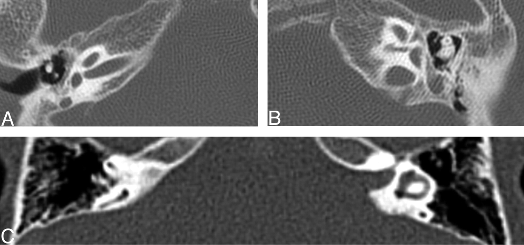
- Fig. 11.
Three axial CT images demonstrating examples of petrosquamosal sinuses (white arrows), a form of emissary vein anomaly.
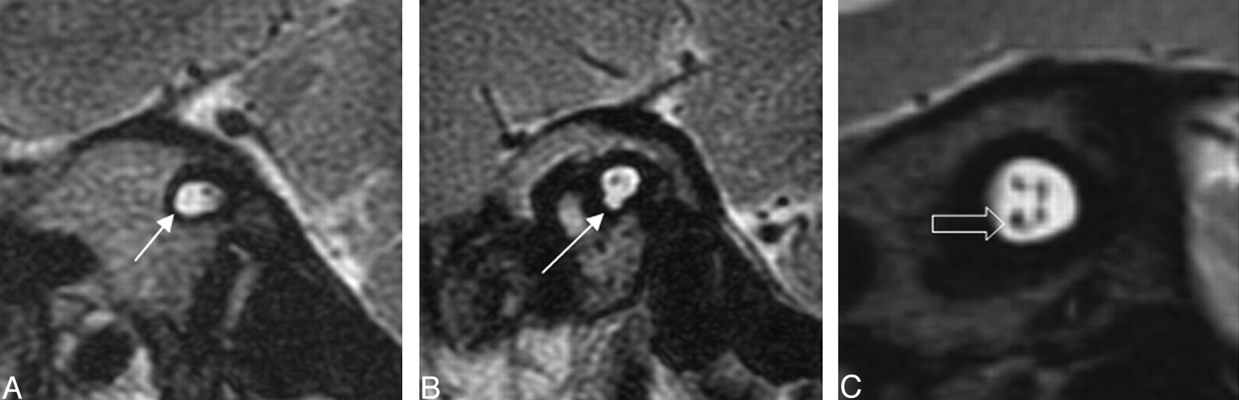
- Fig. 12.
Oblique sagittal T2-weighted images that demonstrate lack of a normal-appearing cochlear nerve (white arrow) in the 2 left images. The third image demonstrates a normal-appearing 4-nerve bundle of facial, cochlear (open arrow), and vestibular nerves.


Tables
Major Criterion Minor Criterion +Coloboma +Heart defect +Choanal atresia −Orofacial cleft +Characteristic ear anomalies +Genital hypoplasia +Cranial nerve dysfunction (facial palsy, vestibular dysfunction, swallowing difficulties) +Growth deficiency +Developmental delay −Tracheo-esophageal fistula +Distinct facial appearance Note:—CHARGE indicates Coloboma, Heart defects, choanal Atresia, mental Retardation, Genitourinary, and Ear anomalies; +, pertinent positive finding; −, pertinent negative finding. A CHARGE diagnosis is indicated by 4 major criteria or 3 major and 3 minor criteria. Exclude other conditions such as velocardiofacial syndrome and DiGeorge sequence using FISH test to exclude 22q11 deletion.
Patient No./Age (mo/sex) Side IAC Cochlea Vest VA VA angle Semi-circular canals Aperture Basal Apical LSCC PSCC SSCC 1/UTI/UTI R Normal Trapped Normal Normal Dys Present Reversed Absent Absent Absent L Normal Trapped Normal Normal Normal Present Normal Absent Absent Absent 2/UTI/UTI R Normal UTI Normal Hypo Hypo UTI UTI Absent Absent Absent L Normal UTI Normal Hypo Dys UTI UTI Absent Absent Absent 3/4/F R Normal Trapped Normal Hypo Hypo Present Reversed Absent Absent Absent L Normal Trapped Normal Hypo Hypo Present Reversed Absent Absent Absent 4 R Normal Trapped Normal Hypo Dys Present Reversed Absent Absent Absent L Normal Trapped Normal Hypo Dys Present Normal Absent Absent Absent 5/UTI/UTI R Normal Trapped Normal Normal Dys Large Normal Absent Absent Absent L Normal Trapped Normal dys Normal Normal Present Normal Absent Absent Absent 6/264/F R Normal Trapped Modio dys Hypo Normal Present Normal Absent Absent Absent L Small Trapped Modio Hypo Normal Large Normal Absent Absent Absent 7/65/UTI R Normal UTI UTI UTI UTI UTI UTI UTI UTI UTI L Normal UTI Enlarged Enlarged Dys UTI UTI Absent Absent Absent 8/UTI/UTI R Normal UTI Enlarged Enlarged UTI UTI UTI Absent Absent Absent L Normal UTI Enlarged Enlarged UTI UTI UTI Absent Absent Absent 9/3/M R Small Trapped Normal Hypo Hypo Present Reversed Absent Absent Absent L Small Trapped Normal Hypo Hypo Present Reversed Absent Absent Absent 10/4/M R Normal Trapped Normal Hypo Hypo Large Reversed Absent Absent Absent L Normal Trapped Normal Hypo Hypo Present Reversed Absent Absent Absent 11/16/F R Normal Trapped Normal Hypo Hypo Present Reversed Absent Absent Absent L Normal Trapped Normal Hypo Hypo Present Reversed Absent Absent Absent 12/60/M R Normal Trapped Normal Hypo Normal Large Reversed Absent Absent Absent L Normal Trapped Normal Hypo Normal Present Reversed Absent Absent Absent 13/72/M R Normal Trapped Normal Hypo Normal Large Normal Absent Absent Absent L Normal Trapped Normal Hypo Normal Present Normal Absent Absent Absent Note:—IAC indicates internal auditory canal; Vest, vestibule; VA, vestibular aqueduct; LSCC, lateral semicircular canal; SSCC, superior semicircular canal; PSCC, posterior semicircular canal; UTI, unable to identify; Dys, dysplastic; Modio, modiolus; Hypo, hypoplastic.
Patient No. Middle Ear Findings Tegmen Round Window Over Window Cavity Size Ossicles Ankylosis 1 R Small Dys Yes Dehiscent Normal Aplasia L Small Dys Yes Dehiscent Normal Aplasia 2 R Small Dys Yes Normal Normal Aplasia L Small Dys Yes Normal Normal Aplasia 3 R Small Dys Yes Normal Normal Aplasia L Small Dys Yes Normal Normal Aplasia 4 R Small Dys Yes Normal Normal Aplasia L Small Dys Yes Normal Normal Aplasia 5 R Small Dys Yes Normal Normal Aplasia L Small Dys Yes Normal Normal Aplasia 6 R Normal Normal No Normal Small Aplasia L Small Normal No Normal Small Aplasia 7 R Normal UTI UTI UTI UTI UTI L Normal Dys Yes Normal Normal Aplasia 8 R Normal Dys Yes Normal UTI Normal L Normal Dys Yes Normal UTI Normal 9 R Small Dys Yes Normal UTI UTI L Small Dys Yes Normal UTI UTI 10 R Small Dys Yes Normal Aplasia Aplasia L Small Dys Yes Normal Small Aplasia 11 R Small Dys Yes Normal Aplasia Aplasia L Small Dys Yes Normal Aplasia Aplasia 12 R Small Dys Yes Normal Aplasia Aplasia L Small Dys Yes Normal Aplasia Aplasia 13 R Small Dys Yes Normal Normal Aplasia L Small Dys Yes Normal Normal Aplasia Note:—UTI indicates unable to identify; Dys, dysplastic.
Patient No. Side Labyrinth Facial Nerve Venous Anomaly Juglar Frist Genu Tympanic Mastoid 1 R Posterior Posterior Prolapsed Normal L Posterior Posterior Prolapsed Normal PSS Diverticulum 2 R Normal Normal UTI UTI L Normal Normal UTI UTI 3 R Posterior Posterior Prolapsed Normal L Posterior Posterior Prolapsed Normal 4 R Posterior Posterior Normal Normal L Posterior Posterior Normal Normal 5 R Posterior Posterior Normal Normal L Posterior Posterior Normal Normal 6 R Posterior Posterior Normal Normal L Posterior Posterior Normal Normal 7 R UTI UTI UTI UTI L Posterior Posterior Normal Normal 8 R Posterior Posterior Normal Normal L Posterior Posterior Normal Normal 9 R Posterior Posterior Normal Normal L Posterior Posterior Normal Normal 10 R Posterior Posterior Normal Normal L Posterior Posterior Normal Normal PSS 11 R Posterior Posterior Normal Normal L Posterior Posterior Normal Normal 12 R Posterior Posterior Normal Normal L Posterior Posterior Normal Normal 13 R Posterior Posterior Normal Normal L Posterior Posterior Normal Normal PSS Diverticulum Note:—PSS indicates petrosquamosal sinus; UTI, unable to identify.
In this issue


Jump to section
Related Articles
Cited By...
- Coronal Clival Cleft in CHARGE Syndrome: Fetal MRI Series
- Persistent Trigeminal Artery: A Novel Imaging Finding in CHARGE Syndrome
- The Forgotten Second Window: A Pictorial Review of Round Window Pathologies
- Imaging of Clival Hypoplasia in CHARGE Syndrome and Hypothesis for Development: A Case-Control Study
- Clival Malformations in CHARGE Syndrome
- Head and Neck MRI Findings in CHARGE Syndrome
- Radiologic and Audiologic Findings in the Temporal Bone of Patients with CHARGE Syndrome
- Pediatric Sensorineural Hearing Loss, Part 2: Syndromic and Acquired Causes
- Superior Semicircular Canal Dehiscence: Congenital or Acquired Condition?