Advertisement

Main menu

  • Home
  • Content
    • Current Issue
    • Accepted Manuscripts
    • Article Preview
    • Past Issue Archive
    • Video Articles
    • AJNR Case Collection
    • Case of the Week Archive
    • Case of the Month Archive
    • Classic Case Archive
  • Special Collections
    • AJNR Awards
    • Low-Field MRI
    • Alzheimer Disease
    • ASNR Foundation Special Collection
    • Photon-Counting CT
    • View All
  • Multimedia
    • AJNR Podcasts
    • AJNR SCANtastic
    • Trainee Corner
    • MRI Safety Corner
    • Imaging Protocols
  • For Authors
    • Submit a Manuscript
    • Submit a Video Article
    • Submit an eLetter to the Editor/Response
    • Manuscript Submission Guidelines
    • Statistical Tips
    • Fast Publishing of Accepted Manuscripts
    • Graphical Abstract Preparation
    • Imaging Protocol Submission
    • Author Policies
  • About Us
    • About AJNR
    • Editorial Board
    • Editorial Board Alumni
  • More
    • Become a Reviewer/Academy of Reviewers
    • Subscribers
    • Permissions
    • Alerts
    • Feedback
    • Advertisers
    • ASNR Home

User menu

  • Alerts
  • Log in

Search

  • Advanced search
American Journal of Neuroradiology

American Journal of Neuroradiology

ASHNR American Society of Functional Neuroradiology ASHNR American Society of Pediatric Neuroradiology ASSR
  • Alerts
  • Log in

Advanced Search

  • Home
  • Content
    • Current Issue
    • Accepted Manuscripts
    • Article Preview
    • Past Issue Archive
    • Video Articles
    • AJNR Case Collection
    • Case of the Week Archive
    • Case of the Month Archive
    • Classic Case Archive
  • Special Collections
    • AJNR Awards
    • Low-Field MRI
    • Alzheimer Disease
    • ASNR Foundation Special Collection
    • Photon-Counting CT
    • View All
  • Multimedia
    • AJNR Podcasts
    • AJNR SCANtastic
    • Trainee Corner
    • MRI Safety Corner
    • Imaging Protocols
  • For Authors
    • Submit a Manuscript
    • Submit a Video Article
    • Submit an eLetter to the Editor/Response
    • Manuscript Submission Guidelines
    • Statistical Tips
    • Fast Publishing of Accepted Manuscripts
    • Graphical Abstract Preparation
    • Imaging Protocol Submission
    • Author Policies
  • About Us
    • About AJNR
    • Editorial Board
    • Editorial Board Alumni
  • More
    • Become a Reviewer/Academy of Reviewers
    • Subscribers
    • Permissions
    • Alerts
    • Feedback
    • Advertisers
    • ASNR Home
  • Follow AJNR on Twitter
  • Visit AJNR on Facebook
  • Follow AJNR on Instagram
  • Join AJNR on LinkedIn
  • RSS Feeds

AJNR Awards, New Junior Editors, and more. Read the latest AJNR updates

OtherHead and Neck Imaging

Incidental Giant Arachnoid Granulation

P. Kan, E.A. Stevens and W.T. Couldwell
American Journal of Neuroradiology August 2006, 27 (7) 1491-1492;
P. Kan
  • Find this author on Google Scholar
  • Find this author on PubMed
  • Search for this author on this site
E.A. Stevens
  • Find this author on Google Scholar
  • Find this author on PubMed
  • Search for this author on this site
W.T. Couldwell
  • Find this author on Google Scholar
  • Find this author on PubMed
  • Search for this author on this site
  • Article
  • Figures & Data
  • Info & Metrics
  • Responses
  • References
  • PDF
Loading

Abstract

SUMMARY: Arachnoid granulations may expand the dural sinuses or inner table of the skull. Although usually incidental, giant arachnoid granulations that are of sufficient size to fill the lumen of a dural sinus and cause local dilation or filling defects can rarely cause symptoms due to sinus obstruction leading to venous hypertension. This 31-year-old man presented with a 3-month history of progressive bifrontal headaches and a giant arachnoid granulation at the posterior superior sagittal sinus. Intrasinus pressure measurements showed no significant pressure difference across the lesion to explain the headaches, which were then treated medically. Dural sinus pressure measurement, in certain cases of giant arachnoid granulations, can be used to exclude the lesion as the cause of the patient’s symptoms.

Arachnoid granulations are growths of arachnoid membrane into the dural sinuses through which CSF enters the venous system.1 They normally measure a few millimeters but may grow to fill and dilate the dural sinuses or expand the inner table of the skull.1–3 They are often discovered as incidental findings in the transverse and posterior superior sagittal sinus,1–3 though they have rarely been reported to cause symptoms from venous hypertension secondary to partial sinus occlusion.2 We present a case of a patient who presented with progressive headaches and a giant arachnoid granulation involving his posterior superior sagittal sinus. Intrasinus pressure measurements across the lesion were performed to exclude the lesion as the cause of the patient’s symptoms.

Case Report

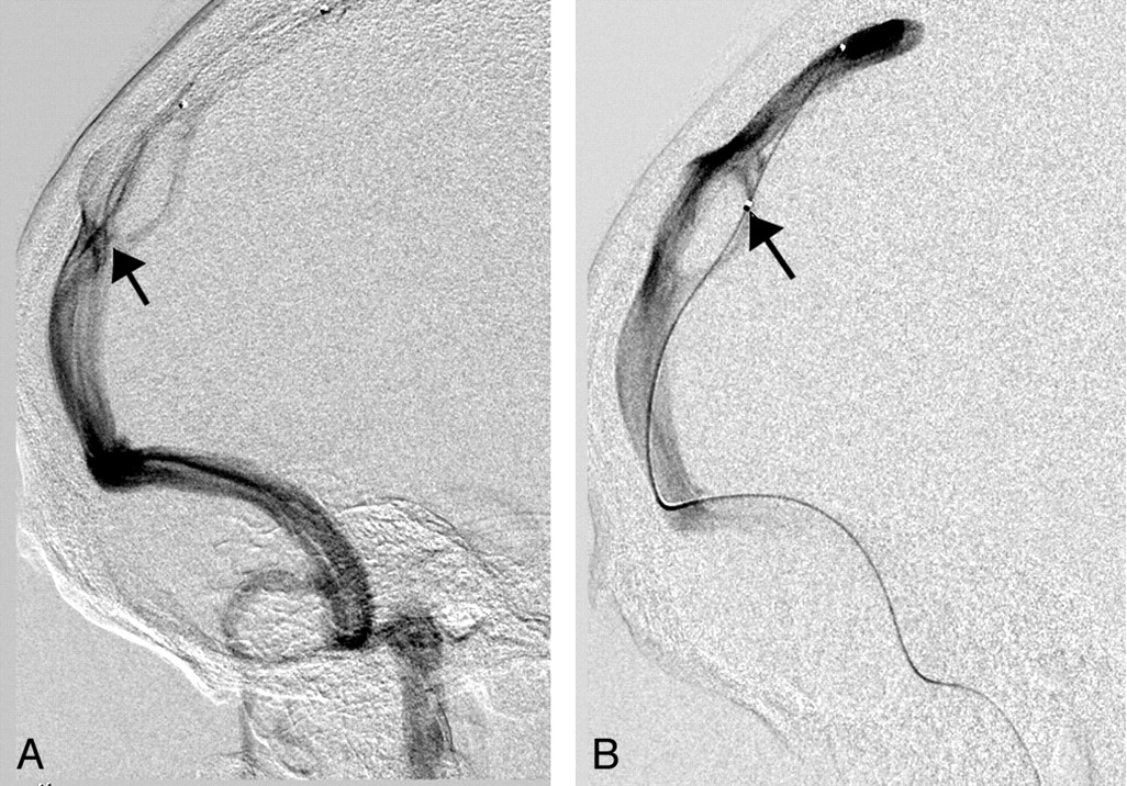
A 31-year-old man presented with a 3-month history of progressive bifrontal headaches. Neurologic examination revealed normal visual fields and acuity. Funduscopic examination showed sharp disk margins. Imaging studies including CT, MR imaging, and CT angiography were performed. Findings demonstrated a giant arachnoid granulation in the posterior third of the superior sagittal sinus, expanding the surrounding inner table (Fig 1A, -B). Lumbar puncture was performed, demonstrating clear CSF with no pleocytosis. Opening pressure was high normal. Because of the possibility of relative venous obstruction producing venous hypertension and headaches, cerebral angiography was performed. The venous phase of the angiogram revealed an enlarged superior sagittal sinus involving the posterior third of the sinus, with an apparent filling defect dividing into 3 separate channels that flowed around the filling defect and reconstituted distally (Fig 2A). Intrasinus pressure measurements showed no evidence of significant pressure difference across the lesion within all 3 dural venous limbs to account for the patient’s symptoms (Fig 2B). The headaches were treated symptomatically with medical therapy.

Fig. 1.
  • Download figure
  • Open in new tab
  • Download powerpoint
Fig. 1.

Axial (A) and sagittal (B) CT angiograms show a giant arachnoid granulation in the posterior superior sagittal sinus (arrow). In B, the expansion of the inner skull table is visible.

Fig. 2.
  • Download figure
  • Open in new tab
  • Download powerpoint
Fig. 2.

A, Angiogram shows the superior sagittal sinus (arrow) dividing into 3 separate channels that flow around the filling defect and reconstitute distally. B, Angiogram of the superior sagittal sinus shows a microcatheter (arrow) traversing through the right venous channel to measure pressure across the lesion within the right venous channel.

Discussion

Arachnoid granulations or villi are growths of arachnoid membrane into the dural sinuses, through which the CSF enters the venous system from the subarachnoid space.1 Arachnoid villi are microscopic, whereas arachnoid granulations represent distended villi and are visible to the naked eye.4 Arachnoid granulations increase in numbers and enlarge with age in response to increased CSF pressure from the subarachnoid space and are usually quite apparent by 4 years of age.5,6 They normally measure a few millimeters but may grow to expand the inner table of the skull, most commonly around midline in the posterior frontal or anterior parietal area.7 Occasionally, they even expand into the diploic space and eventually involve the outer table, mimicking osteolytic lesions.6–8 Although there is no consensus in the literature, we refer to arachnoid granulations as “giant” when they are of sufficient size to fill the lumen of a dural sinus and cause local dilation or filling defects.

Although they are often discovered as incidental findings in the transverse and posterior superior sagittal sinus,1–3 they can grow to fill and dilate the dural sinuses, causing symptoms of increased intracranial pressure from venous hypertension secondary to partial sinus occlusion.2 In our patient, the lesion appeared to impinge on the posterior third of the superior sagittal sinus. To determine whether the lesion was causing venous outflow obstruction and venous hypertension, we performed intrasinus pressure measurements across the lesion through each dural channel. In patients with pseudotumor with venous sinus obstruction as the cause of increased intracranial pressure, the proximal venous pressure was found to be as high as 36 mm Hg, with a corresponding pressure gradient of 25 mm Hg.9 The presence of normal venous pressures (∼15 mm Hg) and the lack of significant differential pressure across the lesion within all dural venous limbs (<5 mm Hg) in our patient ruled out venous hypertension and excluded the lesion as the cause of the patient’s symptoms.

Imaging of giant arachnoid granulations is often suggestive of partial sinus occlusion. Although largely incidental, giant arachnoid granulations can rarely cause symptoms due to sinus obstruction leading to venous hypertension. Dural sinus pressure measurement across the lesion is a novel and valuable approach to determine if the lesion is symptomatic because of venous obstruction and hypertension. Normal venous pressure with no significant differential pressure across the lesion can safely exclude the lesion as the cause of the patient’s symptoms.

Acknowledgments

We thank Kristin Kraus for her editorial assistance in preparing this manuscript.

References

  1. ↵
    Chin SC, Chen CY, Lee CC, et al. Giant arachnoid granulation mimicking dural sinus thrombosis in a boy with headache: MRI. Neuroradiology 1998;40:181–83
    CrossRefPubMed
  2. ↵
    Arjona A, Delgado F, Fernandez-Romero E. Intracranial hypertension secondary to giant arachnoid granulations. J Neurol Neurosurg Psychiatry 2003;74:418
    FREE Full Text
  3. ↵
    Mamourian AC, Towfighi J. MR of giant arachnoid granulation: a normal variant presenting as a mass within the dural venous sinus. AJNR Am J Neuroradiol 1995;16:901–04
    Abstract
  4. ↵
    Potts DG, Reilly KF, Deonarine V. Morphology of the arachnoid villi and granulations. Radiology 1972;105:333–41
    CrossRefPubMed
  5. ↵
    LeGross Clark WE. On the pacchionian granulations. J Anat 1920;55:40–48
    PubMed
  6. ↵
    Grossman CB, Potts DG. Arachnoid granulations: radiology and anatomy. Radiology 1974;113:95–100
    CrossRefPubMed
  7. ↵
    Branan R, Wilson CB. Arachnoid granulations simulating osteolytic lesions of the calvarium. AJR Am J Roentgenol 1976;127:523–25
    PubMed
  8. ↵
    [No authors listed] Case records of the Massachusetts General Hospital: weekly clinicopathological exercises. Case 42-1984. A 29-year-old woman with a lytic lesion of a parietal bone. N Engl J Med 1984;311:1036–43
    PubMed
  9. ↵
    Owler BK, Parker G, Halmagyi GM, et al. Pseudotumor cerebri syndrome: venous sinus obstruction and its treatment with stent placement. J Neurosurg 2003;98:1045–55
    PubMed
  • Received June 17, 2005.
  • Accepted after revision August 21, 2005.
  • Copyright © American Society of Neuroradiology
View Abstract
PreviousNext
Back to top

In this issue

American Journal of Neuroradiology: 27 (7)
American Journal of Neuroradiology
Vol. 27, Issue 7
August 2006
  • Table of Contents
  • Index by author
Advertisement
Print
Download PDF
Email Article

Thank you for your interest in spreading the word on American Journal of Neuroradiology.

NOTE: We only request your email address so that the person you are recommending the page to knows that you wanted them to see it, and that it is not junk mail. We do not capture any email address.

Enter multiple addresses on separate lines or separate them with commas.
Incidental Giant Arachnoid Granulation
(Your Name) has sent you a message from American Journal of Neuroradiology
(Your Name) thought you would like to see the American Journal of Neuroradiology web site.
CAPTCHA
This question is for testing whether or not you are a human visitor and to prevent automated spam submissions.
Cite this article
P. Kan, E.A. Stevens, W.T. Couldwell
Incidental Giant Arachnoid Granulation
American Journal of Neuroradiology Aug 2006, 27 (7) 1491-1492;

Citation Manager Formats

  • BibTeX
  • Bookends
  • EasyBib
  • EndNote (tagged)
  • EndNote 8 (xml)
  • Medlars
  • Mendeley
  • Papers
  • RefWorks Tagged
  • Ref Manager
  • RIS
  • Zotero
0 Responses
Respond to this article
Share
Bookmark this article
Incidental Giant Arachnoid Granulation
P. Kan, E.A. Stevens, W.T. Couldwell
American Journal of Neuroradiology Aug 2006, 27 (7) 1491-1492;
del.icio.us logo Twitter logo Facebook logo Mendeley logo
  • Tweet Widget
  • Facebook Like
  • Google Plus One

Jump to section

  • Article
    • Abstract
    • Case Report
    • Discussion
    • Acknowledgments
    • References
  • Figures & Data
  • Info & Metrics
  • Responses
  • References
  • PDF

Related Articles

  • No related articles found.
  • PubMed
  • Google Scholar

Cited By...

  • Giant arachnoid granulation with a thrombosed dural arteriovenous fistula
  • "Giant" Arachnoid Granulations Just Like CSF?: NOT!!
  • Crossref
  • Google Scholar

This article has not yet been cited by articles in journals that are participating in Crossref Cited-by Linking.

More in this TOC Section

  • ASL Sensitivity for Head and Neck Paraganglioma
  • Post SRS Peritumoral Hyperintense Signal of VSs
  • Temporal Evolution of Vestibular schwannoma
Show more Head and Neck Imaging

Similar Articles

Advertisement

Indexed Content

  • Current Issue
  • Accepted Manuscripts
  • Article Preview
  • Past Issues
  • Editorials
  • Editor's Choice
  • Fellows' Journal Club
  • Letters to the Editor
  • Video Articles

Cases

  • Case Collection
  • Archive - Case of the Week
  • Archive - Case of the Month
  • Archive - Classic Case

More from AJNR

  • Trainee Corner
  • Imaging Protocols
  • MRI Safety Corner
  • Book Reviews

Multimedia

  • AJNR Podcasts
  • AJNR Scantastics

Resources

  • Turnaround Time
  • Submit a Manuscript
  • Submit a Video Article
  • Submit an eLetter to the Editor/Response
  • Manuscript Submission Guidelines
  • Statistical Tips
  • Fast Publishing of Accepted Manuscripts
  • Graphical Abstract Preparation
  • Imaging Protocol Submission
  • Evidence-Based Medicine Level Guide
  • Publishing Checklists
  • Author Policies
  • Become a Reviewer/Academy of Reviewers
  • News and Updates

About Us

  • About AJNR
  • Editorial Board
  • Editorial Board Alumni
  • Alerts
  • Permissions
  • Not an AJNR Subscriber? Join Now
  • Advertise with Us
  • Librarian Resources
  • Feedback
  • Terms and Conditions
  • AJNR Editorial Board Alumni

American Society of Neuroradiology

  • Not an ASNR Member? Join Now

© 2025 by the American Society of Neuroradiology All rights, including for text and data mining, AI training, and similar technologies, are reserved.
Print ISSN: 0195-6108 Online ISSN: 1936-959X

Powered by HighWire