Advertisement

Main menu

  • Home
  • Content
    • Current Issue
    • Accepted Manuscripts
    • Article Preview
    • Past Issue Archive
    • Video Articles
    • AJNR Case Collection
    • Case of the Week Archive
    • Case of the Month Archive
    • Classic Case Archive
  • Special Collections
    • AJNR Awards
    • Low-Field MRI
    • Alzheimer Disease
    • ASNR Foundation Special Collection
    • Photon-Counting CT
    • View All
  • Multimedia
    • AJNR Podcasts
    • AJNR SCANtastic
    • Trainee Corner
    • MRI Safety Corner
    • Imaging Protocols
  • For Authors
    • Submit a Manuscript
    • Submit a Video Article
    • Submit an eLetter to the Editor/Response
    • Manuscript Submission Guidelines
    • Statistical Tips
    • Fast Publishing of Accepted Manuscripts
    • Graphical Abstract Preparation
    • Imaging Protocol Submission
    • Author Policies
  • About Us
    • About AJNR
    • Editorial Board
    • Editorial Board Alumni
  • More
    • Become a Reviewer/Academy of Reviewers
    • Subscribers
    • Permissions
    • Alerts
    • Feedback
    • Advertisers
    • ASNR Home

User menu

  • Alerts
  • Log in

Search

  • Advanced search
American Journal of Neuroradiology

American Journal of Neuroradiology

ASHNR American Society of Functional Neuroradiology ASHNR American Society of Pediatric Neuroradiology ASSR
  • Alerts
  • Log in

Advanced Search

  • Home
  • Content
    • Current Issue
    • Accepted Manuscripts
    • Article Preview
    • Past Issue Archive
    • Video Articles
    • AJNR Case Collection
    • Case of the Week Archive
    • Case of the Month Archive
    • Classic Case Archive
  • Special Collections
    • AJNR Awards
    • Low-Field MRI
    • Alzheimer Disease
    • ASNR Foundation Special Collection
    • Photon-Counting CT
    • View All
  • Multimedia
    • AJNR Podcasts
    • AJNR SCANtastic
    • Trainee Corner
    • MRI Safety Corner
    • Imaging Protocols
  • For Authors
    • Submit a Manuscript
    • Submit a Video Article
    • Submit an eLetter to the Editor/Response
    • Manuscript Submission Guidelines
    • Statistical Tips
    • Fast Publishing of Accepted Manuscripts
    • Graphical Abstract Preparation
    • Imaging Protocol Submission
    • Author Policies
  • About Us
    • About AJNR
    • Editorial Board
    • Editorial Board Alumni
  • More
    • Become a Reviewer/Academy of Reviewers
    • Subscribers
    • Permissions
    • Alerts
    • Feedback
    • Advertisers
    • ASNR Home
  • Follow AJNR on Twitter
  • Visit AJNR on Facebook
  • Follow AJNR on Instagram
  • Join AJNR on LinkedIn
  • RSS Feeds

AJNR Awards, New Junior Editors, and more. Read the latest AJNR updates

OtherPediatric Neuroimaging

Short Echo Time MR Spectroscopic Imaging for Neonatal Pediatric Imaging

D.H. Kim, A.J. Barkovich and D.B. Vigneron
American Journal of Neuroradiology June 2006, 27 (6) 1370-1372;
D.H. Kim
  • Find this author on Google Scholar
  • Find this author on PubMed
  • Search for this author on this site
A.J. Barkovich
  • Find this author on Google Scholar
  • Find this author on PubMed
  • Search for this author on this site
D.B. Vigneron
  • Find this author on Google Scholar
  • Find this author on PubMed
  • Search for this author on this site
  • Article
  • Figures & Data
  • Info & Metrics
  • Responses
  • References
  • PDF
Loading

Abstract

SUMMARY: A short echo time (30 milliseconds) MR spectroscopic imaging pulse sequence was implemented for applications of neonatal brain imaging. Multiple spatial saturation bands were used to eliminate strong signals originating from the subcutaneous lipids to enable volumetric region coverage. Metabolite signal intensity–to-noise ratio ≤40 was acquired in 9 minutes of scan time over an 8 × 8 × 8 spatial matrix with 1 cm3 isoresolution.

MR spectroscopy has been shown to be useful in providing clinically important assessment of neonatal brain metabolism.1–3 Metabolite concentration levels have also been studied with MR spectroscopy during early brain development to better understand the rapidly ongoing biochemical changes that occur at this stage of life.4–6 Several studies have used MR spectroscopic imaging (MRSI) for neonatal and pediatric imaging applications.7,8 Although single-voxel MR spectroscopy techniques have been successfully implemented in both long echo time (TE; >130 milliseconds) and short TE (<40 milliseconds) acquisitions, MRSI methods have mainly been limited to long TE studies for reasons of technical feasibility. In this study, we demonstrate the applicability of a short TE MRSI sequence, targeted for neonatal brain examinations, by using a newly derived short TE MRSI protocol to acquire volumetric datasets from a group of neonates.

Description of Technique

All studies were performed on a 1.5T Signa EchoSpeed system (GE Healthcare, Milwaukee, Wis) by using an MR imaging–compatible incubator with a specialized neonatal head coil.9 For the MRSI acquisition, the TE was set to 30 milliseconds with a repetition time of 1 second. An 8 × 8 × 8 voxel phase-encoding scheme was used to collect the data. The effective voxel size was kept at 1 cm3 with isotropic resolution. Very selective saturation pulses were placed to eliminate signals from subcutaneous lipids.10 Three saturation pulses were played out per band at 6 different locations for a total of 18 saturation pulses providing robust saturation. To reduce eddy current effects from crusher gradients commonly encountered in short TE imaging, the length of the crusher gradients was shortened to 2.5 milliseconds. This shortening resulted in a crusher gradients area of 67 mT/m milliseconds, which is within the range suggestive for in vivo spectroscopy.11 Phantom studies with these set of crushers showed neither significant eddy current induced artifacts nor spurious echoes because of simulated echoes.

Short TE MRSI data were acquired from 10 neonates who had MR imaging examinations as part of a neonatal study protocol. The study protocol was approved by our institutional review board, and parental consent was obtained for all infants. Of these neonates, 4 were born prematurely and were studied at adjusted ages (gestational age added to postnatal age) between 27 and 34 weeks (mean at study, 30.5 ± 3.2 week). Six neonates born at term were imaged because of suspected neurologic damage from congenital heart disease; the mean corrected age for these infants at the time of the MR study was 41 ± 1.4 weeks. Sedation was performed by using pentobarbital (Nembutal) only after parental consent and only when necessary (eg, when the baby woke up and started moving). As a result, only those infants with congenital heart disease required sedation. To validate the technique, quantitative analysis was performed by using the linear combination model-fitting procedure and metabolites from the premature group and term births were compared.12

Results and Discussion

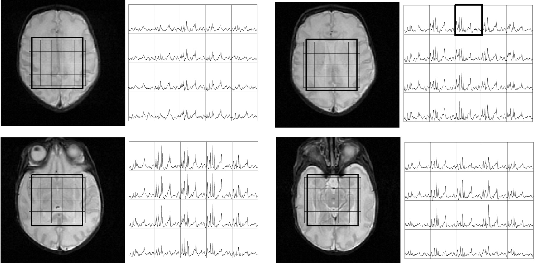
Figure 1 shows volumetric spectra obtained from a 31-week age-adjusted premature neonate. Four representative sections of spectroscopic data are shown along with the anatomic images. Spatially varying metabolite components can be seen. In general, individual metabolite levels are higher near the deep gray matter, which is in agreement with previous studies of early brain development. An analysis of the signal intensity–to-noise ratio (SNR) of the choline (Cho) peak showed values ≤40. The metabolite assignments of each peak are given in Fig 2, which was extracted from the emphasized voxel in Fig 1. Metabolites such as myo-inositol (mIns) and glutamate/glutamine (Glx) can be seen in addition to the metabolites normally observed for long TE acquisitions. The lactate (Lac) doublet peak at 1.3 parts per million (ppm) is well illustrated, though care must be taken in the interpretation because this region contains signal intensity contributions from macromolecules as well. Macromolecules also occupy small portions of the 2.3-ppm region whereas glycine overlaps with the mI molecule at 3.15 ppm.13 The Lac doublet peak was well resolved for several of the preterm infants, which confirmed previous observations of Lac in the white matter for premature infants.14

Fig 1.
  • Download figure
  • Open in new tab
  • Download powerpoint
Fig 1.

Volumetric dataset from a preterm neonate imaged at 31-week adjusted age. Shown are the T2 weighted images along with the spectra between the 4.0–1.0-ppm region. Regional variation is seen with the highest metabolite levels within the deep gray matter.

Fig 2.
  • Download figure
  • Open in new tab
  • Download powerpoint
Fig 2.

Representative spectra from a voxel in Fig 1. The labeled metabolites correspond to (1) NAA, (2) Cr, (3) Cho, (4) mIns, (5) Lac, (6) Glx, and (7) macromolecular baseline in the 2.0–1.0-ppm region.

Figure 3 shows representative short TE spectra typically obtained from the labeled regions for a prematurely born infant and a term infant. In general, the level of metabolites followed the previously described maturation process, with the deep gray matter maturing early and the frontal and temporal white matter maturing last. High levels of mIns were seen in the calcarine gray matter for premature infants. This is particularly interesting because topologic and temporal changes of mIns have not been well studied. mIns and its transporter are believed to play an important role in normal fetal brain development, though reasons for the elevated mIns in the neonatal brain and its function are not well understood. Previous studies in neonatal encephalopathy suggest increased mIns metabolites in the basal ganglia region.15 A clear benefit of the short TE sequence is the ability to help understand the function and distribution of these short T2 components.

Fig 3.
  • Download figure
  • Open in new tab
  • Download powerpoint
Fig 3.

Spectra extracted from a 33-week and a 40-week age-adjusted infant. Corresponding regions are indicated for the spectra. In general, the deep gray matter regions show higher metabolite levels indicating earlier development. Note the high levels of Cho in all spectra, which is typical of neonates. Finally, note the elevated mIns level in the calcarine gray matter of the 33-week baby.

Finally, Table 1 shows quantitative metabolite ratios obtained. Statistically significant differences were mostly obtained from regions in the deep gray matter corresponding to rapid changes in these regions. Elevated Cho/creatine (Cr) ratios were seen in preterm births in comparison with term births in regions of the basal ganglia, thalamus, and calcarine gray matter (P < .005). Decreases in the Cho concentration in this age group have been documented elsewhere.6 Elevated mIns/Cr ratios were also seen in the basal ganglia, thalamus (P < .005), and the frontal white matter (P < .05), which illustrates mIns decreasing with age. Overall, N-acetylaspartate (NAA) values increased with age suggestive of increased neuronal activity. No significant differences were found for Glx.

View this table:
  • View inline
  • View popup
Table 1:

Quantitative metabolite ratios for preterm neonates (group 1) and term infants (group 2)

This study shows the feasibility of acquiring 3D MRSI data from most parts of the neonatal brain at a 1 cm3 nominal spatial resolution with an TE of 30 milliseconds in 9 minutes. The ability to robustly acquire short TE spectra from the brain has great potential in helping to understand regional and temporal variations of long and short T2 metabolites during normal brain development and in helping to diagnose causes of abnormal development. The use of repeated saturation pulses to eliminate lipid signals is a significant factor, because aliasing artifacts can be dramatically reduced.

To verify the technical feasibility, quantitative analysis, and statistical comparisons were performed for 2 groups—premature and term-born infants—which showed metabolite distributions in accordance with previous studies. Because the patients examined were not completely normal, however, these numbers should not be considered normative data. Indeed, even if early development of the patients were normal, it is likely that premature birth and congenital heart defects both have some effect on normal brain development. Previous studies of infants with congenital heart disease have indicated reduced NAA/Cho ratios compared with age-matched controls for these infants.16 These effects must be taken into consideration if the results were to be used for clinical diagnosis.

Acknowledgments

This study was supported by grants from the National Institutes of Health (NIH R01 NS40117 and NIH R01 NS046432). This study was carried out in part in the Pediatric Clinical Research Center, University of California, San Francisco, with funds provided by the National Center for Research Resources (5 M01 RR-01271), US Public Health Service.

References

  1. ↵
    Groenendaal F, Veenhoven RH, van der Grond J et al. Cerebral lactate and N-acetyl-sapartate/choline ratios in asphyxiated full-term neonates demonstrated in vivo using proton MR spectrsocopy. Pediatr Res 1994;35:148–51
    PubMed
  2. Penrice J, Cady EB, Lorek A, et al. Proton magnetic resonance spectroscopy of the brain in normal preterm and term infants, and early changes after perinatal hepoxia-ischemia. Pediatr Res 1996;40:6–14
    PubMed
  3. ↵
    Barkovich AJ, Westmark KD, Bedi HS, et al. Proton spectroscopy and diffusion imaging on the first day of life after perinatal asphyxia: preliminary report. AJNR Am J Neuroradiol 2001;22:1786–94
    Abstract/FREE Full Text
  4. ↵
    van der Knaap MS, van der Grond J, van Rijen PC, et al. Age-dependent changes in localized proton and phosphorus MR spectroscopy of the brain. Radiology 1990;176:509–15.
    PubMed
  5. Toft PB, Leth H, Lou HC, et al. Metabolite concentrations in the developing brain estimated with proton MR spectroscopy. J Magn Reson Imaging 1994;4:674–80
    PubMed
  6. ↵
    Kreis R, Hofmann L, Kuhlmann B et al. Brain metabolite composition during early human brain development as measured by quantitative in vivo 1H MR spectroscopy. Magn Reson Med 2002;48:949–58
    CrossRefPubMed
  7. ↵
    Vigneron DB, Barkovich AJ, Noworolski SM, et al. Three-dimensional proton MR spectroscopic imaging of premature and term neonates. AJNR Am J Neuroradiol 2001;22:1424–33
    Abstract/FREE Full Text
  8. ↵
    Horska A, Kaufmann WE, Brant LJ, et al. In vivo quantitative proton MRSI study of brain development from childhood to adolescence. J Magn Reson Imaging 2002;15:137–43
    CrossRefPubMed
  9. ↵
    Dumoulin CL, Rohling KW, Piel JE, et al. Magnetic resonance imaging compatible neonate incubator. Magn Reson Eng 2002;15:117–28
  10. ↵
    Tran TK, Vigneron DB, Sailasuta N, et al. Very selective suppression pulses for clinical MRSI studies of brain and prostate cancer. Magn Reson Med 2000;43:23–33
    CrossRefPubMed
  11. ↵
    Ernst T, Chang L. Elimination of artifacts in short echo time 1H MR spectroscopy of the frontal lobe. Magn Reson Med 1996;36:462–68
    PubMed
  12. ↵
    Provencher SW. Estimation of metabolite concentration from localized in vivo proton NMR spectra. Magn Reson Med 1993;30:672–79
    CrossRefPubMed
  13. ↵
    Govindaraju V, Young K, Maudsley AA. Proton NMR chemical shifts and coupling constants for brain metabolites. NMR Biomed 2000;13:129–53
    CrossRefPubMed
  14. ↵
    Leth H, Toft PB, Pryds O et al. Brain lactate in preterm and growth-retarded neonates. Acta Paediatr 1995;84:495–99
    PubMed
  15. ↵
    Robertson NJ, Lewis RH, Cowan FM, et al. Early increases in brain myo-inositol measured by proton MR spectroscopy in term infants with neonatal encephalopathy. Ped Research 2001;50:692–700
    PubMed
  16. ↵
    Miller SP, McQuillen PS, Vigneron DB, et al. Preoperative brain injury in neonates with transposition of the great arteries. Ann Thorac Surg 2004;77:1698–706
    CrossRefPubMed
  • Received January 4, 2006.
  • Accepted after revision January 9, 2006.
  • Copyright © American Society of Neuroradiology
View Abstract
PreviousNext
Back to top

In this issue

American Journal of Neuroradiology: 27 (6)
American Journal of Neuroradiology
Vol. 27, Issue 6
June 2006
  • Table of Contents
  • Index by author
Advertisement
Print
Download PDF
Email Article

Thank you for your interest in spreading the word on American Journal of Neuroradiology.

NOTE: We only request your email address so that the person you are recommending the page to knows that you wanted them to see it, and that it is not junk mail. We do not capture any email address.

Enter multiple addresses on separate lines or separate them with commas.
Short Echo Time MR Spectroscopic Imaging for Neonatal Pediatric Imaging
(Your Name) has sent you a message from American Journal of Neuroradiology
(Your Name) thought you would like to see the American Journal of Neuroradiology web site.
CAPTCHA
This question is for testing whether or not you are a human visitor and to prevent automated spam submissions.
Cite this article
D.H. Kim, A.J. Barkovich, D.B. Vigneron
Short Echo Time MR Spectroscopic Imaging for Neonatal Pediatric Imaging
American Journal of Neuroradiology Jun 2006, 27 (6) 1370-1372;

Citation Manager Formats

  • BibTeX
  • Bookends
  • EasyBib
  • EndNote (tagged)
  • EndNote 8 (xml)
  • Medlars
  • Mendeley
  • Papers
  • RefWorks Tagged
  • Ref Manager
  • RIS
  • Zotero
0 Responses
Respond to this article
Share
Bookmark this article
Short Echo Time MR Spectroscopic Imaging for Neonatal Pediatric Imaging
D.H. Kim, A.J. Barkovich, D.B. Vigneron
American Journal of Neuroradiology Jun 2006, 27 (6) 1370-1372;
del.icio.us logo Twitter logo Facebook logo Mendeley logo
  • Tweet Widget
  • Facebook Like
  • Google Plus One

Jump to section

  • Article
    • Abstract
    • Description of Technique
    • Results and Discussion
    • Acknowledgments
    • References
  • Figures & Data
  • Info & Metrics
  • Responses
  • References
  • PDF

Related Articles

  • No related articles found.
  • PubMed
  • Google Scholar

Cited By...

  • Neurometabolite Mapping Highlights Elevated Myo-inositol Profiles within the Developing Brain in Down Syndrome
  • An MR-compatible neonatal incubator
  • Crossref
  • Google Scholar

This article has not yet been cited by articles in journals that are participating in Crossref Cited-by Linking.

More in this TOC Section

  • FRACTURE MR in Congenital Vertebral Anomalies
  • Neuroimaging Delineation and Progression of SLSMD
  • fetal brain development of 10 weeks gestation
Show more PEDIATRIC NEUROIMAGING

Similar Articles

Advertisement

Indexed Content

  • Current Issue
  • Accepted Manuscripts
  • Article Preview
  • Past Issues
  • Editorials
  • Editor's Choice
  • Fellows' Journal Club
  • Letters to the Editor
  • Video Articles

Cases

  • Case Collection
  • Archive - Case of the Week
  • Archive - Case of the Month
  • Archive - Classic Case

More from AJNR

  • Trainee Corner
  • Imaging Protocols
  • MRI Safety Corner
  • Book Reviews

Multimedia

  • AJNR Podcasts
  • AJNR Scantastics

Resources

  • Turnaround Time
  • Submit a Manuscript
  • Submit a Video Article
  • Submit an eLetter to the Editor/Response
  • Manuscript Submission Guidelines
  • Statistical Tips
  • Fast Publishing of Accepted Manuscripts
  • Graphical Abstract Preparation
  • Imaging Protocol Submission
  • Evidence-Based Medicine Level Guide
  • Publishing Checklists
  • Author Policies
  • Become a Reviewer/Academy of Reviewers
  • News and Updates

About Us

  • About AJNR
  • Editorial Board
  • Editorial Board Alumni
  • Alerts
  • Permissions
  • Not an AJNR Subscriber? Join Now
  • Advertise with Us
  • Librarian Resources
  • Feedback
  • Terms and Conditions
  • AJNR Editorial Board Alumni

American Society of Neuroradiology

  • Not an ASNR Member? Join Now

© 2025 by the American Society of Neuroradiology All rights, including for text and data mining, AI training, and similar technologies, are reserved.
Print ISSN: 0195-6108 Online ISSN: 1936-959X

Powered by HighWire