Advertisement

Main menu

  • Home
  • Content
    • Current Issue
    • Accepted Manuscripts
    • Article Preview
    • Past Issue Archive
    • Video Articles
    • AJNR Case Collection
    • Case of the Week Archive
    • Case of the Month Archive
    • Classic Case Archive
  • Special Collections
    • AJNR Awards
    • Low-Field MRI
    • Alzheimer Disease
    • ASNR Foundation Special Collection
    • Photon-Counting CT
    • View All
  • Multimedia
    • AJNR Podcasts
    • AJNR SCANtastic
    • Trainee Corner
    • MRI Safety Corner
    • Imaging Protocols
  • For Authors
    • Submit a Manuscript
    • Submit a Video Article
    • Submit an eLetter to the Editor/Response
    • Manuscript Submission Guidelines
    • Statistical Tips
    • Fast Publishing of Accepted Manuscripts
    • Graphical Abstract Preparation
    • Imaging Protocol Submission
    • Author Policies
  • About Us
    • About AJNR
    • Editorial Board
    • Editorial Board Alumni
  • More
    • Become a Reviewer/Academy of Reviewers
    • Subscribers
    • Permissions
    • Alerts
    • Feedback
    • Advertisers
    • ASNR Home

User menu

  • Alerts
  • Log in

Search

  • Advanced search
American Journal of Neuroradiology

American Journal of Neuroradiology

ASHNR American Society of Functional Neuroradiology ASHNR American Society of Pediatric Neuroradiology ASSR
  • Alerts
  • Log in

Advanced Search

  • Home
  • Content
    • Current Issue
    • Accepted Manuscripts
    • Article Preview
    • Past Issue Archive
    • Video Articles
    • AJNR Case Collection
    • Case of the Week Archive
    • Case of the Month Archive
    • Classic Case Archive
  • Special Collections
    • AJNR Awards
    • Low-Field MRI
    • Alzheimer Disease
    • ASNR Foundation Special Collection
    • Photon-Counting CT
    • View All
  • Multimedia
    • AJNR Podcasts
    • AJNR SCANtastic
    • Trainee Corner
    • MRI Safety Corner
    • Imaging Protocols
  • For Authors
    • Submit a Manuscript
    • Submit a Video Article
    • Submit an eLetter to the Editor/Response
    • Manuscript Submission Guidelines
    • Statistical Tips
    • Fast Publishing of Accepted Manuscripts
    • Graphical Abstract Preparation
    • Imaging Protocol Submission
    • Author Policies
  • About Us
    • About AJNR
    • Editorial Board
    • Editorial Board Alumni
  • More
    • Become a Reviewer/Academy of Reviewers
    • Subscribers
    • Permissions
    • Alerts
    • Feedback
    • Advertisers
    • ASNR Home
  • Follow AJNR on Twitter
  • Visit AJNR on Facebook
  • Follow AJNR on Instagram
  • Join AJNR on LinkedIn
  • RSS Feeds

AJNR Awards, New Junior Editors, and more. Read the latest AJNR updates

Research ArticleNeurointervention

Silent Ischemia after Neuroprotected Percutaneous Carotid Stenting: A Diffusion-Weighted MRI Study

P. Piñero, A. González, A. Mayol, E. Martínez, J.R. González-Marcos, F. Boza, A. Cayuela and A. Gil-Peralta
American Journal of Neuroradiology June 2006, 27 (6) 1338-1345;
P. Piñero
  • Find this author on Google Scholar
  • Find this author on PubMed
  • Search for this author on this site
A. González
  • Find this author on Google Scholar
  • Find this author on PubMed
  • Search for this author on this site
A. Mayol
  • Find this author on Google Scholar
  • Find this author on PubMed
  • Search for this author on this site
E. Martínez
  • Find this author on Google Scholar
  • Find this author on PubMed
  • Search for this author on this site
J.R. González-Marcos
  • Find this author on Google Scholar
  • Find this author on PubMed
  • Search for this author on this site
F. Boza
  • Find this author on Google Scholar
  • Find this author on PubMed
  • Search for this author on this site
A. Cayuela
  • Find this author on Google Scholar
  • Find this author on PubMed
  • Search for this author on this site
A. Gil-Peralta
  • Find this author on Google Scholar
  • Find this author on PubMed
  • Search for this author on this site
  • Article
  • Figures & Data
  • Info & Metrics
  • Responses
  • References
  • PDF
Loading

Article Figures & Data

Figures

  • Tables
  • Fig. 1.
    • Download figure
    • Open in new tab
    • Download powerpoint
    Fig. 1.

    A, MR angiography of the right common carotid artery before carotid artery stent (CAS) placement, showing a severe stenosis of the proximal internal carotid artery.

    B, Diffusion-weighted image (DWI) obtained at the same time shows no abnormalities.

    C, DWI after CAS with a new ipsilateral lesion in the deep territory of the ipsilateral middle cerebral artery.
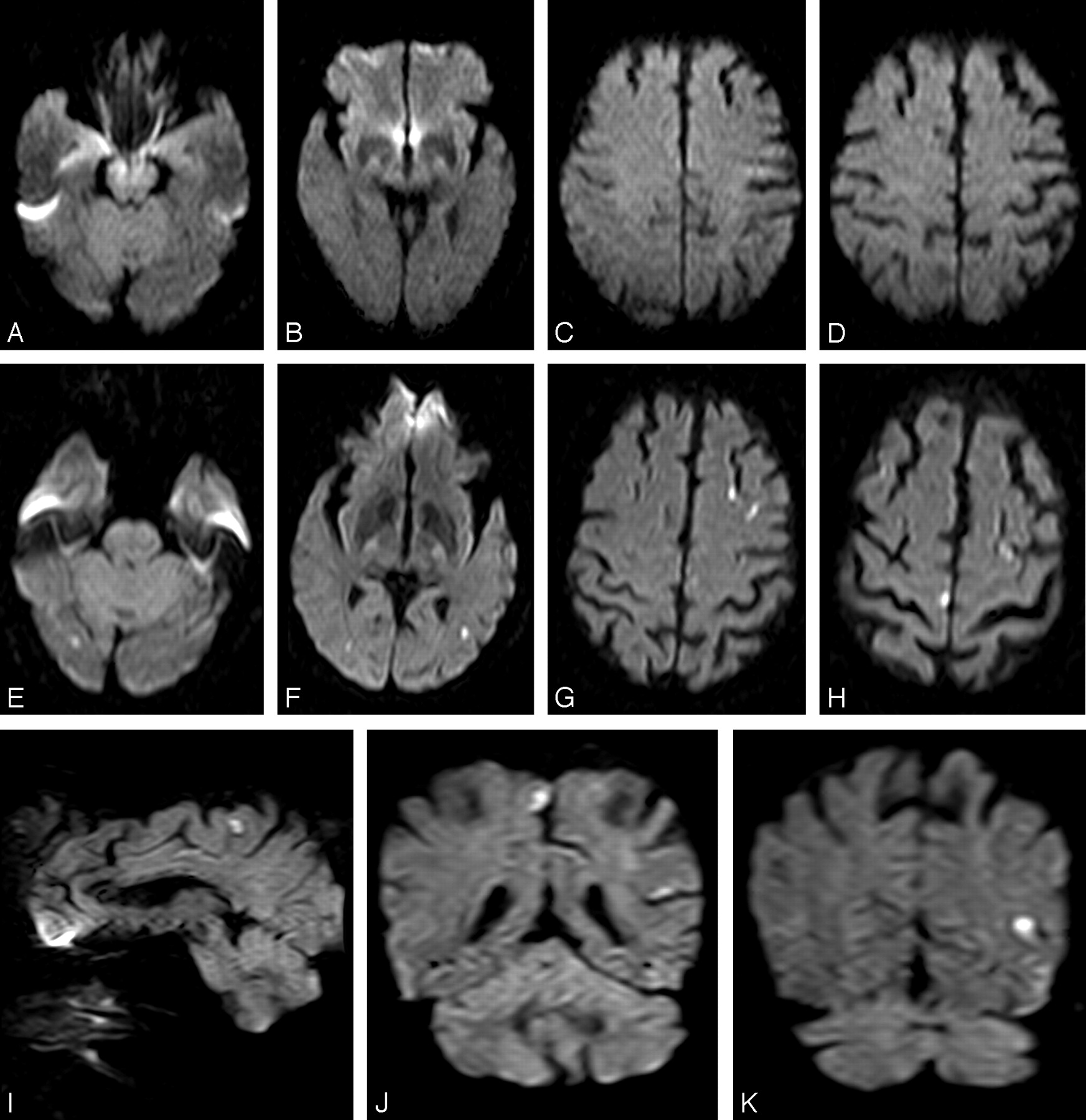
  • Fig. 2.
    • Download figure
    • Open in new tab
    • Download powerpoint
    Fig. 2.

    A–D, Normal axial diffusion-weighted image (DWI).

    E–K, Post-carotid artery stent placement DWI (axial, sagittal, and coronal) with multiple and bilateral cortical new ischemic lesions in the territory of both middle cerebral artery and contralateral anterior and posterior arteries.
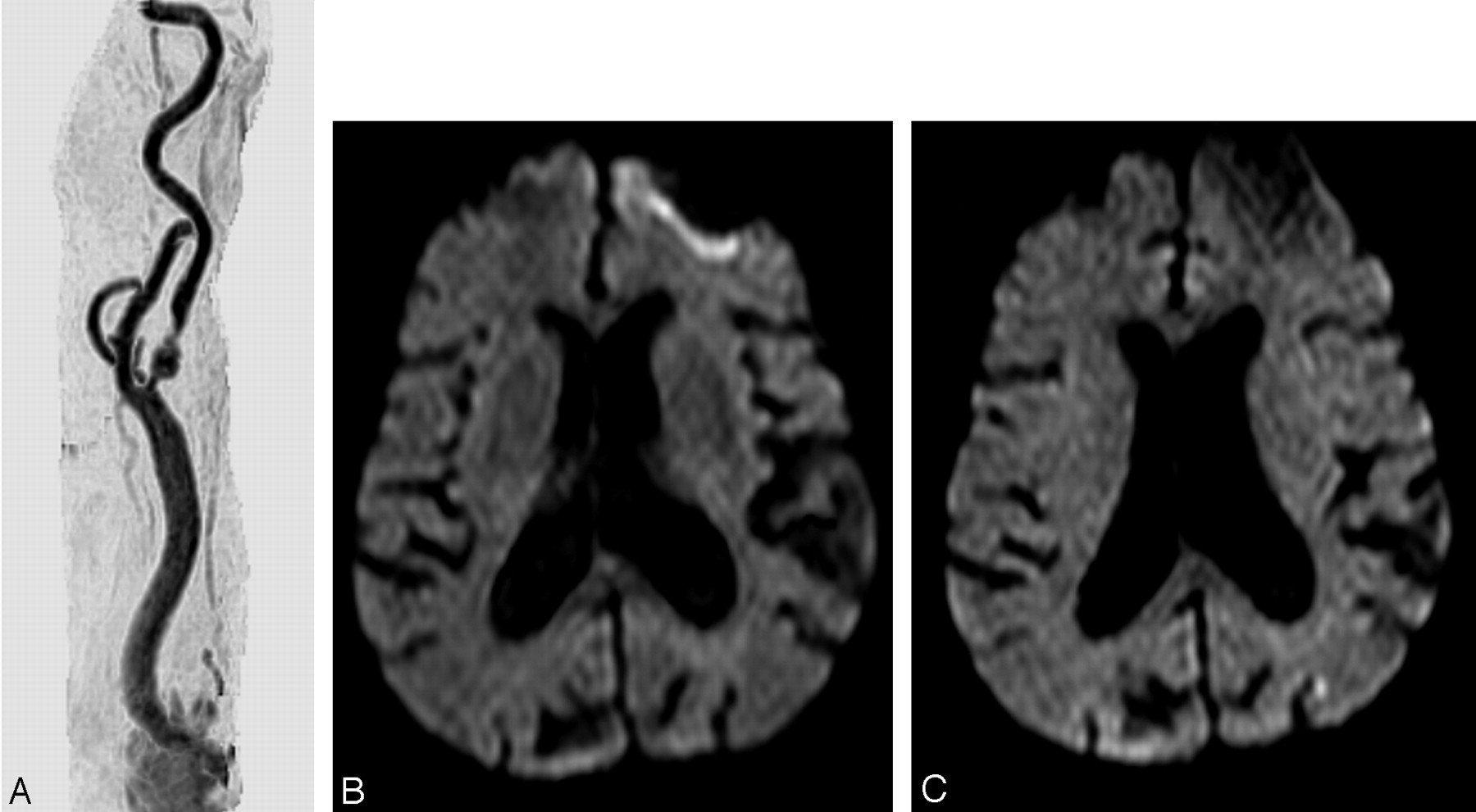
  • Fig. 3.
    • Download figure
    • Open in new tab
    • Download powerpoint
    Fig. 3.

    A, MR angiography of the right common carotid artery before carotid artery stent placement (CAS) with a tandem lesion in the proximal internal carotid artery.

    B and C, Diffusion-weighted image before (B) and after CAS (C), showing a new lesion <5 mm in the superficial territory of the contralateral middle cerebral artery.
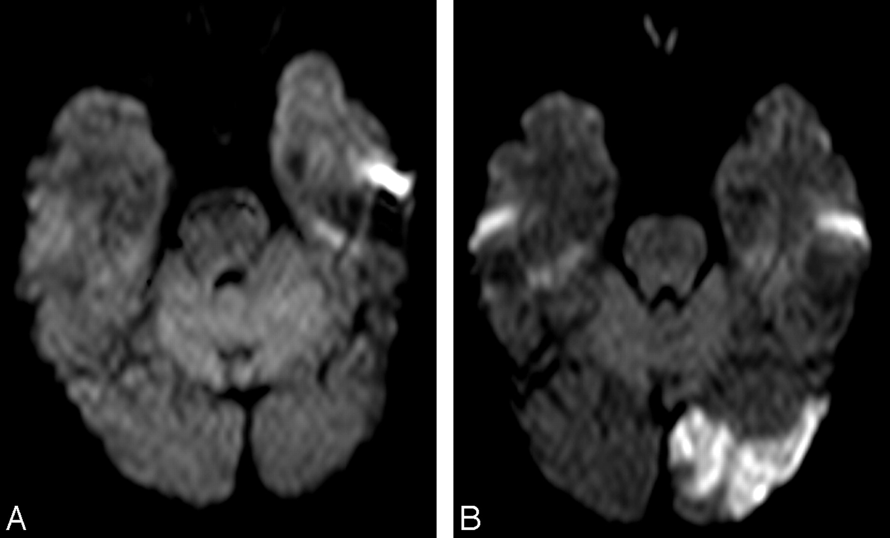
  • Fig. 4.
    • Download figure
    • Open in new tab
    • Download powerpoint
    Fig. 4.

    A, Right common carotid artery MR angiography before carotid artery stent placement (CAS). Ulcerated stenosis of the proximal internal carotid artery (ICA).

    B–D, Axial diffusion-weighted images (B and C) and apparent diffusion coefficient map (D). Before CAS, ischemic focal lesion >10 mm in the left caudate nucleus, contralateral to the treated ICA.

    E and F, After CAS, a new lesion is shown in the contralateral posterior fossa.

  • Fig. 5.
    • Download figure
    • Open in new tab
    • Download powerpoint
    Fig. 5.

    A, Normal diffusion-weighted image before carotid artery stent placement.

    B, After a successful procedure, a new ischemic cortical lesion >10 mm is seen in the contralateral posterior artery territory.

Tables

  • Figures
    • View popup
    Table 1:

    Baseline characteristics of patients

    n (%)
    No. of patients162 (100)
    Men131 (80.9)
    Women31 (19.1)
    Mean age/range (y)68.5 (33–86)
    Symptomatic patients122 (75.3)
       TIA/Amaurosis fugax64 (39.5)
       Minor stroke58 (35.8)
    Asymptomatic patients40 (24.7)
    Vascular risks factors
       Diabetes79 (48.8)
       Hypertesion127 (78.4)
       Hyperlipidemia99 (61.1)
       Cigarette smoking81 (50)
       Coronary artery disease53 (32.7)
       Peripheral vascular disease49 (30.2)
    • Note:—TIA indicates transient ischemic attacks.

    • View popup
    Table 2:

    Angiographic characteristics of patients

    n (%)
    Left ICA stenosis70 (43.2)
    Right ICA stenosis92 (56.8)
    % Stenosis (treated vessel)
       70–8559 (36.4)
       86–9979 (48.8)
       >9924 (14.8)
    % Contralateral stenosis
       <70112 (69.1)
       70–9924 (14.8)
       10026 (16.1)
    Ulcerated plaque71 (44.1)
    Calcified plaque80 (49.4)
    Functioning AcomA122 (75.3)
    Functioning PcomA72 (47.1)
    Intracranial lesion13 (8)
    Plaque
       <1 cm48 (29.6)
       ≥1 cm114 (70.4)
    Position
       <0.5 mm130 (80.1)
       ≥0.5 mm32 (19.9)
    Dissection post-TPA25 (15.4)
    Transient symptomatic vasospasm42 (25.9)
    • Note:—ICA indicates internal carotid artery; AcomA, anterior communicating artery; PcomA, posterior communicating artery; TPA, transluminal percutaneous angioplasty.

    • View popup
    Table 3:

    Microembolic signals (MES) detected in phases of carotid angioplasty and stenting (CAS) as determined by transcranial Doppler

    Phase of CASNo. of MES
    Median(P25–P75)
    16(3–11)
    227(13–47)
    30(0–1)
    Total58(27–94)
    • Note:—Of 95 cases, 88 (92.6%) were positive, 7 (7.4%) were negative.

    • View popup
    Table 4:

    Characteristics of ischemic lesions after carotid angioplasty and stenting

    No. of new lesions58 (28 patients)
    No. of single lesions15 (15 patients)
    No. of multiple lesions43 (13 patients)
    Vascular distribution
       Ipsilateral MCA territory19 (67.9)
       Contralateral MCA1 (3.6)
       Posterior fossa4 (14.3)
       Contralateral MCA + posterior fossa1 (3.6)
       Ipsilateral MCA + contralateral MCA3 (10.6)
    Location
       Cortical14 (50)
       Subcortical1 (3.6)
       Cortical + subcortical5 (17.9)
       Border zone3 (10.7)
       Deep wm3 (10.7)
       Cortical + deep wm1 (3.6)
       Cortical + subcortical + deep wm1 (3.6)
    Size
       <5 mm16 (57.1)
       5–10 mm8 (28.6)
       >10 mm3 (10.7)
       <5 and >10 mm1 (3.6)
    Anatomic location
       Frontal3 (10.7)
       Parietal12 (42.9)
       Temporal1 (3.6)
       Occipital2 (7.1)
       Basal ganglia4 (14.3)
       Frontal + parietal + occipital2 (7.1)
       Parietal + occipital + basal ganglia1 (3.6)
       Cerebellum3 (10.7)
    • Note:—MCA indicates middle cerebral artery; wm, white matter. Values in parentheses are percentages.

PreviousNext
Back to top

In this issue

American Journal of Neuroradiology: 27 (6)
American Journal of Neuroradiology
Vol. 27, Issue 6
June 2006
  • Table of Contents
  • Index by author
Advertisement
Print
Download PDF
Email Article

Thank you for your interest in spreading the word on American Journal of Neuroradiology.

NOTE: We only request your email address so that the person you are recommending the page to knows that you wanted them to see it, and that it is not junk mail. We do not capture any email address.

Enter multiple addresses on separate lines or separate them with commas.
Silent Ischemia after Neuroprotected Percutaneous Carotid Stenting: A Diffusion-Weighted MRI Study
(Your Name) has sent you a message from American Journal of Neuroradiology
(Your Name) thought you would like to see the American Journal of Neuroradiology web site.
CAPTCHA
This question is for testing whether or not you are a human visitor and to prevent automated spam submissions.
Cite this article
P. Piñero, A. González, A. Mayol, E. Martínez, J.R. González-Marcos, F. Boza, A. Cayuela, A. Gil-Peralta
Silent Ischemia after Neuroprotected Percutaneous Carotid Stenting: A Diffusion-Weighted MRI Study
American Journal of Neuroradiology Jun 2006, 27 (6) 1338-1345;

Citation Manager Formats

  • BibTeX
  • Bookends
  • EasyBib
  • EndNote (tagged)
  • EndNote 8 (xml)
  • Medlars
  • Mendeley
  • Papers
  • RefWorks Tagged
  • Ref Manager
  • RIS
  • Zotero
0 Responses
Respond to this article
Share
Bookmark this article
Silent Ischemia after Neuroprotected Percutaneous Carotid Stenting: A Diffusion-Weighted MRI Study
P. Piñero, A. González, A. Mayol, E. Martínez, J.R. González-Marcos, F. Boza, A. Cayuela, A. Gil-Peralta
American Journal of Neuroradiology Jun 2006, 27 (6) 1338-1345;
del.icio.us logo Twitter logo Facebook logo Mendeley logo
  • Tweet Widget
  • Facebook Like
  • Google Plus One

Jump to section

  • Article
    • Abstract
    • Methods
    • Results
    • Discussion
    • Conclusion
    • Footnotes
    • References
  • Figures & Data
  • Info & Metrics
  • Responses
  • References
  • PDF

Related Articles

  • No related articles found.
  • PubMed
  • Google Scholar

Cited By...

  • Progressive changes in cerebral perfusion after carotid stenting: a dynamic susceptibility contrast perfusion weighted imaging study
  • Can Doppler Flow Parameters of Carotid Stenosis Predict the Occurrence of New Ischemic Brain Lesions Detected by Diffusion-Weighted MR Imaging after Filter-Protected Internal Carotid Artery Stenting?
  • Periprocedural Hemodynamic Depression Is Associated With a Higher Number of New Ischemic Brain Lesions After Stenting in the International Carotid Stenting Study-MRI Substudy
  • Strategies of Clopidogrel Load and Atorvastatin Reload to Prevent Ischemic Cerebral Events in Patients Undergoing Protected Carotid Stenting: Results of the Randomized ARMYDA-9 CAROTID (Clopidogrel and Atorvastatin Treatment During Carotid Artery Stenting) Study
  • Unprotected Carotid Artery Stenting in Symptomatic Patients with High-Grade Stenosis: Results and Long-Term Follow-Up in a Single-Center Experience
  • New Ischemic Brain Lesions on Diffusion-Weighted MRI after Carotid Artery Stenting with Filter Protection: Frequency and Relationship with Plaque Morphology
  • Are Distal Protection Devices 'Protective' During Carotid Angioplasty and Stenting?
  • Internal Carotid Artery Stenting in Patients with Near Occlusion: 30-Day and Long-Term Outcome
  • Ischemic Cerebrovascular Complications of Cardiac Procedures
  • Volume and Composition of Emboli in Neuroprotected Stenting of the Carotid Artery
  • Assessing Carotid Revascularization: Should We Abandon the Neurological Examination?
  • New Brain Lesions After Carotid Stenting Versus Carotid Endarterectomy: A Systematic Review of the Literature
  • Risk Factors and Neurological Consequences of Syncopes Induced by Internal Carotid Artery Angioplasty
  • Response to Letter by Wong and Poon
  • New MRI Brain Lesions as Surrogate Outcome for Carotid Stenting With and Without Cerebral Protection
  • Advances in Interventional Neuroradiology 2006
  • Crossref
  • Google Scholar

This article has not yet been cited by articles in journals that are participating in Crossref Cited-by Linking.

More in this TOC Section

  • Contour Neurovascular System: Five Year Follow Up
  • Effect of SARS-CoV2 on Endovascular Thrombectomy
  • Flow diversion for distal circulation aneurysms
Show more NEUROINTERVENTION

Similar Articles

Advertisement

Indexed Content

  • Current Issue
  • Accepted Manuscripts
  • Article Preview
  • Past Issues
  • Editorials
  • Editor's Choice
  • Fellows' Journal Club
  • Letters to the Editor
  • Video Articles

Cases

  • Case Collection
  • Archive - Case of the Week
  • Archive - Case of the Month
  • Archive - Classic Case

More from AJNR

  • Trainee Corner
  • Imaging Protocols
  • MRI Safety Corner
  • Book Reviews

Multimedia

  • AJNR Podcasts
  • AJNR Scantastics

Resources

  • Turnaround Time
  • Submit a Manuscript
  • Submit a Video Article
  • Submit an eLetter to the Editor/Response
  • Manuscript Submission Guidelines
  • Statistical Tips
  • Fast Publishing of Accepted Manuscripts
  • Graphical Abstract Preparation
  • Imaging Protocol Submission
  • Evidence-Based Medicine Level Guide
  • Publishing Checklists
  • Author Policies
  • Become a Reviewer/Academy of Reviewers
  • News and Updates

About Us

  • About AJNR
  • Editorial Board
  • Editorial Board Alumni
  • Alerts
  • Permissions
  • Not an AJNR Subscriber? Join Now
  • Advertise with Us
  • Librarian Resources
  • Feedback
  • Terms and Conditions
  • AJNR Editorial Board Alumni

American Society of Neuroradiology

  • Not an ASNR Member? Join Now

© 2025 by the American Society of Neuroradiology All rights, including for text and data mining, AI training, and similar technologies, are reserved.
Print ISSN: 0195-6108 Online ISSN: 1936-959X

Powered by HighWire