
Article Figures & Data
Figures
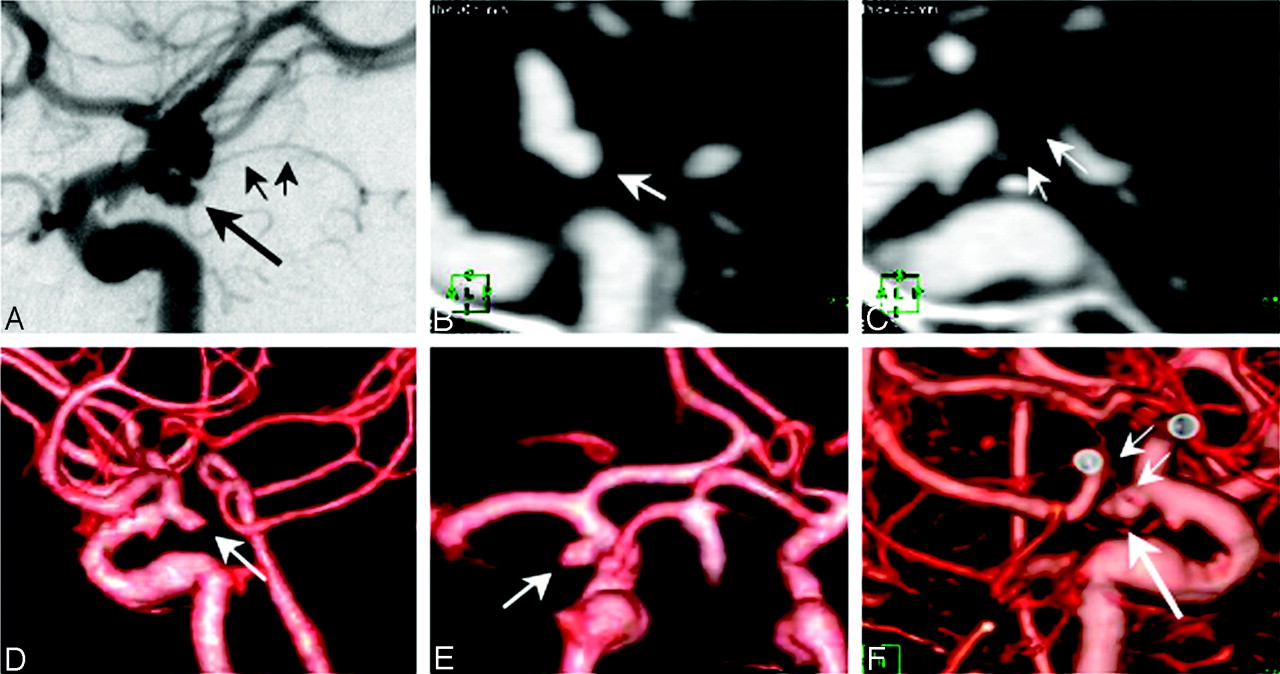
- Fig. 1.
Case 11: Patient with 3 ophthalmic artery (OphA), 1 carotid cave, and 1 cavernous aneurysm. A, Intraarterial angiogram, oblique view, showing the right OphA aneurysm (long arrow) and the right carotid cave aneurysm (short arrow).
B, Intraarterial angiogram, lateral view, showing the right OphA aneurysm (long arrow), the right carotid cave aneurysm (arrowhead), and the right cavernous aneurysm (short arrow).
C, Multiplanar reformatted (MPR) image, sagittal view, showing the right OphA aneurysm (long arrow) and OphA branching at the aneurysm neck (short arrow).
D, MPR image, sagittal view, which did not clearly visualize the right cavernous aneurysm.
E, MPR image, sagittal view, which did not clearly visualize the right carotid cave aneurysm.
F, Nonsubtracted (NS) 3D computed tomographic angiography (3DCTA), superior view, showing the right OphA aneurysm (arrow), but the right carotid cave and cavernous aneurysms were not visualized.
G, Volume-subtracted (VS)-3DCTA, superior view, clearly showing the right OphA (long arrow) and the right cavernous aneurysm (short arrow).
H, VS-3DCTA, oblique view, clearly showing the right OphA aneurysm (long arrow), the right carotid cave aneurysm (short arrow), the right cavernous aneurysm (double arrows), and OphA branching at aneurysm neck (arrowhead).
- Fig. 2.
Case 8: Patient with 1 carotid cave aneurysm and infundibular widening of the posterior communicating artery (PcomA).
A, Intraarterial angiogram, anteroposterior view, showing the left carotid cave aneurysm in the inferomedial direction (arrow).
B, Nonsubtracted (NS) 3D computed tomographic angiography (3DCTA), anteroposterior view, which did not show the left carotid cave aneurysm.
C, Volume subtraction (VS)-3DCTA, anteroposterior view, clearly showing the left carotid cave aneurysm with inferomedial direction (arrow).
D, Intra-arterial angiogram, lateral view, showing an infundibular widening (long arrow) of the left PcomA and a PcomA (short arrow) originating from the top of the infundibular widening.
E, VS-3DCTA, medial view, showing the left carotid cave aneurysm in the inferomedial direction (arrow) and the PcomA (arrowhead) originating from the top of the infundibular widening.
F, NS-3DCTA, posterior view, showing the infundibular widening of the left PcomA and the PcomA (arrow) originating from the top of the infundibular widening.
- Fig. 3.
Case 20: Patient with 1 anterior choroidal artery (AchoA) aneurysm. A, Intra-arterial angiogram, lateral view, showing the left AchoA aneurysm (long arrow) originating at the junction of the AchoA (short arrows) and the internal carotid artery (ICA).
B, Multiplanar reformatted (MPR) image, sagittal view, showing the left AchoA aneurysm (arrow).
C, MPR image, sagittal view, showing AchoA branching at aneurysm neck (arrows).
D, Volume subtracted (VS)-3D computed tomographic angiography (3DCTA), lateral view, showing the left AchoA aneurysm (arrow).
E, VS-3DCTA, posterior view, showing the left AchoA aneurysm with a posterolateral direction (arrow).
F, VS-3DCTA, medial view, clearly showing the left AchoA aneurysm (long arrow) and AchoA branching at the aneurysm neck (short arrows).


Tables
- Table 1:
Summary of 25 patients with internal carotid artery aneurysms evaluated by digital subtraction angiography (DSA) and computed tomogrAchAaphic angiography (CTA)
Patient No./Age (y)/Sex Location on DSA Detection of Aneurysm Sac Size (mm) Neck Size (mm) Direction of Aneurysm Branching Artery VS-CTA NS-CTA MPR CTA MPR DSA CTA MPR DSA CTA DSA CTA MPR DSA 1/75/F OphA 3.8 3.6 4.0 3.5 3.6 3.5 Anterosuperior Anterosuperior OphA OphA OphA 2/70/F Cave × 5.0 4.3 5.0 4.5 3.2 3.0 Posteromedial Posteromedial None None None 3/65/M AchoA 4.8 5.1 4.7 4.7 2.8 3.4 Posterolateral Posterolateral AchoA AchoA AchoA 4/67/F Cave × 6.5 6.3 6.7 5.4 4.9 4.8 Posteromedial Posteromedial None None None 5/79/F Cavernous 24.4 24.8 25.7 11.2 10.6 10.1 Lateral Lateral None None None 6/69/F AchoA 6.6 6.1 6.5 3.1 2.7 2.4 Posterolateral Posterolateral AchoA AchoA AchoA 7/61/F PcomA 5.7 5.5 5.4 2.8 2.2 2.2 Posterolateral Posterolateral PcomA PcomA PcomA AchoA × 2.3 2.4 2.2 * 1.5 Posterolateral Posterolateral AchoA AchoA 8/55/F Cave × × 3.6 3.6 3.0 * 2.7 Inferomedial Inferomedial None None 9/51/F AchoA × 2.0 2.0 1.5 * 1.5 Posterolateral Posterolateral AchoA AchoA 10/49/M SHA × 3.7 4.0 3.8 3.4 3.1 3.2 Medial Medial None None None 11/62/F OphA 9.5 10.5 10.0 5.1 4.1 4.3 Anterosuperior Anterosuperior OphA OphA OphA Cave × × 2.7 * 3.1 1.9 * 1.6 Inferomedial Inferomedial None None Cavernous × × 2.6 * 3.0 2.6 * 2.0 Superior Superior None None 12/55/M PcomA 6.8 6.4 6.5 3.4 3.3 2.8 Posterolateral Posterolateral PcomA PcomA PcomA 13/59/F Cave × 3.5 3.6 3.3 3.1 3.0 1.4 Posteromedial Posteromedial None None None 14/38/F SHA 5.8 6.0 5.6 3.3 2.9 2.9 Medial Medial None None None 15/48/F Cave × 5.0 4.8 5.2 3.5 3.0 3.2 Medial Medial None None None 16/53/F OphA 7.5 8.0 8.3 6.7 5.7 3.9 Superior Superior OphA OphA OphA 17/62/F PcomA 5.0 4.7 4.8 3.2 3.0 3.1 Posterolateral Posterolateral PcomA PcomA PcomA 18/64/F PcomA 5.6 5.8 5.9 2.9 2.9 2.8 Posterior Posterior PcomA PcomA PcomA 19/73/F PcomA 7.6 8.0 7.9 4.0 3.6 3.7 Posterolateral Posterolateral PcomA PcomA PcomA 20/65/F AchoA 4.5 4.6 4.4 2.8 3.3 1.9 Posterolateral Posterolateral AchoA AchoA AchoA 21/60/F AchoA 6.1 6.5 6.3 3.9 4.3 3.1 Posterolateral Posterolateral AchoA AchoA AchoA 22/58/F PcomA 4.3 4.1 4.0 3.1 2.9 2.6 Posterolateral Posterolateral PcomA PcomA PcomA AchoA 6.1 6.3 6.1 4.9 4.6 4.7 Lateral Lateral AchoA AchoA AchoA 23/57/M Cave × 5.1 5.5 5.3 3.6 3.4 3.1 Posteromedial Posteromedial None None None 24/51/F Cave × 4.1 4.0 4.2 3.5 3.0 3.0 Medial Medial None None None 25/31/F OphA 4.3 3.2 3.3 4.2 3.3 2.3 Superior Superior OphA OphA OphA Note:—VS indicates volume subtraction; NS, nonsubtraction; MPR, 2D multiplanar reformatted imaging; OphA, ophthalmic artery; AchoA, anterior choroidal artery; SHA, superior hypophyseal artery.
Location of Aneurysm Detection of Aneurysms DSA VS-CTA NS-CTA MPR Anterior choroidal artery 7 7 7 5 Posterior communicating artery 6 6 6 6 Superior hypohyseal artery 2 2 1 2 Ophthalmic artery 4 4 4 4 Carotid cave 8 8 0 6 Carotid cavernous 2 2 1 1 Total 29 29 19 24 Note:—DSA indicates digital subtraction angiography; VS-CTA, volume subtraction computed tomographic angiography; NS-CTA, nonsubtraction computed tomographic angiography; MPR, 2D multiplaner reformatted imaging.
Size of Aneurysm (mm) DSA CTA <3 2 4 3–5 13 12 5.1–10 13 12 >10 1 1 Total 29 29 Note:—DSA indicates digital subtraction angiography; CTA, computed tomographic angiography.
Branching Artery DSA CTA MPR Anterior choroidal artery 7 7 6 Posterior communicating artery 6 6 6 Ophthalmic artery 4 4 4 Total 17 17 16 Note:—DSA indicates digital subtraction angiography; CTA, computed tomographic angiography; MPR, 2D multiplanar reformatted imaging.
In this issue


Jump to section
Related Articles
Cited By...
- Surveillance of Unruptured Intracranial Saccular Aneurysms Using Noncontrast 3D-Black-Blood MRI: Comparison of 3D-TOF and Contrast-Enhanced MRA with 3D-DSA
- Dose reduction and image quality in CT angiography for cerebralaneurysm with various tube potentials and current settings
- Feasibility of Flat Panel Angiographic CT after Intravenous Contrast Agent Application in the Postoperative Evaluation of Patients with Clipped Aneurysms
- Three-dimensional reconstruction of a fibro-osseous lesion using binary images transformed from histopathological images