
Article Figures & Data
Figures
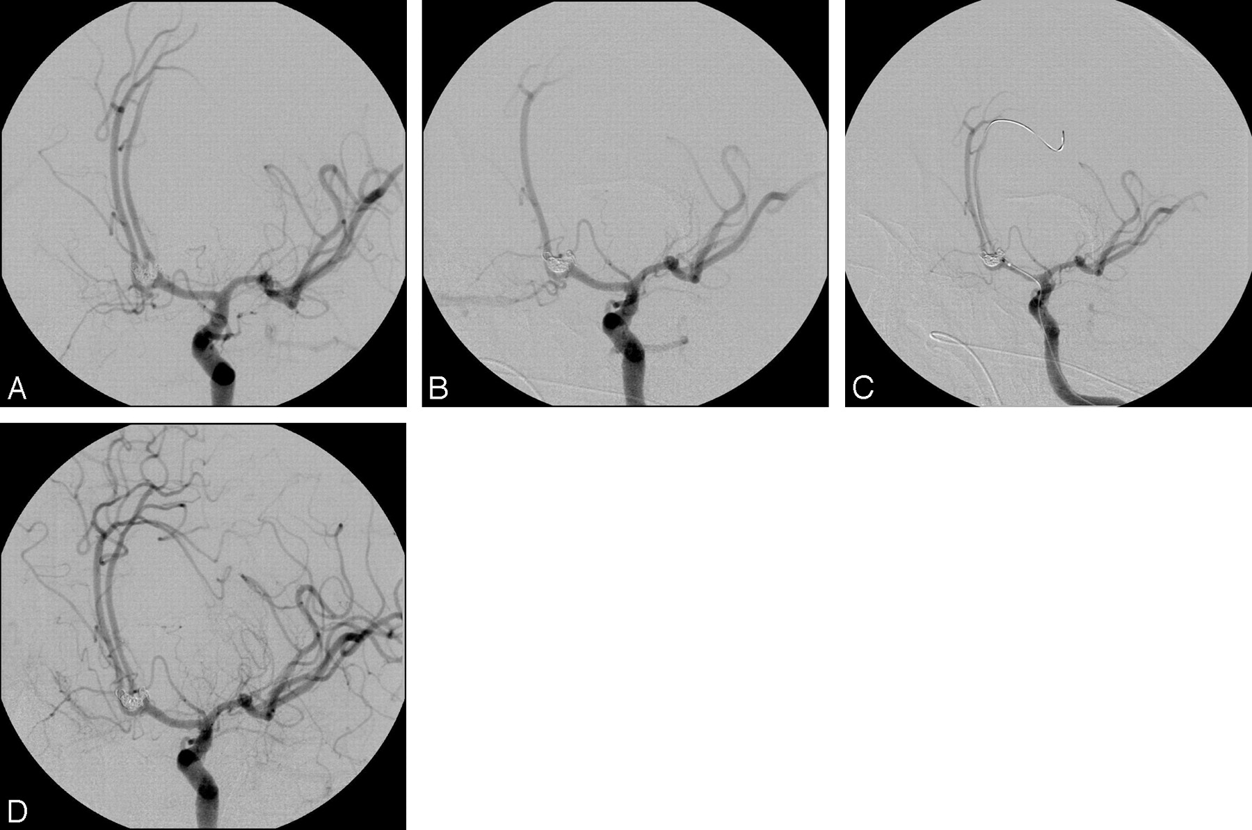
- Fig. 1.
This 49-year-old female patient was admitted to the hospital in good clinical condition (WFNS 1). A 5 × 4-mm aneurysm of the basilar tip involving the right superior cerebellar artery (SCA) was found on diagnostic angiography (A). Despite care taken not to compromise the ostium of the SCA with the coil package, the SCA was occluded during the coiling procedure (B). After administration of tirofiban, the vessel reopened within 15 minutes (C). Follow-up angiography 4 months later confirmed patency of the SCA, and the aneurysm remained occluded (D).
- Fig. 2.
Local thrombosis controlled by tirofiban. This 56-year-old female patient presented with a subarachnoidal hemorrhage and focal deficits (WFNS 3). Multiple aneurysms were found on diagnostic angiography; however, the pericallosal aneurysm was determined to be the symptomatic one. During the interventional procedure, a local thrombus was detected in the left pericallosal artery (arrow, A). Tirofiban was administered and patency of the vessel was restored (B). Outcome of the patient was excellent; there were no focal or generalized deficits (mRS 0).
- Fig. 3.
Extensive thrombosis and thromboembolism in part controlled by tirofiban. This 64-year-old male patient (10) presented with an extensive subarachnoidal hemorrhage (Fisher IV) and suffered a severe rebleeding during transfer from an outside hospital (WFNS V). The bilobar aneurysm of the anterior communicating branch had broad-based contact to the parent vessel (A). Because of the poor clinical status, a surgical approach was excluded. During the interventional procedure, an occlusion of the left pericallosal artery was detected (B). Tirofiban and aspirin were administered; however, patency of the vessel was not restored after 30 minutes. After mechanical assistance with the use of a microwire and various microcatheters (C), the pericallosal artery was partially recanalized (D). However, the patient had focal deficits (mRS 4).


Tables
Summary of anatomic and clinical data
Patient No./Age (y) Aneurysm Location Fisher Grade Dome Size Max (mm) Neck Size Max (mm) Dome/Neck Ratio Affected Vessel Patency after Tirofiban Admission (WFNS) Outcome (MRS) Aspirin Previously (mg) 1/78 AcomA 3 8 5 1.6 A2 bilateral Recanalized 1 0 500 2/49 AcomA 2 6 3 2.0 AcomA Recanalized 1 0 500 3/44 AcomA 2 8 4 2.0 A1, A2 right Recanalized 1 0 500 4/35 ACI 2 11 6 1.8 Local thrombus Dissolved 1 0 No 5/40 ACI 2 10 5 2.0 C1 Recanalized 2 0 No 6/49 Basil 3 5 3 1.7 SCA Recanalized 1 0 No 7/42 PCA 1 5 3 1.7 P1 Recanalized 1 0 500 8/56 Pericallosal artery 3 6 2.5 2.4 Local thrombus Dissolved 3 0 500 9/60 PcomA 2 8 3 2.7 Local thrombus Dissolved 3 2 500 10/64 AcomA 4 6 3 2.0 A2 left Partly recanalized* 5 4 500 11/57 AcomA 3 7 4 1.8 A1, A2 right Recanalized 2 5 500 12/55 PcomA 2 8 6 1.3 PcomA Recanalized 5 5 500 13/58 PcomA 2 5 3 1.7 Local thrombus Dissolved 5 5 500 14/63 Basil 18 5 3.6 P1 Recanalized* 5 5 500 15/47 AcomA 3 6 3 2.0 Local thrombus Dissolved 5 6 No 16/50 MCA 3 12 4 3.0 M2 Not recanalized 5 6 No Median 52.9 3.0 7.5 3.5 2.0 2.5 1.0 SD 10.7 0.7 3.4 1.2 0.6 1.8 2.6 Min 35.1 1.0 5.0 2.5 1.3 1 0 Max 77.7 4.0 18.0 6.0 3.6 5 6 Note:—AcomA indicates anterior communicating artery; ACI, acute cerebral infarction; Basil, basilar tip; PCA, posterior cerebral artery; MCA, middle cerebral artery; mean indicates average number of all of the above; SD indicates standard deviation; PcomA indicates posterior communicating artery; SCA, superior cerebellar artery.
* With assistance of mechanical thrombus fragmentation.
In this issue


Jump to section
Related Articles
Cited By...
- Rescue mechanical thrombectomy using a retrievable stent for thromboembolic occlusion occurring during coil embolization of ruptured intracranial aneurysms
- Rescue Treatment of Thromboembolic Complications during Endovascular Treatment of Cerebral Aneurysms: A Meta-Analysis
- Intra-Arterial Infusion of a Glycoprotein IIb/IIIa Antagonist for the Treatment of Thromboembolism During Coil Embolization of Intracranial Aneurysm: A Comparison of Abciximab and Tirofiban
- Rescue Treatment of Thromboembolic Complications During Endovascular Treatment of Cerebral Aneurysms
- Intra-arterial abciximab for the treatment of thrombus formation during coil embolization of intracranial aneurysms
- Abciximab Is a Safe Rescue Therapy in Thromboembolic Events Complicating Cerebral Aneurysm Coil Embolization: Single Center Experience in 42 Cases and Review of the Literature
- Coiling of basilar tip aneurysms: Results in 154 consecutive patients with emphasis on recurrent haemorrhage and re-treatment during mid- and long-term follow-up
- Advances in Interventional Neuroradiology 2006