
Article Figures & Data
Figures
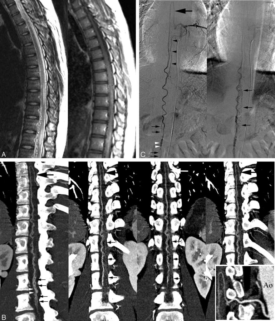
- Fig 1.
22-year-old man with intradural perimedullary SCAVF.
A, Sagittal fast spin-echo T2-weighted MR image (left) and postgadolinium T1-weighted image (right) show multiple enlarged pial vessels along the surface of the cord. Intrinsic increased signal intensity centrally within the spinal cord (left) and abnormal enhanced cord (right) extend from the lower thoracic levels to the conus medullaris.
B, Oblique coronal multiprojection volume-reconstruction images with different plane projections show the fistula (left, white arrowheads [type A, perimedullary SCAVF]) at the lower L2 spine level supplied by the mildly enlarged anterior spinal artery (large arrow). Multiple engorged outflow veins draining both cephalic and caudal directions are also noted (small arrows). Curved planar reformation image (right bottom) delineates the aorta (Ao) and anterior spinal artery feeder from the left T8 intercostal artery.
C, Conventional angiography of the left T8 intercostal artery in early (left) and late (right) phases, anteroposterior view, shows similar depiction of Fig 1B. The posterior spinal artery (black arrowheads) was also injected via the left T8 intercostal artery but did not supply the fistula.
- Fig 2.
55-year-old man with SDAVF.
A, Sagittal fast spin-echo T2-weighted MR (left) and postgadolinium T1-weighted images (right) show multiple enlarged pial vessels along the surface of the cord. Intrinsic increased signal intensity centrally within the spinal cord (left) and abnormal enhanced cord (right) extend from midthoracic levels to the conus medullaris.
B, Sagittal (upper left) multiplanar reconstruction image shows multiple engorged perimedullary veins (arrows) along the anterior and posterior surfaces of the spinal cord. Transverse MIP image (upper right) shows the fistula (large arrows) fed by the radiculomeningeal branch of the left T12 intercostal artery and engorged perimedullary veins (small arrows). Oblique coronal multiplanar reconstruction image (lower left) reveals the fistula (large arrows) and extensive engorgement of perimedullary veins (small arrows). Curved planar reformation image (lower right) along the left T12 intercostal artery and SDAVF delineates the aorta (Ao), intercostal artery (large white arrows), fistula (black arrows), and multiple engorged perimedullary veins (small white arrows).
C, Conventional angiography of the left T12 intercostal artery obtained in the arteriovenous phase, anteroposterior view, shows findings similar to those in Fig 2B.


In this issue


Jump to section
Related Articles
Cited By...
- Application of Spinal Subtraction and Bone Background Fusion CTA in the Accurate Diagnosis and Evaluation of Spinal Vascular Malformations
- Dilated Vein of the Filum Terminale on MRI: A Marker for Deep Lumbar and Sacral Dural and Epidural Arteriovenous Fistulas
- Bone-Subtracted Spinal CT Angiography Using Nonrigid Registration for Better Visualization of Arterial Feeders in Spinal Arteriovenous Fistulas
- Time-Resolved Contrast-Enhanced MR Angiography of Spinal Vascular Malformations
- Safety of spinal angiography: Complication rate analysis in 302 diagnostic angiograms