
Article Figures & Data
Figures
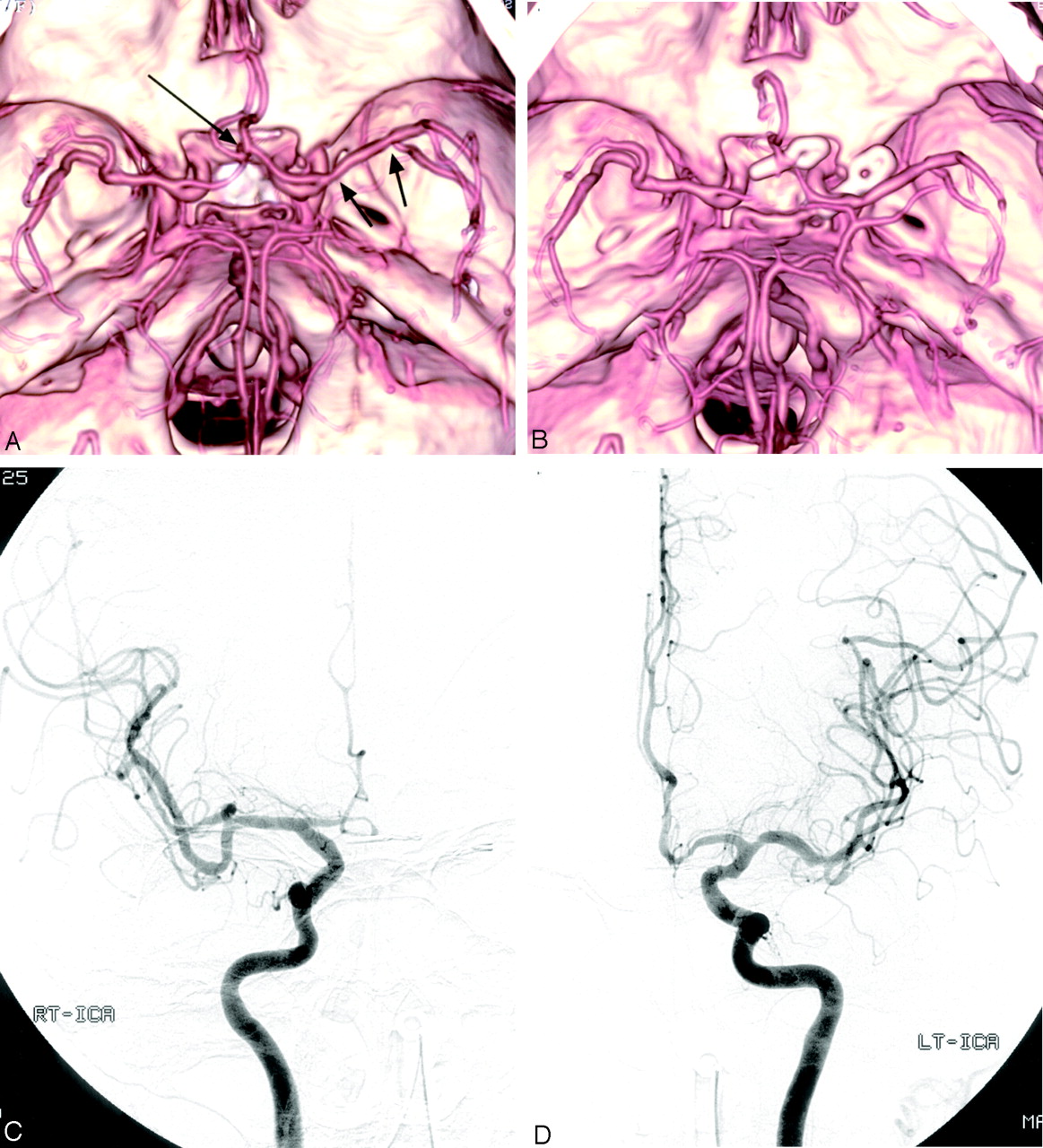
- Fig 1.
Case 6, a 73-year-old woman with SAH.
A, Preoperative MDCTA image, superior view, shows an anterior communicating artery aneurysm (long arrow). Left posterior communicating artery aneurysm was well visualized on other projections of MDCTA images (not shown). Note the multiple focal stenoses in the left M1 segment (arrows).
B, Postoperative MDCTA image, obtained 7 days after surgery, shows clipping of the aneurysms and multiple spasms of bilateral A1 and A2 segments. Note total occlusion of right A1 segment, as well as no change of stenoses in the left M1 segment consistent with preexisting atherosclerotic stenoses.
C and D, Postoperative right (C) and left (D) carotid angiograms, anteroposterior view, confirm vasospasm involving bilateral anterior cerebral arteries. Note grade 3 spasm (50%–99% luminal narrowing) in the right A1 segment, which was overestimated as grade 4 spasm (total occlusion) on MDCTA (B).
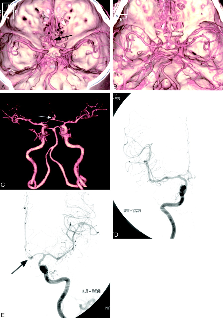
- Fig 2.
Case 5, a 42-year-old man with SAH.
A, Preoperative MDCTA image, superior view, shows an anterior communicating artery aneurysm (arrow).
B, Postoperative MDCTA image, obtained 5 days after surgery, shows clipping of the aneurysms and multiple spasms of bilateral A1, A2, left M1, and M2 segments.
C, The small remnant aneurysm (arrow) after aneurysm clipping is depicted on the anteroposterior MDCTA image with automated segmentation.
D and E, Postoperative right (D) and left (E) carotid angiograms, anteroposterior view, confirm vasospasm involving bilateral anterior cerebral and left middle cerebral arteries. The small remnant aneurysm (arrow) is also noted.


Tables
Grade Definition 0 No spasm 1 Mild spasm (<25% luminal narrowing) 2 Moderate spasm (25–50% luminal narrowing) 3 Severe spasm (51–99% luminal narrowing) 4 Total occlusion Patient No./Age (y)/Sex Site of Aneurysm Vasospasm on DSA Site Grade 1/40/M AcomA Bil ICA, Rt A1 2 Bil A2, M1, M2, Lt A1 3 2/72/F Rt ICA bifurcation Lt A1 1 Rt M2 2 Bil A2 3 Rt A1 4 3/55/M Rt MCA bifurcation Lt A1 2 Rt A1, Lt M2 3 4/67/F AcomA Bil A2, Lt M2 3 5/42/M AcomA Bil ICA 1 Bil A1, A2, Lt M1 3 6/73/F AcomA Lt A2 1 Lt PcomA Bil A1, Rt A2 3 7/33/M AcomA Rt ICA 1 Lt M1 2 Bil A1, A2, Rt M1 3 8/69/F Lt PcomA Lt P1 1 9/46/M AcomA Bil M1 1 Bil A1, A2 3 10/52/M BA tip BA, Rt P1 1 11/61/M Rt PcomA Rt ICA 1 Rt A1, A2, M1, P1, P2 2 12/39/F Lt PcomA Lt ICA 1 Lt A1, M1, M2 2 Lt P1, P2 3 13/67/F AcomA Lt M1, M2 1 Rt MCA bifurcation Bil ICA 2 Bil A2, Lt A1, Rt M1, M2 3 Rt A1 4 14/44/M Rt MCA bifurcation Rt ICA 1 Rt M1, M2 2 15/47/F Lt PICA Lt. VA 1 16/47/F Rt PcomA None 17/65/F Lt PcomA None Note.— Bil indicates bilateral; Lt, left; Rt, right; AComA, anterior communicating artery; MCA, middle cerebral artery; ICA, internal carotid artery; PcomA, posterior communicating artery; PICA, posterior inferior cerebellar artery; A1, first segment of anterior cerebral artery; A2, second segment of anterior cerebral artery; M1, first segment of middle cerebral artery; M2, second segment of middle cerebral artery; P1, first segment of posterior cerebral artery; P2, second segment of posterior cerebral artery; VA, vertebral artery; BA, basilar artery.
- Table 3:
Vasospasm (grades 0–4) of the cerebral arteries (251 segments) as determined with DSA and multidetector row CT angiography in 17 patients
Arterial Segment (No. of Segment)* Grade of Vasospasm No. of Arteries Overestimated by CTA No. of Arteries Underestimated by CTA r† 0 1 2 3 4 DSA CTA DSA CTA DSA CTA DSA CTA DSA CTA ICA (34) 24 24 6 6 4 4 0 0 0 0 1.00 A1 (32) 14 14 1 0 4 5 11 9 2 4 4 1 0.9623 A2 (34) 17 17 1 1 1 0 15 14 0 2 3 0.9702 M1 (34) 22 22 3 2 4 5 5 5 0 0 1 0.9978 M2 (34) 25 25 1 0 3 3 5 6 0 0 2 0.9958 P1 (32) 28 28 2 2 1 1 1 1 0 0 1.00 P2 (34) 32 32 0 0 1 0 1 2 0 0 1 0.9995 VBA (17) 15 15 2 2 0 0 0 0 0 0 1.00 Total (251) 177 177 16 13 18 18 38 35 2 6 11 1 0.9966 Note.— Data are number of segments. Grade 0 indicates no spasm; grade 1, <25%; grade 2, 25%–50%; grade 3, 51%–99% luminal reduction; grade 4, total occlusion. DSA indicates digital subtraction angiography; CTA, multidetector row CT angiography.
* See Table 2 for definitions of segments.
† r, Spearman correlation coefficient.
- Table 4:
Diagnostic performance of multidetector row CT angiography compared with DSA in the detection of hemodynamically significant vasospasm (51%–100% of luminal narrowing) according to the anatomic district in 17 patients (251 segments)
Sensitivity (%) Specificity (%) PPV (%) NPV (%) DA (%) P Value Proximal locations (ICA, M1, A1, P1, VA, and BA) 94.7 99.2 94.7 99.2 98.7 <.001 Distal locations (A2, M2, P2, and their branches) 100 96.3 87.5 100 97.1 <.001 Total 97.5 98.1 90.7 99.5 98.0 <.001 Note.— PPV indicates positive predictive value; NPV, negative predictive value; DA, diagnostic accuracy; ICA, internal carotid artery; M1, first segment of middle cerebral artery; M2, second segment of middle cerebral artery; A1, first segment of anterior cerebral artery; A2, second segment of anterior cerebral artery; P1, first segment of posterior cerebral artery; P2, second segment of posterior cerebral artery; VA, vertebral artery; BA, basilar artery.
Differences between proximal and distal locations are statistically nonsignificant.
In this issue


Jump to section
Related Articles
Cited By...
- Reliability of CT Angiography in Cerebral Vasospasm: A Systematic Review of the Literature and an Inter- and Intraobserver Study
- Haptoglobin phenotype predicts the development of focal and global cerebral vasospasm and may influence outcomes after aneurysmal subarachnoid hemorrhage
- Diagnostic Accuracy of CT Angiography and CT Perfusion for Cerebral Vasospasm: A Meta-Analysis
- Diagnostic Threshold Values of Cerebral Perfusion Measured With Computed Tomography for Delayed Cerebral Ischemia After Aneurysmal Subarachnoid Hemorrhage
- Diagnosing Delayed Cerebral Ischemia With Different CT Modalities in Patients With Subarachnoid Hemorrhage With Clinical Deterioration
- Multidetector Row CT Angiography in Spontaneous Lobar Intracerebral Hemorrhage: A Prospective Comparison with Conventional Angiography