
Article Figures & Data
Figures
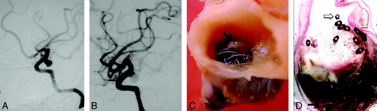
- Fig 1.
Ruptured aneurysm of the PComA removed 2 months after treatment with GDCs (case 10, Tables 1 and 2).
A, Digital subtraction angiography (DSA) before treatment demonstrates PComA aneurysm.
B, DSA immediately following treatment demonstrates minimal neck remnant.
C, Gross pathology, demonstrating free coils covered by an incomplete fibrin layer (arrow).
D, Microscopic section (H&E stain, low-power magnification, 2×) demonstrates unorganized thrombus in the aneurysm sac (arrow) and exposed coils within the neck (open arrow).
- Fig 2.
Ruptured aneurysm of the MCA removed surgically 3 years after treatment with GDCs (case 9, Tables 1 and 2).
A, DSA before treatment.
B, DSA immediately following treatment with standard GDCs, demonstrating neck remnant (broken arrow).
C, DSA 3 years after treatment demonstrates aneurysm recanalization (broken arrow).
D, Gross pathology demonstrating partially exposed coils within the neck (arrow) and coils protruding through the thin wall of the aneurysm dome.
E, Histologic section (H&E stain, low-power magnification, 2×) of the same specimen. Most of the aneurysm sac is filled with organized thrombus, but a large empty space is also seen (arrow).
F, Higher power magnification (20×) demonstrates attenuated fibrocellular tissue (arrow), an empty space (open arrow), and residual unorganized thrombus (broken arrow) within the same aneurysm.
- Fig 3.
Ruptured aneurysm of the AComA, treated with Matrix coils and removed during surgery 6 months later (case 18, Tables 1 and 2).
A, DSA before treatment demonstrates ruptured AComA aneurysm (arrow) and a small incidental aneurysm at the pericallosal artery (open arrow).
B, DSA immediately after treatment demonstrating a small neck remnant (arrow).
C, DSA, 6 months later, demonstrates growing neck remnant (arrow). Two incidental aneurysms (one on the left MCA and the other on the left pericallosal artery) were clipped, and the AComA aneurysm was clipped and removed during surgery.
D, Gross pathology of the surgical specimen. The coils within the neck (arrow) are covered by a thick tissue layer. The wall of the aneurysm is very thin.
E, Microscopic section of the specimen (H&E stain, low magnification, 2×). The aneurysm cavity is filled with fibrocellular tissue without any residual blood clot or empty spaces.
F, Higher magnification (10×) H&E stain demonstrates coils embedded in fibrocellular granulation tissue with multiple neocapillaries (arrowheads).
G, Higher power view (20×) demonstrates collagen deposition (arrow), smooth muscle cells (broken arrow), and small blood vessels (arrowheads).
H, Leukocyte invasion (arrow) represents granulation tissue (20×, H&E stain).


Tables
Clinical, morphologic, and histologic characteristics of aneurysms
Patient No./Age (y)/Sex Indication H&H Location Size (mm) Type of Coils Used No. of Coils Used Length (cm) of Coils Used Angiographic Results Coil Compaction Reason for Death Implant Time 1/38/M SAH 4 ICA 8 Matrix, GDC 6 60 Compl N/A Vasospasm 8 d 2/44/F SAH 4 AComA 6 GDC 3 30 Compl Yes N/A 36 mo 3/43/F SAH 4 AComA 8 GDC 3 24 RN N/A Vasospasm 8 d 4/39/M SAH 4 PComA 5 GDC 5 30 RN Yes N/A 36 mo 5/39/F SAH 4 Peric 5 GDC 4 22 Compl N/A SAH 18 d 6/56/M SAH 3 MCA 5 GDC 2 6 Compl N/A io rupt 5 d 7/16/M SAH 5 PICA 4 GDC 3 8 Compl N/A SAH 3 d 8/59/F SAH 4 AComA 4 GDC 3 14 Compl N/A SAH 5 d 9/43/M SAH 3 MCA 5 GDC 5 36 RN Yes N/A 35 mo 10/39/F SAH 5 PComA 6 GDC 3 22 RN N/A SAH 2 mo 11/17/M SAH 5 PComA 10 GDC 5 61 RA N/A SAH 3d 12/33/M ICH 5 AComA 3 GDC 2 7 RN N/A SAH 11 d 13/27/F SAH 2 AComA 5 GDC 4 14 Compl N/A Vasospasm 9 d 14/52/F SAH 3 MCA 4 GDC 4 13 Compl N/A io rupt 2 d 15/49/F SAH 3 VA 4 GDC 3 12 RN N/A io rupt 12 d 16/65/F SAH 2 PICA 3 GDC 2 6 RN N/A io rupt 1 d 17/67/F SAH 3 MCA 3 GDC 1 4 Compl N/A SAH 3 mo 18/44/F SAH 1 AComA 8 Matrix, GDC 6 55 RN Yes N/A 6 mo Note.—HH indicates Hunt and Hess grade; SAH, subarachnoid hemorrhage; ICH, intracerebral hemorrhage; ICA, internal carotid artery; AComA, anterior communicating arter; PComA, posterior communicating artery; Peric, pericallosal artery; MCA, middle cerebral artery; PICA, posterior inferior cerebellar artery; VA, vertebral artery; Compl, complete occlusion; RN, residual neck; RA, residual aneurysm; io rupt, intraoperative aneurysm rupture.
Clinical, morphologic, and histologic characteristics of aneurysms (cont.)
Patient No. Neck Identified Layer Covering Coils at Neck Content of the Aneurysm Sack Blood Unorganized Thrombus Void Spaces Foreign Body Giant Cells Leukocyte Infiltration Macrophages Fibrocellular Reaction Neocapillaries Collagen Formation 1 0 N/A 1 1 0 1 1 1 0 0 0 2 1 Fibrotic 0 1 1 1 0 0 1 1 1 3 1 Fibrin 1 1 0 1 1 1 0 0 0 4 1 Fibrotic 0 1 1 1 1 0 1 1 1 5 1 Fibrin 1 0 0 0 0 0 0 0 0 6 0 N/A 1 0 0 0 0 1 0 0 0 7 0 N/A 1 0 1 0 0 1 0 0 0 8 1 None 1 0 1 1 0 1 0 0 0 9 1 Fibrotic 1 1 1 1 0 1 1 1 1 10 1 Fibrin 1 1 0 1 0 1 0 0 1 11 1 Fibrin 1 1 0 0 1 1 0 0 0 12 1 Fibrin 1 0 0 1 0 1 0 0 0 13 0 N/A 1 0 0 0 0 0 0 0 0 14 0 N/A 1 1 0 0 1 1 0 0 0 15 0 N/A 1 1 0 0 0 1 0 0 0 16 0 N/A 1 1 0 0 1 1 0 0 0 17 1 Fibrotic 1 1 0 1 0 1 0 0 1 18 0 N/A 0 0 0 1 1 1 1 1 1
In this issue


Jump to section
Related Articles
Cited By...
- Wall Enhancement of Coiled Intracranial Aneurysms Is Associated with Aneurysm Recanalization: A Cross-Sectional Study
- Aneurysm wall cellularity affects healing after coil embolization: assessment in a rat saccular aneurysm model
- Endothelialization following Flow Diversion for Intracranial Aneurysms: A Systematic Review
- Perianeurysmal vasogenic oedema (PAVO) following aneurysm embolisation: a unique case of asymptomatic long-term progression and review of the literature
- Risk Factor Analysis of Recanalization Timing in Coiled Aneurysms: Early versus Late Recanalization
- Evolution of Flow-Diverter Endothelialization and Thrombus Organization in Giant Fusiform Aneurysms after Flow Diversion: A Histopathologic Study
- Immunohistochemical analysis of a ruptured basilar top aneurysm autopsied 22 years after embolization with Guglielmi detachable coils
- Mechanisms of Healing in Coiled Intracranial Aneurysms: A Review of the Literature
- Immunohistochemical analysis of a ruptured basilar top aneurysm autopsied 22 years after embolization with Guglielmi detachable coils
- In Memoriam: The Matrix Coil
- A history of detachable coils: 1987-2012
- Healing of saccular aneurysms following platinum coil embolization: lack of improved efficacy with vitamin C supplementation
- New Generation of Flow Diverter (Surpass) for Unruptured Intracranial Aneurysms: A Prospective Single-Center Study in 37 Patients
- Bioactivity and bioinactivity: two sides of the same coin
- Computerized Assessment of Angiographic Occlusion Rate and Coil Density in Embolized Human Cerebral Aneurysms
- Stent-Assisted Reconstructive Endovascular Repair of Cranial Fusiform Atherosclerotic and Dissecting Aneurysms: Long-Term Clinical and Angiographic Follow-Up