Advertisement

Main menu

  • Home
  • Content
    • Current Issue
    • Accepted Manuscripts
    • Article Preview
    • Past Issue Archive
    • Video Articles
    • AJNR Case Collection
    • Case of the Week Archive
    • Case of the Month Archive
    • Classic Case Archive
  • Special Collections
    • AJNR Awards
    • Low-Field MRI
    • Alzheimer Disease
    • ASNR Foundation Special Collection
    • Photon-Counting CT
    • View All
  • Multimedia
    • AJNR Podcasts
    • AJNR SCANtastic
    • Trainee Corner
    • MRI Safety Corner
    • Imaging Protocols
  • For Authors
    • Submit a Manuscript
    • Submit a Video Article
    • Submit an eLetter to the Editor/Response
    • Manuscript Submission Guidelines
    • Statistical Tips
    • Fast Publishing of Accepted Manuscripts
    • Graphical Abstract Preparation
    • Imaging Protocol Submission
    • Author Policies
  • About Us
    • About AJNR
    • Editorial Board
    • Editorial Board Alumni
  • More
    • Become a Reviewer/Academy of Reviewers
    • Subscribers
    • Permissions
    • Alerts
    • Feedback
    • Advertisers
    • ASNR Home

User menu

  • Alerts
  • Log in

Search

  • Advanced search
American Journal of Neuroradiology

American Journal of Neuroradiology

ASHNR American Society of Functional Neuroradiology ASHNR American Society of Pediatric Neuroradiology ASSR
  • Alerts
  • Log in

Advanced Search

  • Home
  • Content
    • Current Issue
    • Accepted Manuscripts
    • Article Preview
    • Past Issue Archive
    • Video Articles
    • AJNR Case Collection
    • Case of the Week Archive
    • Case of the Month Archive
    • Classic Case Archive
  • Special Collections
    • AJNR Awards
    • Low-Field MRI
    • Alzheimer Disease
    • ASNR Foundation Special Collection
    • Photon-Counting CT
    • View All
  • Multimedia
    • AJNR Podcasts
    • AJNR SCANtastic
    • Trainee Corner
    • MRI Safety Corner
    • Imaging Protocols
  • For Authors
    • Submit a Manuscript
    • Submit a Video Article
    • Submit an eLetter to the Editor/Response
    • Manuscript Submission Guidelines
    • Statistical Tips
    • Fast Publishing of Accepted Manuscripts
    • Graphical Abstract Preparation
    • Imaging Protocol Submission
    • Author Policies
  • About Us
    • About AJNR
    • Editorial Board
    • Editorial Board Alumni
  • More
    • Become a Reviewer/Academy of Reviewers
    • Subscribers
    • Permissions
    • Alerts
    • Feedback
    • Advertisers
    • ASNR Home
  • Follow AJNR on Twitter
  • Visit AJNR on Facebook
  • Follow AJNR on Instagram
  • Join AJNR on LinkedIn
  • RSS Feeds

AJNR Awards, New Junior Editors, and more. Read the latest AJNR updates

Research ArticleHead and Neck Imaging

Optic Nerve Hyperintensity on T2-Weighted Images among Patients with Pituitary Macroadenoma: Correlation with Visual Impairment

A.M. Tokumaru, I. Sakata, H. Terada, S. Kosuda, H. Nawashiro and M. Yoshii
American Journal of Neuroradiology February 2006, 27 (2) 250-254;
A.M. Tokumaru
  • Find this author on Google Scholar
  • Find this author on PubMed
  • Search for this author on this site
I. Sakata
  • Find this author on Google Scholar
  • Find this author on PubMed
  • Search for this author on this site
H. Terada
  • Find this author on Google Scholar
  • Find this author on PubMed
  • Search for this author on this site
S. Kosuda
  • Find this author on Google Scholar
  • Find this author on PubMed
  • Search for this author on this site
H. Nawashiro
  • Find this author on Google Scholar
  • Find this author on PubMed
  • Search for this author on this site
M. Yoshii
  • Find this author on Google Scholar
  • Find this author on PubMed
  • Search for this author on this site
  • Article
  • Figures & Data
  • Info & Metrics
  • Responses
  • References
  • PDF
Loading

Article Figures & Data

Figures

  • Tables
  • Fig 1.
    • Download figure
    • Open in new tab
    • Download powerpoint
    Fig 1.
    • Download figure
    • Open in new tab
    • Download powerpoint
    Fig 1.

    Degree of optic chiasmal compression. A, No compression to optic chiasma (−). B, Compression of less than half of the optic chiasm (+). C, Compression with marked thinning (++).

  • Fig 2.
    • Download figure
    • Open in new tab
    • Download powerpoint
    Fig 2.

    Correlation between abnormal signal intensity of optic nerve and tumor size.

  • Fig 3.
    • Download figure
    • Open in new tab
    • Download powerpoint
    Fig 3.

    Correlation between abnormal signal intensity of optic nerve and optic chiasm compression.
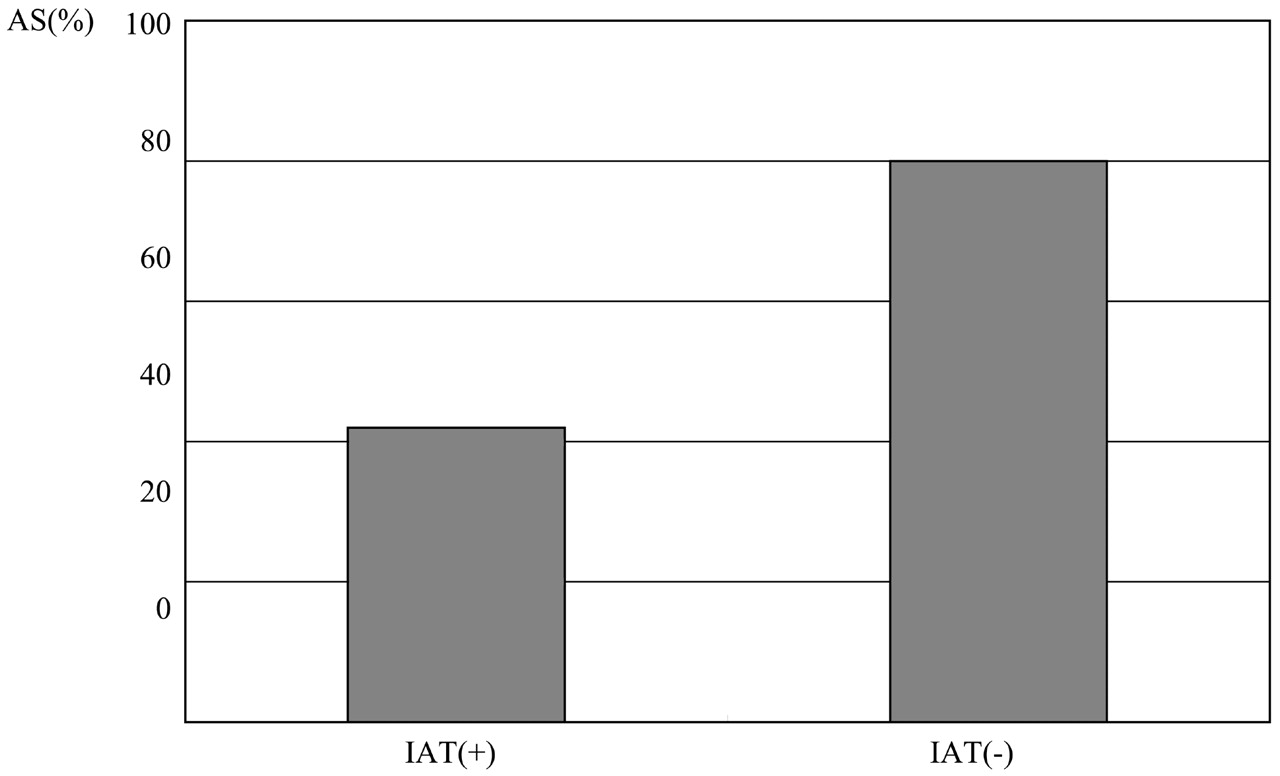
  • Fig 4.
    • Download figure
    • Open in new tab
    • Download powerpoint
    Fig 4.

    Correlation between abnormal signal intensity of optic nerve and improvement after treatment of VA.
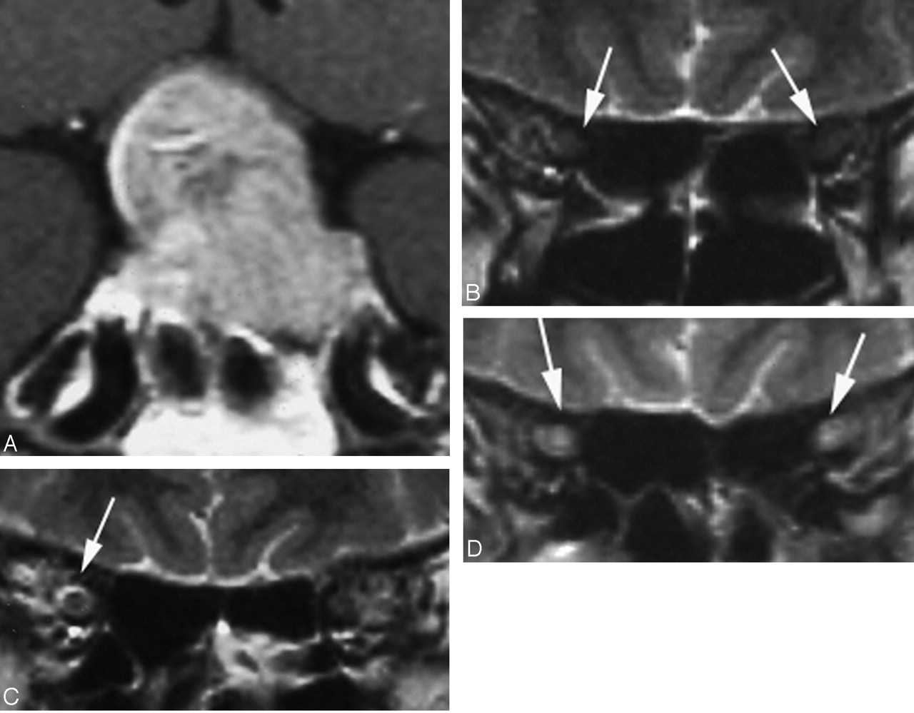
  • Fig 5.
    • Download figure
    • Open in new tab
    • Download powerpoint
    Fig 5.

    Case 3, a 42-year-old woman whose disease duration was 6 months. Right VA disturbance was recognized (right VA = 0.02; left VA = 1.5). A, Pituitary macroadenoma markedly compressed the optic chiasm especially the right side (white arrows). B–E, Hyperintensity was recognized in the right optic nerve on T2-weighted image (arrows). F–H, Hyperintensiy in the right optic nerve lasted for 2 years after the tumor reduction (arrows).
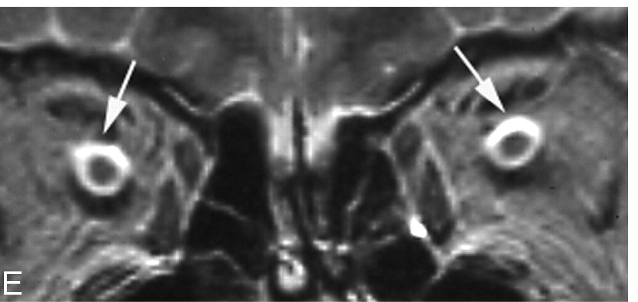
  • Fig 6.
    • Download figure
    • Open in new tab
    • Download powerpoint
    Fig 6.
    • Download figure
    • Open in new tab
    • Download powerpoint
    Fig 6.

    Case 17, a 58-year-old man, whose disease duration from the initial examination to the operation was 26 months. VA was not stable, though fixed VA disturbance was not recognized on the initial examination. A, Pituitary adenoma compressed the optic chiasm. B, Hyperintensiy of the optic nerve was not shown (arrows) on the initial examination. C, Right-side perioptic subarachnoid space dilated slightly on the initial examination (arrow). D and E, Tumor reduction was performed 26 months after the initial examination. Hyperintensity was shown in the bilateral optic nerve ventral to the optic chiasm (D, white arrows) and bilateral perioptic subarachnoid space dilated markedly (E, white arrows), probably representing atrophic change of the optic nerves.

Tables

  • Figures
  • Optic tract hyperintensity on T2-weighted images among patients with pituitary macroadenoma: correlation with visual impairment

    Patient No./Age (y)/SexDurationSize (mm)OCASVADVFDPathologyIAT
    1/36/M2 mo18−−−+
    2/61/M9 y23.7+−+ (rt 0.7, lt blind)+PA−
    3/42/F16 mo26.5++++ (rt 0.02, lt 1.5)+PA (corticotro)−
    4/38/F10 mo14−−−+
    5/40/M6 mo35++++ (rt 0.07, lt 0.05)+PA (null)−
    6/55/F4 y46++++ (rt 0.06, lt 0.06)+PA (chromophobe)−
    7/65/F2 mo13−−−+PA (null)
    8/74/M18 mo16.4+++ (rt 0.7, lt 0.4)+PA (null)−
    9/54/F3 mo27+++ (rt 0.06, lt 0.1)+PA (nonfunctioning)+ (rt 1.2, lt 0.8)
    10/65/M2 mo13−++ (rt light sense)+Pituitary carcinoma−
    11/59/F14 mo35++++ (rt 0.3, lt 0.9)+PA (null)−
    12/55/M3 mo22++++ (rt 0.5, lt 0.1)+PA+ (rt 1.0, lt 1.0)
    13/41/F19 mo25++++ (hand sense)+PA (null)−
    14/37/F3 mo16−−−+
    15/70/F6 mo18+++ (rt 0.1, lt blind)−
    16/27/M6 mo15−++ (finger sense)+PA (chromophobe)+ (rt 0.7, lt 0.9)
    17/58/M26 mo24++++ (rt 1.5, lt 0.4)−PA (null)−
    18/21/F8 mo30+−−+PA (GH)−
    19/50/F10 y51++++ (rt 0.03, lt 0.1)+PA(FSH-LH)−
    20/74/F1.5 mo18−++ (lt light sense)−PA (prolactinoma)+ (rt 0.8, lt 0.4)
    21/66/F2 mo15−−−−PA (null)
    22/63/M2 mo23.7++++ (rt 0.1, lt 0.7)+PA (chromophobe)+ (rt 0.7, lt 0.8)
    23/54/F3 mo13−−−−PA
    24/56/F3 mo20−−−−PA
    25/45/M2 mo16−−−+PA (chromophobe)
    26/36/M2 mo12.2−−−−
    27/50/M3 mo16++++ (rt 0.9, lt 0.3)+PA (GH)+ (rt 0.9, lt 0.7)
    • Note.—OC indicates optic chiasm compression; AS, abnormal signal in optic nerve; VAD, visual acuity disturbance; VFD, visual field disturbance; IAT, improvement of visual acuity after treatment; PA, pituitary adenoma; FSH-LH, follicle stimulating hormone-luteinizing hormone; GH, growth hormone.

PreviousNext
Back to top

In this issue

American Journal of Neuroradiology: 27 (2)
American Journal of Neuroradiology
Vol. 27, Issue 2
February, 2006
  • Table of Contents
  • Index by author
Advertisement
Print
Download PDF
Email Article

Thank you for your interest in spreading the word on American Journal of Neuroradiology.

NOTE: We only request your email address so that the person you are recommending the page to knows that you wanted them to see it, and that it is not junk mail. We do not capture any email address.

Enter multiple addresses on separate lines or separate them with commas.
Optic Nerve Hyperintensity on T2-Weighted Images among Patients with Pituitary Macroadenoma: Correlation with Visual Impairment
(Your Name) has sent you a message from American Journal of Neuroradiology
(Your Name) thought you would like to see the American Journal of Neuroradiology web site.
CAPTCHA
This question is for testing whether or not you are a human visitor and to prevent automated spam submissions.
Cite this article
A.M. Tokumaru, I. Sakata, H. Terada, S. Kosuda, H. Nawashiro, M. Yoshii
Optic Nerve Hyperintensity on T2-Weighted Images among Patients with Pituitary Macroadenoma: Correlation with Visual Impairment
American Journal of Neuroradiology Feb 2006, 27 (2) 250-254;

Citation Manager Formats

  • BibTeX
  • Bookends
  • EasyBib
  • EndNote (tagged)
  • EndNote 8 (xml)
  • Medlars
  • Mendeley
  • Papers
  • RefWorks Tagged
  • Ref Manager
  • RIS
  • Zotero
0 Responses
Respond to this article
Share
Bookmark this article
Optic Nerve Hyperintensity on T2-Weighted Images among Patients with Pituitary Macroadenoma: Correlation with Visual Impairment
A.M. Tokumaru, I. Sakata, H. Terada, S. Kosuda, H. Nawashiro, M. Yoshii
American Journal of Neuroradiology Feb 2006, 27 (2) 250-254;
del.icio.us logo Twitter logo Facebook logo Mendeley logo
  • Tweet Widget
  • Facebook Like
  • Google Plus One

Jump to section

  • Article
    • Abstract
    • Patients and Methods
    • Results
    • Discussion
    • References
  • Figures & Data
  • Info & Metrics
  • Responses
  • References
  • PDF

Related Articles

  • No related articles found.
  • PubMed
  • Google Scholar

Cited By...

  • White matter changes linked to visual recovery after nerve decompression
  • Crossref
  • Google Scholar

This article has not yet been cited by articles in journals that are participating in Crossref Cited-by Linking.

More in this TOC Section

  • FDG Uptake and Thyroid Cancer Post-BRAF Therapy
  • Normal Facial Nerve Enhancement on VIBE Sequence
  • Lipochoristomas and inner ear signal
Show more Head and Neck Imaging

Similar Articles

Advertisement

Indexed Content

  • Current Issue
  • Accepted Manuscripts
  • Article Preview
  • Past Issues
  • Editorials
  • Editor's Choice
  • Fellows' Journal Club
  • Letters to the Editor
  • Video Articles

Cases

  • Case Collection
  • Archive - Case of the Week
  • Archive - Case of the Month
  • Archive - Classic Case

More from AJNR

  • Trainee Corner
  • Imaging Protocols
  • MRI Safety Corner

Multimedia

  • AJNR Podcasts
  • AJNR Scantastics

Resources

  • Turnaround Time
  • Submit a Manuscript
  • Submit a Video Article
  • Submit an eLetter to the Editor/Response
  • Manuscript Submission Guidelines
  • Statistical Tips
  • Fast Publishing of Accepted Manuscripts
  • Graphical Abstract Preparation
  • Imaging Protocol Submission
  • Evidence-Based Medicine Level Guide
  • Publishing Checklists
  • Author Policies
  • Become a Reviewer/Academy of Reviewers
  • News and Updates

About Us

  • About AJNR
  • Editorial Board
  • Editorial Board Alumni
  • Alerts
  • Permissions
  • Not an AJNR Subscriber? Join Now
  • Advertise with Us
  • Librarian Resources
  • Feedback
  • Terms and Conditions
  • AJNR Editorial Board Alumni

American Society of Neuroradiology

  • Not an ASNR Member? Join Now

© 2025 by the American Society of Neuroradiology All rights, including for text and data mining, AI training, and similar technologies, are reserved.
Print ISSN: 0195-6108 Online ISSN: 1936-959X

Powered by HighWire