Advertisement

Main menu

  • Home
  • Content
    • Current Issue
    • Accepted Manuscripts
    • Article Preview
    • Past Issue Archive
    • Video Articles
    • AJNR Case Collection
    • Case of the Week Archive
    • Case of the Month Archive
    • Classic Case Archive
  • Special Collections
    • AJNR Awards
    • Low-Field MRI
    • Alzheimer Disease
    • ASNR Foundation Special Collection
    • Photon-Counting CT
    • View All
  • Multimedia
    • AJNR Podcasts
    • AJNR SCANtastic
    • Trainee Corner
    • MRI Safety Corner
    • Imaging Protocols
  • For Authors
    • Submit a Manuscript
    • Submit a Video Article
    • Submit an eLetter to the Editor/Response
    • Manuscript Submission Guidelines
    • Statistical Tips
    • Fast Publishing of Accepted Manuscripts
    • Graphical Abstract Preparation
    • Imaging Protocol Submission
    • Author Policies
  • About Us
    • About AJNR
    • Editorial Board
    • Editorial Board Alumni
  • More
    • Become a Reviewer/Academy of Reviewers
    • Subscribers
    • Permissions
    • Alerts
    • Feedback
    • Advertisers
    • ASNR Home

User menu

  • Alerts
  • Log in

Search

  • Advanced search
American Journal of Neuroradiology

American Journal of Neuroradiology

ASHNR American Society of Functional Neuroradiology ASHNR American Society of Pediatric Neuroradiology ASSR
  • Alerts
  • Log in

Advanced Search

  • Home
  • Content
    • Current Issue
    • Accepted Manuscripts
    • Article Preview
    • Past Issue Archive
    • Video Articles
    • AJNR Case Collection
    • Case of the Week Archive
    • Case of the Month Archive
    • Classic Case Archive
  • Special Collections
    • AJNR Awards
    • Low-Field MRI
    • Alzheimer Disease
    • ASNR Foundation Special Collection
    • Photon-Counting CT
    • View All
  • Multimedia
    • AJNR Podcasts
    • AJNR SCANtastic
    • Trainee Corner
    • MRI Safety Corner
    • Imaging Protocols
  • For Authors
    • Submit a Manuscript
    • Submit a Video Article
    • Submit an eLetter to the Editor/Response
    • Manuscript Submission Guidelines
    • Statistical Tips
    • Fast Publishing of Accepted Manuscripts
    • Graphical Abstract Preparation
    • Imaging Protocol Submission
    • Author Policies
  • About Us
    • About AJNR
    • Editorial Board
    • Editorial Board Alumni
  • More
    • Become a Reviewer/Academy of Reviewers
    • Subscribers
    • Permissions
    • Alerts
    • Feedback
    • Advertisers
    • ASNR Home
  • Follow AJNR on Twitter
  • Visit AJNR on Facebook
  • Follow AJNR on Instagram
  • Join AJNR on LinkedIn
  • RSS Feeds

AJNR Awards, New Junior Editors, and more. Read the latest AJNR updates

Research ArticleSpine Imaging and Spine Image-Guided Interventions

Plasma Radio-Frequency–Based Diskectomy for Treatment of Cervical Herniated Nucleus Pulposus: Feasibility, Safety, and Preliminary Clinical Results

G. Bonaldi, F. Baruzzi, A. Facchinetti, P. Fachinetti and S. Lunghi
American Journal of Neuroradiology November 2006, 27 (10) 2104-2111;
G. Bonaldi
  • Find this author on Google Scholar
  • Find this author on PubMed
  • Search for this author on this site
F. Baruzzi
  • Find this author on Google Scholar
  • Find this author on PubMed
  • Search for this author on this site
A. Facchinetti
  • Find this author on Google Scholar
  • Find this author on PubMed
  • Search for this author on this site
P. Fachinetti
  • Find this author on Google Scholar
  • Find this author on PubMed
  • Search for this author on this site
S. Lunghi
  • Find this author on Google Scholar
  • Find this author on PubMed
  • Search for this author on this site
  • Article
  • Figures & Data
  • Info & Metrics
  • Responses
  • References
  • PDF
Loading

Article Figures & Data

Figures

  • Tables
  • Fig 1.
    • Download figure
    • Open in new tab
    • Download powerpoint
    Fig 1.

    A, The 19-gauge cannula with an internal mandrel is positioned against the anterior surface of the annulus fibrosus. The cannula is held by a surgical forceps to minimize x-ray exposure of the surgeon’s hand.

    B, Cannula placement as observed under fluoroscopy.

    C and D, The cannula is advanced into the disk, and the SpineWand device (D, arrow) is introduced into the nucleus pulposus via the cannula.

    E, The device is activated and then rotated 360°(E) to create a spheric void by means of the loop-shaped active electrode (arrow). Between 2 and 4 voids are ablated in a linear direction to create a channel.

    F, After the first channel into the disk is completed, the device is repositioned to a different part of the nucleus, with the placement depending on the topography of the herniation. For left-sided herniations, the first channel is made in an oblique direction, from the right anterolateral entry point toward the left posterolateral herniation; the other channel is made on the midline and directed toward the posterior profile of the disk. For a right-sided lesion, the first channel is directed obliquely toward the center of the disk; the second channel is directed toward the right paramedian, along the medial surface of the uncal process to reach the herniation in the posterior aspect of the disk.

    G, A schematic drawing shows the entry route, with the clinician’s fingers pushing the trachea across the midline while protecting the neurovascular bundle.

  • Fig 2.
    • Download figure
    • Open in new tab
    • Download powerpoint
    Fig 2.

    A, MR imaging at baseline for a 34-year-old woman who underwent percutaneous plasma radio-frequency–based diskectomy for a disk (C6–7) herniation compressing the spinal cord and causing clinical signs of myelopathy.

    B, A 7-week MR imaging follow-up shows regression of both herniation and cord compression; the patient is almost asymptomatic.

    C, An MR image at 9-month follow-up shows almost complete regression of the disk herniation; the patient is completely asymptomatic.

  • Fig 3.
    • Download figure
    • Open in new tab
    • Download powerpoint
    Fig 3.

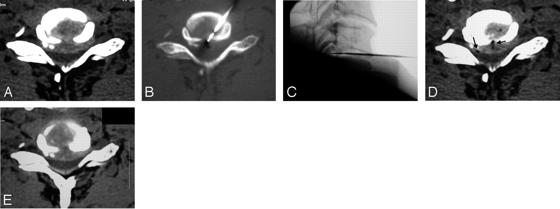
    A, Bilateral disk herniation at C6–7 encroaching the spinal canal. The SpineWand device was placed into position by using CT-guidance and switching to direct fluoroscopy for the disk ablation procedure. This allowed activation of the plasma-field energy directly inside the herniation.

    B and C, The loop-shaped active electrode was inserted beyond the posterior limit of the vertebral body into the spinal canal.

    D, At the end of the procedure, the gas generated from the tissue excision is evident inside the disk.

    E, The 5-month follow-up image shows only minimal or questionable reduction of disk size; despite this, the patient reports good clinical improvement (Macnab 3) (see text for discussion).

  • Fig 4.
    • Download figure
    • Open in new tab
    • Download powerpoint
    Fig 4.

    A, Lateral disk herniation compressing the nerve root.

    B, The cannula and SpineWand device are positioned under CT guidance and then safely and precisely directed toward the herniation.

    C, Note gas from tissue excision diffusing inside the herniation itself.

    D, A 4-month CT follow-up shows partial regression of the lesion; the patient reports a definite clinical improvement (Macnab 3).

  • Fig 5.
    • Download figure
    • Open in new tab
    • Download powerpoint
    Fig 5.

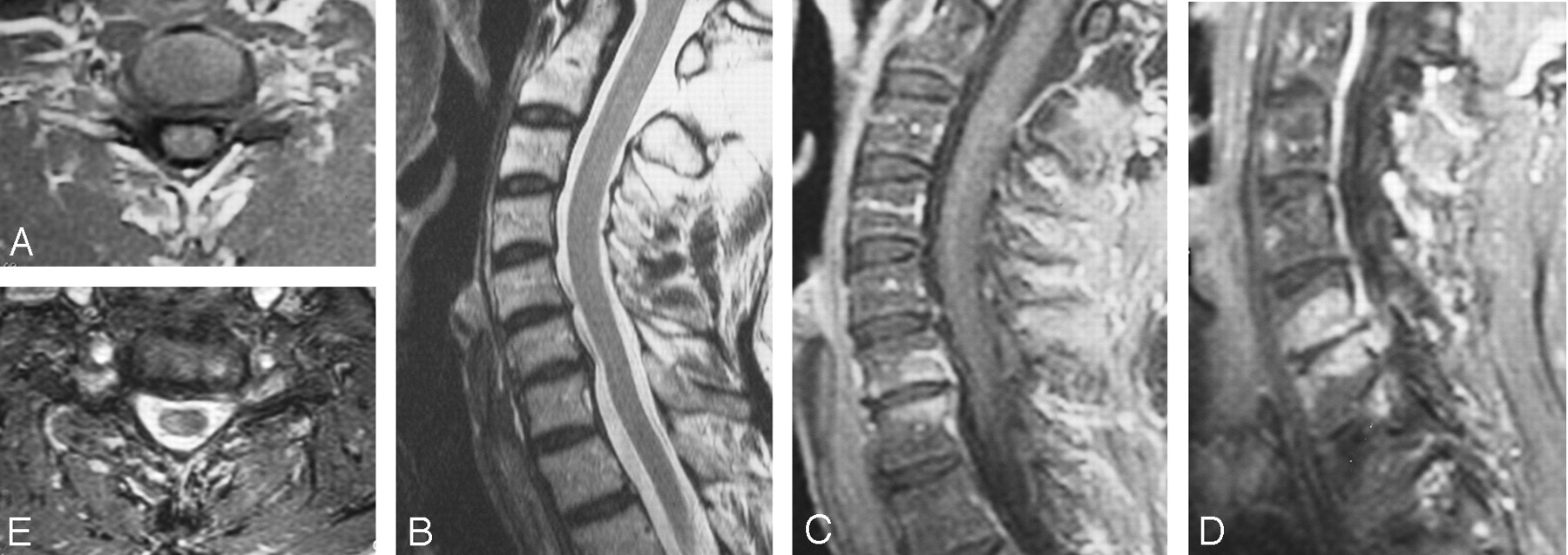
    A and B, The C6–7 disk before undergoing the ablation procedure because of a mostly lateral left intraforaminal herniation (A) with the associated mild central protrusion shown in a sagittal MR image (B).

    C and D, MR imaging follow-up at 2 months shows hypo-T1, hyper-T2 modifications of the adjacent vertebral bodies and evidence of an inflammatory condition.

    E, Partial reduction of the herniation is observed at the 2-month follow-up. The patient is completely asymptomatic despite only partial reduction of the disk herniation (see text for discussion).

Tables

  • Figures
    • View popup
    Table 1:

    Modified Macnab criteria used to grade overall clinical outcome

    GradeDefinition
    ExcellentComplete resolution of all symptoms and free of pain; no restriction of mobility; able to return to normal work and activities
    GoodMarked reduction of pain with the patient generally satisfied, returning to work or usual daytime activities, and taking analgesics seldom or not at all
    FairSome improved functional capacity; still handicapped and/or unemployed
    PoorContinued objective symptoms of root involvement; additional operative intervention needed at index level, irrespective of repeated operations or length of postoperative follow-up
    WorseClinical symptoms considered worse than before receiving procedure
    • View popup
    Table 2:

    Clinical outcomes according to the modified Macnab criteria at months 2 and 6

    ExcellentGoodFair or PoorTotal
    Macnab Grade: 2 Months25 (45%)19 (35%)11 (20%)55 (100%)
    Macnab Grade: 6 Months
        Excellent224026 (47%)
        Good311418 (33%)
        Fair or Poor0358 (15%)
        Not yet available0123 (5%)
    • View popup
    Table 3:

    Regression of disk herniation findings in patients who underwent postoperative MR imaging (n = 15)

    Postoperative Clinical Outcome (Macnab Grade)
    Regression of Disk HerniationGood or ExcellentPoor
        Yes (n = 6)51
        No (n = 8)53
    Diskitis complication (n = 1)1
    • View popup
    Table 4:

    Outcomes at month 2 stratified by selected demographic characteristics

    All (n = 55)Excellent (n = 25)Excellent or Good (n = 44)Fair or Poor (n = 11)P-value*
    Male55%52% (13)†57% (25)45% (5).574
    >45 years old31%24% (6)27% (12)45% (5).995
    Herniation direction
        Central27%24% (6)27% (12)27% (3).981
        Right42%40% (10)41% (18)45% (5).993
        Left35%36% (9)32% (14)27% (3).865
    >1 Disk treated38%24% (6)34% (15)55% (6).838
    Related pathology24%28% (7)23% (10)‡27% (3)§.803
    • Note:—Significance of variables determined using forward-entry binary logistic regression.

    • † (No. of patients).

    • ‡ Including: multiple sclerosis (good), osteophyte and partially calcified herniation (good), osteophytes (1 case excellent, 1 case good), carpal tunnel syndrome (2 cases, excellent), C5–C6 calcified disk herniation (excellent), celiac disease and anorexia (excellent), Scleroderma-Hashimoto thyroiditis (excellent), pulmonary embolism (excellent).

    • § Including: osteophytes with retrolisthesis C5–C6 (1 case), L5–S1 disk herniation already operated on 3 times (1 case), L4–L5 disk herniation (1 case).

PreviousNext
Back to top

In this issue

American Journal of Neuroradiology: 27 (10)
American Journal of Neuroradiology
Vol. 27, Issue 10
November 2006
  • Table of Contents
  • Index by author
Advertisement
Print
Download PDF
Email Article

Thank you for your interest in spreading the word on American Journal of Neuroradiology.

NOTE: We only request your email address so that the person you are recommending the page to knows that you wanted them to see it, and that it is not junk mail. We do not capture any email address.

Enter multiple addresses on separate lines or separate them with commas.
Plasma Radio-Frequency–Based Diskectomy for Treatment of Cervical Herniated Nucleus Pulposus: Feasibility, Safety, and Preliminary Clinical Results
(Your Name) has sent you a message from American Journal of Neuroradiology
(Your Name) thought you would like to see the American Journal of Neuroradiology web site.
CAPTCHA
This question is for testing whether or not you are a human visitor and to prevent automated spam submissions.
Cite this article
G. Bonaldi, F. Baruzzi, A. Facchinetti, P. Fachinetti, S. Lunghi
Plasma Radio-Frequency–Based Diskectomy for Treatment of Cervical Herniated Nucleus Pulposus: Feasibility, Safety, and Preliminary Clinical Results
American Journal of Neuroradiology Nov 2006, 27 (10) 2104-2111;

Citation Manager Formats

  • BibTeX
  • Bookends
  • EasyBib
  • EndNote (tagged)
  • EndNote 8 (xml)
  • Medlars
  • Mendeley
  • Papers
  • RefWorks Tagged
  • Ref Manager
  • RIS
  • Zotero
0 Responses
Respond to this article
Share
Bookmark this article
Plasma Radio-Frequency–Based Diskectomy for Treatment of Cervical Herniated Nucleus Pulposus: Feasibility, Safety, and Preliminary Clinical Results
G. Bonaldi, F. Baruzzi, A. Facchinetti, P. Fachinetti, S. Lunghi
American Journal of Neuroradiology Nov 2006, 27 (10) 2104-2111;
del.icio.us logo Twitter logo Facebook logo Mendeley logo
  • Tweet Widget
  • Facebook Like
  • Google Plus One

Jump to section

  • Article
    • Abstract
    • Materials and Methods
    • Results
    • Discussion
    • Conclusion
    • Acknowledgments
    • Footnotes
    • References
  • Figures & Data
  • Info & Metrics
  • Responses
  • References
  • PDF

Related Articles

  • No related articles found.
  • PubMed
  • Google Scholar

Cited By...

  • No citing articles found.
  • Crossref
  • Google Scholar

This article has not yet been cited by articles in journals that are participating in Crossref Cited-by Linking.

More in this TOC Section

  • Management Outcomes For VO Spine Biopsy
  • Cone Beam CT Myelography
  • Diagnostic Value of Brain WMH in SIH
Show more SPINE IMAGING AND SPINE IMAGE-GUIDED INTERVENTIONS

Similar Articles

Advertisement

Indexed Content

  • Current Issue
  • Accepted Manuscripts
  • Article Preview
  • Past Issues
  • Editorials
  • Editor's Choice
  • Fellows' Journal Club
  • Letters to the Editor
  • Video Articles

Cases

  • Case Collection
  • Archive - Case of the Week
  • Archive - Case of the Month
  • Archive - Classic Case

More from AJNR

  • Trainee Corner
  • Imaging Protocols
  • MRI Safety Corner
  • Book Reviews

Multimedia

  • AJNR Podcasts
  • AJNR Scantastics

Resources

  • Turnaround Time
  • Submit a Manuscript
  • Submit a Video Article
  • Submit an eLetter to the Editor/Response
  • Manuscript Submission Guidelines
  • Statistical Tips
  • Fast Publishing of Accepted Manuscripts
  • Graphical Abstract Preparation
  • Imaging Protocol Submission
  • Evidence-Based Medicine Level Guide
  • Publishing Checklists
  • Author Policies
  • Become a Reviewer/Academy of Reviewers
  • News and Updates

About Us

  • About AJNR
  • Editorial Board
  • Editorial Board Alumni
  • Alerts
  • Permissions
  • Not an AJNR Subscriber? Join Now
  • Advertise with Us
  • Librarian Resources
  • Feedback
  • Terms and Conditions
  • AJNR Editorial Board Alumni

American Society of Neuroradiology

  • Not an ASNR Member? Join Now

© 2025 by the American Society of Neuroradiology All rights, including for text and data mining, AI training, and similar technologies, are reserved.
Print ISSN: 0195-6108 Online ISSN: 1936-959X

Powered by HighWire