
Article Figures & Data
Figures
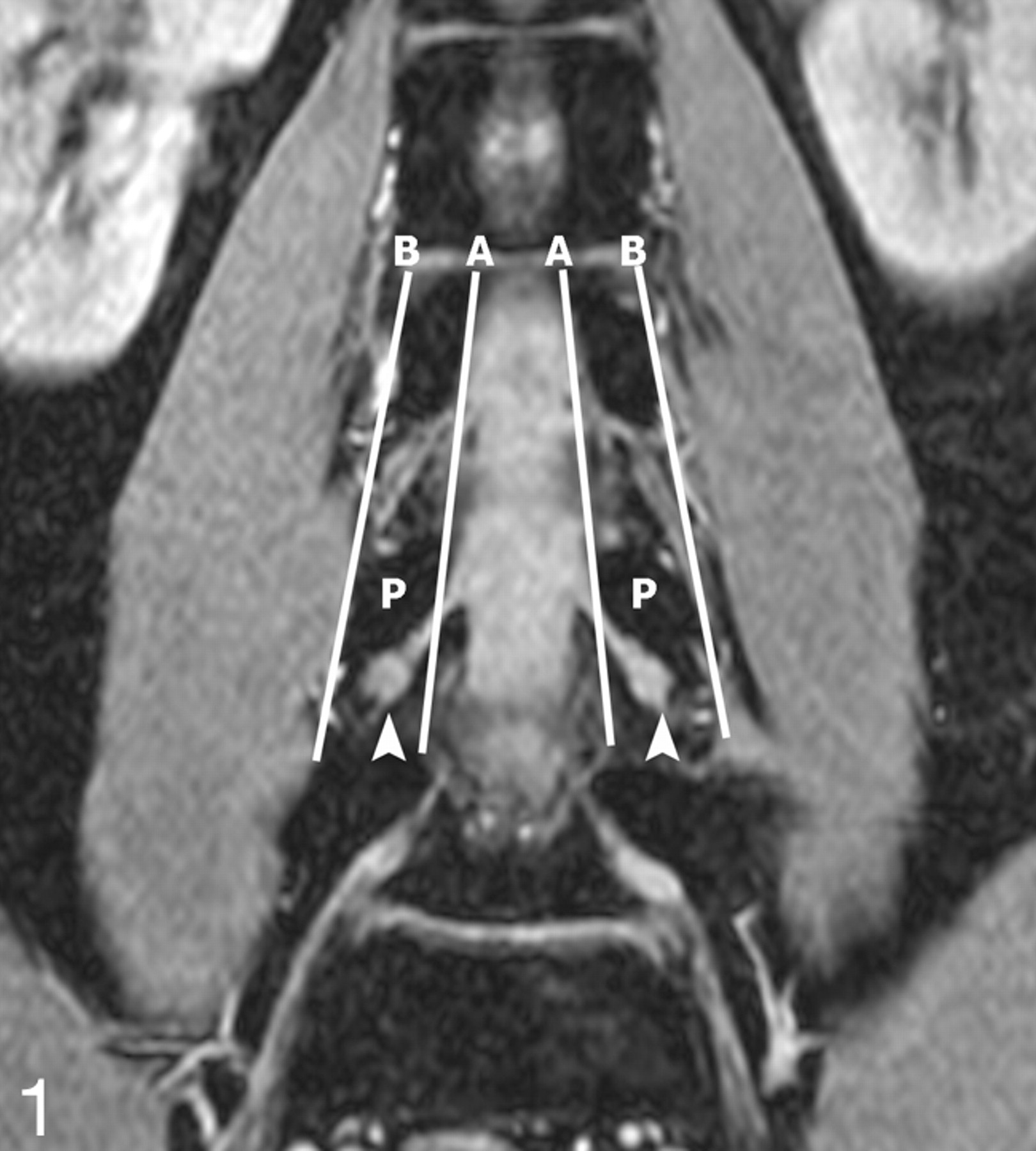
- Fig 1.
Determination of DRG position. The position of the DRG was determined on the original coronal FFE images. A and B are the lines respectively connecting the medial and lateral borders of the pedicles. If the midpoint of the DRG lies proximal to A, it is an intraspinal type; between A and B, a foraminal type; and distal to B, an extraforaminal type. In this 28-year-old volunteer, the L4 DRGs are the foraminal type (arrowheads). P indicates the L4 vertebral pedicles.
- Fig 2.
Lumbar DRGs on coronal MIP imaging. The reconstructed MIP image clearly depicts the DRGs of the spinal nerve bilaterally from L1 to L5, which demonstrates intermediate signal intensity (arrowheads indicate DRGs from L4 to L1; arrows indicate L5 DRGs).
- Fig 3.
Architecture of DRGs. The singular right L5 DRG (arrow) and the biganglia right L4 DRG (arrowheads) are clearly demonstrated on the MIP image with an angle of 24° rotated to the right. The dimension of the DRGs on this plane is maximal, and the interference of adjacent vessels could be removed. Whether in biganglia or singular ganglion, a high intensity rim could be noted.
- Fig 4.
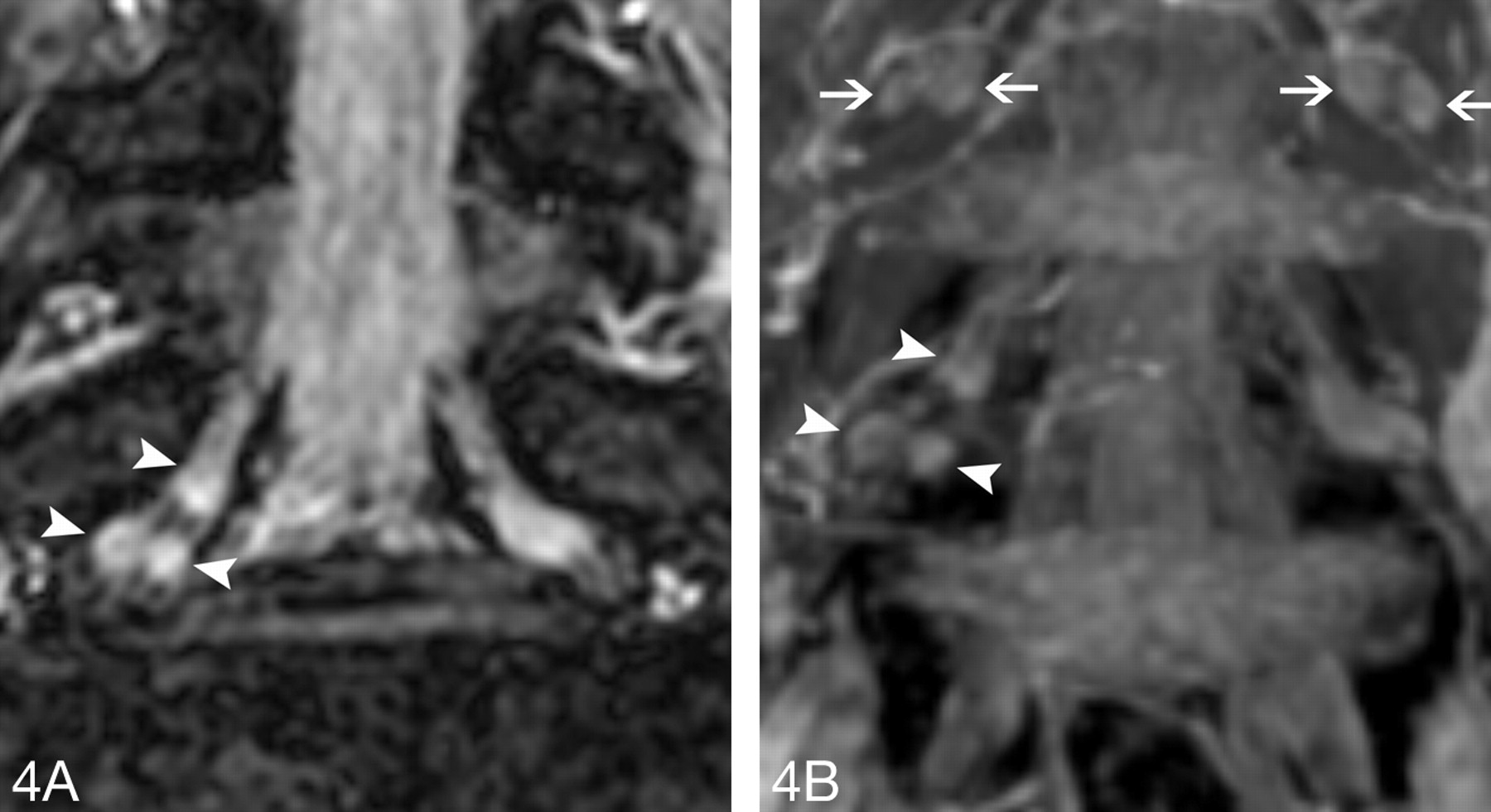
Triganglia architecture of L5. The right L5 DRG, with a composition of 3 separate ganglia, is defined as the triganglia.
A, The original FFE coronal image distinctly demonstrates the triganglia architecture of the right L5 DRG (arrowheads).
B, The MIP coronal image also clearly shows the triganglia architecture of the right L5 DRG (arrowheads). At same time, the biganglia architecture could be found in the L4 DRGs bilaterally (arrows).
- Fig 5.
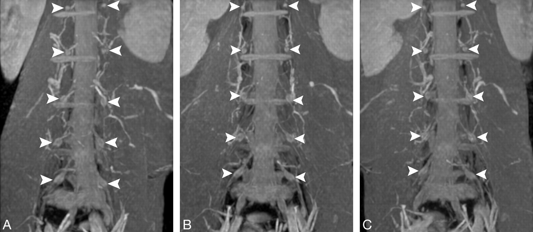
Lumbar DRGs on serial coronal MIP images. All 5 pairs of DRGs of the spinal nerves from L1 to L5 (arrowheads) are well displayed on serial views. Although some segmental and radicular vessels surrounding the DRGs are also displayed, the DRGs could be distinguished from these vessels by the rotation of the serial images with different angulations.
A, Left antero-oblique view.
B, Frontal view.
C, Right antero-oblique view.


Tables
Intraspinal* Foraminal Extraforaminal Total L1 Female 0 (0) 121 (99.2) 1 (0.8) 122 Male 0 (0) 108 (100) 0 (0) 108 Combined 0 (0) 229 (99.6) 1 (0.4) 230 L2 Female 0 (0) 121 (99.2) 1 (0.8) 122 Male 0 (0) 107 (99.1) 1 (0.9) 108 Combined 0 (0) 228 (99.1) 2 (0.9) 230 L3 Female 0 (0) 120 (98.4) 2 (1.6) 122 Male 0 (0) 105 (97.2) 3 (2.8) 108 Combined 0 (0) 225 (97.8) 5 (2.2) 230 L4 Female 0 (0) 122 (100) 0 (0) 122 Male 0 (0) 108 (100) 0 (0) 108 Combined 0 (0) 230 (100) 0 (0) 230 L5 Female 9 (7.4)† 113 (92.6) 0 (0) 122 Male 4 (3.7)† 104 (96.3) 0 (0) 108 Combined 13 (5.7) 217 (94.3) 0 230 Total 13 1129 8 1150 Note:—Data in parentheses are percentages calculated on the basis of the total of each sex. DRG indicates dorsal root ganglion.
* There was statistically significant difference in the distribution of DRG position among the different levels (χ2 = 53.687, P < .0001).
† At level L5, there was no statistical difference in the incidence of intraspinal ganglia of DRG between women and men (Fisher exact test, P = .265).
Level of DRG L5 L4 L3 L2 L1 Width (mm) Female 6.22 ± 0.86 5.75 ± 0.84 5.25 ± 0.91 4.35 ± 0.88 3.23 ± 0.75 Male 6.60 ± 0.93 5.92 ± 1.03 5.49 ± 1.00 4.69 ± 0.86 3.55 ± 0.75 Combined 6.40 ± 0.91 5.83 ± 0.94 5.37 ± 0.96 4.51 ± 0.88 3.38 ± 0.77 P value* 0.025 (2.270)† 0.331 (0.975) 0.189 (1.323) 0.038 (2.099)† 0.026 (2.254)† Length (mm) Female 11.55 ± 2.45 8.47 ± 1.45 7.05 ± 1.41 5.66 ± 1.03 4.14 ± 0.88 Male 11.61 ± 2.02 8.82 ± 1.52 7.36 ± 1.30 6.07 ± 1.17 4.58 ± 0.85 Combined 11.58 ± 2.25 8.64 ± 1.49 7.20 ± 1.36 5.85 ± 1.11 4.35 ± 0.89 P value* 0.884 (0.147) 0.200 (1.288) 0.222 (1.229) 0.044 (2.037)† 0.007 (2.749)† Width/Length Female 0.28 ± 0.06 0.35 ± 0.05 0.38 ± 0.04 0.39 ± 0.04 0.39 ± 0.05 Male 0.29 ± 0.05 0.34 ± 0.06 0.37 ± 0.04 0.39 ± 0.04 0.39 ± 0.04 Combined 0.28 ± 0.05 0.34 ± 0.06 0.39 ± 0.04 0.39 ± 0.04 0.39 ± 0.05 P value* 0.225 (1.221) 0.630 (0.483) 0.739 (0.334) 0.565 (0.577) 0.601 (0.525) Note:—Data are presented as mean ± SD. DRG indicates dorsal root ganglion.
* t test between women and men (t values in parentheses).
† The width and length of the L1 and L2 DRGs, and the width of the L5 DRGs differ statistically between men and women.
Singular Biganglia Triganglia Total P value L1 Female 119 (97.5) 3 (2.5) 0 (0) 122 0.058* Male 100 (92.6) 8 (7.4) 0 (0) 108 Combined 219 (95.2) 11 (4.8) 0 (0) 230 L2 Female 93 (76.3) 27 (22.1) 2 (1.6) 122 0.324 [2.254]† Male 74 (68.5) 33 (30.6) 1 (0.9) 108 Combined 167 (72.6) 60 (26.1) 3 (1.3) 230 L3 Female 66 (54.1) 56 (45.9) 0 (0) 122 0.34* Male 61 (56.5) 45 (41.7) 2 (1.8) 108 Combined 127 (55.2) 101 (43.9) 2 (0.9) 230 L4 Female 52 (42.7) 68 (55.7) 2 (1.6) 122 0.043*‡ Male 61 (56.5) 47 (43.5) 0 (0) 108 Combined 113 (49.1) 115 (50.0) 2 (0.9) 230 L5 Female 101 (82.8) 18 (14.8) 3 (2.4) 122 0.453 [1.586]† Male 95 (88.0) 12 (11.1) 1 (0.9) 108 Combined 196 (85.2) 30 (13.0) 4 (1.8) 230 Total 925 255 10 1150 Note:—Data in parentheses and bracket are percentages calculated on the basis of the total of each sex and χ2 values, respectively. DRG indicates dorsal root ganglion.
* Fisher exact text.
† Likelihood ratio test.
‡ The incidence of the bi- or triganglia differs statistically between female and male patients.
Level of DRGs L1 L2 L3 L4 L5 Ganglia position* NA NA 1.000 NA 1.000 Signal intensity* 0.727 0.453 0.289 0.500 0.500 Width† 0.389 (0.865) 0.476 (0.716) 0.259 (1.134) 0.834 (0.210) 0.206 (1.271) Length† 0.641 (0.467) 1.00 (0) 0.298 (1.044) 0.214 (1.250) 0.860 (0.176) Width/Length† 0.610 (0.512) 0.339 (0.960) 0.751 (0.319) 0.639 (0.470) 0.332 (0.974) Architecture* 0.453 0.065 0.435 0.876 0.407 Note:—Data are calculated P values. DRG indicates dorsal root ganglion.
* McNemar test. NA, the data is not applicable for the McNemar test because only 1, 2, and 0 pair DRGs are different in the position, respectively, at L1, L2, and L4 levels.
† Paired t test. Data in parentheses are t values.


